© Author: A. Olesya Valerievna, candidate of medical sciences, practicing physician, teacher at a medical university, especially for SosudInfo.ru (about the authors)
Peripheral circulation plays a vital role in providing tissues with nutrients, removing metabolic products from them, and delivering oxygen. Microcirculation vessels receive arterial blood from the pulmonary circle and return venous blood, saturated with carbon dioxide and catabolic products.
Peripheral vessels include small arteries and veins, arterioles and venules, capillaries of the microvasculature, which have a small diameter and a specifically designed wall, which allows not only substances, but also cells to penetrate through them. Without this link of microcirculation, normal metabolism and maintenance of tissue functioning would be impossible.
The peripheral bloodstream receives arterial blood from larger arteries, where it is pumped by the heart from the lungs. After passing through the microvasculature, the blood becomes venous, is sent to the veins, reaches the right ventricle of the heart and is sent to the lungs for gas exchange, which also occurs with the direct participation of small arteries and veins.
In addition to the metabolic function, microcirculation is needed to maintain body temperature. In a hot room, in the sun, the body begins to overheat, and then small vessels expand and accelerate the evaporation of liquid. In the cold, the opposite happens: the blood vessels narrow, preventing evaporation and retaining heat.
The peripheral circulation contains the bulk of the blood in the human body and affects blood pressure and heart rate, regulating them so that in the event of trouble, vital organs receive the nutrition they need.
Disorders of peripheral circulatory function include decreased blood flow velocity and thrombosis, embolic syndrome, blood stasis, various types of hyperemia and anemia. These processes can combine with each other, exacerbating ischemic and degenerative processes in tissues.
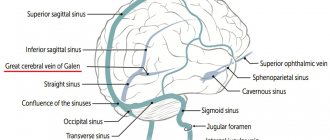
Signs of disturbances in the peripheral blood flow are quite stereotypical and appear not only in the skin, when it is easier to suspect pathology, but also in internal organs, especially those with a developed microcirculation network (liver, kidneys, lungs, brain).
If peripheral circulation is impaired, the cause should be sought and, if possible, eliminated. For this purpose, a variety of medications are used to help normalize coagulation, blood fluidity, and its cellular composition.
How does the microcirculation system work?
The vessels providing peripheral blood flow include:
- Small arteries and arterioles;
- Capillaries;
- Venules and small veins;
- Arteriovenular anastomoses;
- Lymphatic vessels.
Venules, arterioles, capillaries and anastomoses between them constitute the main link of microcirculation, ensuring metabolic processes. Vascular resistance, and therefore blood pressure, is maintained by small arteries, arterioles and precapillary sphincters. The exchange occurs in capillaries and post-capillary venules, and the capacitive part of the blood flow consists of venules and small veins, which contain the largest amount of all human blood.
The connection between the arterial and venous parts of the systemic blood flow is carried out by special anastomoses (shunts), which are activated in case of trouble. Through anastomoses, blood flows from the arterioles directly into the venules, and the microcirculation does not receive enough of it. This mechanism forms the basis for the centralization of blood circulation, necessary for redirecting blood to vital organs (brain, myocardium, kidneys), which is clearly manifested in shock.
Arterioles are small precursor vessels to capillaries. Their peculiarity is the presence of smooth muscle cells in the walls, due to which the vessels are able to contract and relax, changing the diameter of the lumen. Changes in arteriolar diameter can occur both locally and throughout the body. Arterioles provide total peripheral resistance, which determines blood pressure levels.
Capillaries continue into venules , through which blood flows out from the microvasculature. The muscular layer of their walls is much less developed than in the arterioles, therefore the wall of these vessels is thinner and is not able to react with a strong spasm under pathological conditions, but the process of expansion and stagnation here occurs easier and faster.
The intermediate link between the arteriole and venule is the capillary - the thinnest vessel of the human body, which carries out the metabolic role. Transport of substances into tissues and back into capillaries is possible thanks to the single-layer wall of the latter, which consists only of endothelium and can have many pores and fenestrae (in the liver, bone marrow, lymphatic tissue).
The work of peripheral circulation is regulated by the nervous and endocrine systems and depends on the action of vasoactive metabolites and other chemicals. In response to stimulation of sympathetic nerve fibers, microcirculatory vessels constrict due to the action of adrenaline and similar metabolites. Vasodilators (histamine) have the opposite effect.
The expansion of the peripheral vascular network occurs under the influence of the parasympathetic nervous system, the main neurotransmitter in this case is acetylcholine. In addition to nervous regulation, the humoral mechanism plays a major role in vasodilation. Thus, hyperkalemia, excess sodium and magnesium, accumulation of acidic metabolic products (acidosis), inflammatory mediators (histamine, bradykinin) provoke a sharp expansion of the vascular network, while catecholamines (adrenaline), the hormone vasopressin, angiotensin and other substances form vasospasm with a decrease microvasculature capacity.
Humoral mechanisms are realized more slowly than the direct influence on the vascular walls from the nerve fibers. In addition, the venous bed responds better to nervous regulation than the resistive arterial bed.
Anatomy and physiology of the microvasculature
The term microcirculation in the broad sense of the word understands not only blood flow and lymph flow in microvessels, but also metabolic processes occurring through the wall of microvessels, as well as interstitial (extravascular) transport of fluid and the substances, cells and various structures it contains.
Microvessels include:
- vessels of the arteriolar type - arterioles, metarterioles, precapillaries and capillaries with a diameter not exceeding 100 microns;
- vessels of the venular type - postcapillaries, venules, the diameter of which does not exceed 200 microns;
- lymphatic microvessels - lymphatic capillaries, postcapillaries and microvessels with a diameter of no more than 300 microns;
- anastomoses (shunts) connecting two arterioles (arteriolo-arteriolar), two venules (venulo-venular), an arteriole with a venule (arteriolo-venular).
The main difference between microvessels and macrovessels is that, in addition to transport, they perform an exchange function. It should be noted that metabolism occurs through the wall of all microvessels - from arteriole to venule - and not only through the wall of capillaries, as previously assumed. A feature of lymphatic macro- and microvessels is a constant two-way exchange from tissue to vessel and from vessel to tissue throughout the entire length until the final section of the thoracic lymphatic duct flows into the right venous angle and into the right atrium. In this regard, the composition of lymph in each section of the lymphangion (the distance between adjacent valves of the lymphatic vessel) is different. The increased permeability of lymphatic microvessels is explained by the thin structure of their wall, which does not have a continuous basement membrane, and in lymphatic capillaries and post-capillaries there is no basement membrane. Their wall consists of a single row of imbricated endothelial cells. Interendothelial channels freely pass not only individual cells, but also entire conglomerates, which eliminates the need for lymphovenous anastomoses.
One of the important structures of the microvasculature is the precapillary sphincter (Fig. 1), which is a section of the precapillary containing two smooth muscle cells located at the beginning of the precapillary. Only one red blood cell can pass through it without deformation. In narrower capillaries with a diameter of about 5 microns, the red blood cell is necessarily deformed, stretching 3-7 times in length (depending on the speed of blood flow and the pressure gradient in the capillary). The shape of the red blood cell can be used to judge the speed of blood flow in the microvessel. At high speeds, the cells are elongated; at low speeds, the red blood cells take on a more rounded shape (Fig. 2).
The reason why we dwell in detail on the structure of the precapillary sphincter is explained by the importance of its role in the development of pathology. Smooth muscle cells of the precapillary sphincter are hypersensitive to catecholamines (adrenaline, norepinephrine). For example, the sensitivity of a precapillary sphincter smooth muscle cell to adrenaline is 100 times greater compared to a similar arteriole cell with a diameter of 50 μm and 50 times greater compared to a similar arteriole cell with a diameter of 20 μm (see Fig. 1).
This feature of the smooth muscle cells of the precapillary sphincter is of particular importance. Under conditions of acute stress, the release of a significant amount of catecholamines is accompanied by spasm of the coronary and other vessels. Acute pain in the heart area signals the need to take a vasodilator such as nitroglycerin. Everyone knows this: both the patient and the doctor.
A completely different situation develops under conditions of chronic stress, by which we mean conditions accompanied by a person’s long-term dissatisfaction with the reality around him: long-term worries due to the loss of loved ones or loss of a job; conflicts in the family or work team, constant lack of time, insufficient sleep, lack of positive emotions and much more that causes irritation in a person. In these situations, the release of catecholamines will be insignificant: there will be no pain attack, disturbances in central hemodynamics, blood pressure will remain within normal limits. However, the hypersensitive cells of the precapillary sphincter will respond by contracting even to minute doses of catecholamines. Its lumen will decrease, and not a single red blood cell will be able to pass through the precapillary sphincter. The tissues that are supplied with blood through capillary networks will continue to receive plasma flow, but will not receive the oxygen carried by red blood cells. As a consequence of prolonged narrowing of the precapillary sphincter in response to small doses of catecholamines in the blood under conditions of chronic stress, chronic hypoxia of tissues and organs supplied by the capillary networks will occur.
Figure 2. Changes in the shape of an erythrocyte at different blood flow rates in the microvessels of the mesentery of the small intestine of a rat. Elongated red blood cells in the capillary (3) and arteriole (5) at normal blood flow speed; oval shape - with sharply slowed blood flow (4); “square” red blood cells when blood flow slows down (1, 2). Biomicroscopy. Magnification: vol. x 70, approx. x 3.
The future scenario is far from the most optimistic: from a decrease in the functional activity of organs to the development of various diseases, such as cancer. Under hypoxic conditions, cells degrade faster; cells of the immune system perceive them as foreign and phagocytose. The rapid death of cells that have not reached maturity and the increased growth of new ones, which are also soon destroyed, will lead to malignancy of cells and a tumor process.
Types of peripheral circulatory disorders
Pathologies of peripheral circulation include:
- Slowing down or accelerating the flow of fluid through microcirculation vessels;
- Blood shunting with centralization of blood circulation;
- Stasis, sludge phenomenon and thrombosis;
- Plasma impregnation and plasmorrhagia;
- Plethora;
- Embolism;
- Anemia.
An acceleration or decrease in peripheral blood flow usually reflects compensatory reactions aimed at maintaining metabolism in unfavorable conditions. For example, at the beginning of inflammation, blood vessels dilate, and the transport of substances and cells occurs more actively, and then the blood flow slows down to localize the source of pathology. With an increase in body temperature, tachycardia, anemia, blood circulation also becomes more intense.
Heart defects with its insufficiency, hypothermia, congestion are accompanied by a slowdown in blood flow, stagnation, release of the liquid part into the intercellular space, and the formation of edema. These processes already reflect the pathology of peripheral circulation.
Blood shunting is aimed at supplying power to life support systems - the central nervous system, myocardium, and kidneys. This mechanism is most clearly represented during shocks, when blood is discharged from the arteries into the veins, bypassing the microcirculatory bed. Of course, peripheral tissues are “scarred” to a certain extent, but such a forced measure allows for survival.
blood stasis in microcirculatory vessels
Stasis and sludge phenomena occur when there is a violation of the rheological properties of blood, a decrease in peripheral blood flow, metabolic, electrolyte disorders, thrombosis and congestion. Stasis is a stop of blood flow in microcirculation vessels. It has a complex mechanism and depends on a number of reasons (hemocoagulation, blood pressure, blood shunting, the action of toxins, an inflammatory component, etc.), but the main one is an increase in blood cell aggregation. Short-term stasis is reversible, long-term stasis promotes ischemia and necrosis.
The sludge phenomenon is a disturbance of peripheral circulation when blood cells, mainly red blood cells, stick together and formation of cellular and protein aggregates occurs in the lumens of small vessels. It accompanies stasis, continuing it, and manifests itself during an inflammatory reaction, injury, infections, increased blood viscosity, venous and arterial hyperemia, and heart failure.
In parallel with stasis, sludge and dilation of microcirculation vessels, plasma impregnation develops, when the permeable vascular wall is infiltrated by plasma components, and plasmorrhagia with the release of blood components into the surrounding perivascular space. These changes are observed in arterial hypertension, systemic connective tissue diseases, and immunopathological processes.
Thrombosis is intravital blood clotting in the chambers of the heart and the lumens of blood vessels with the formation of dense clots. The main factors of thrombosis are considered to be trauma to the vascular wall, stasis and increased aggregation, which are combined during thrombosis.
Thrombosis is observed in varicose veins, heart failure, arrhythmias, inflammation, severe infections, disseminated intravascular coagulation syndrome, shock, hereditary thrombophilia, venous congestion, implanted heart valves and many other pathological conditions.
Large red, white and mixed clots are formed more often in vessels of large diameter, while in the microvasculature the so-called hyaline thrombi, consisting of destroyed cellular fragments, platelets and fibrin protein, become important.
Hyaline thrombi are formed mainly during disseminated intravascular coagulation, which occurs during shock and terminal conditions. Blockade of peripheral circulation by hyaline thrombi forms the basis of acute multiple organ (liver, renal, respiratory) failure, which can cause death against the background of acute ischemic and necrotic processes in parenchymal organs.
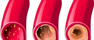
Anemia is a disorder of peripheral circulation when tissues experience a deficiency of arterial supply due to vascular spasm, compression from outside (tourniquet, neoplasm, scar) or obstruction inside (thromb, embolus, fatty protein plaque).
With anemia, the intensity of blood flow in the capillaries decreases, some of the vessels are reduced, blood cells are redistributed, and the vessels contain mainly plasma. In the parenchyma of organs during prolonged ischemia, dystrophic and atrophic phenomena are observed, fibrous tissue grows, and with acute disruption of blood supply, necrosis develops.
Another type of pathology of peripheral circulation is plethora , which can be arterial and venous. The first type is associated with excessive flow of arterial blood into the microcirculatory bed, the second - with insufficient outflow of venous blood.
Pathological arterial hyperemia is characteristic of inflammatory processes, increases in ischemic tissues after restoration of blood circulation, and is observed with a sharp dilation of blood vessels due to a disorder in the nervous regulation of their tone, due to blood redistribution.
Venous congestion is characterized by impaired outflow of venous blood due to thrombosis, heart failure, compression of the veins by a neoplasm, scar tissue, or a tourniquet. Venous blood accumulates in the microcirculation system, fluid sweats into the tissue with the development of edema, dystrophy progresses in the parenchymal elements, and necrosis is possible. Chronic venous hyperemia leads to compaction of organs due to sclerosis and atrophy.
Embolism is the circulation in the bloodstream of elements that are not normally found there. They clog small vessels and disrupt the movement of blood through them. Embolism can be fat (for fractures), gas, air, tissue (for tumors), microbial (at the basis of sepsis).
emboli in the bloodstream
Disturbances of microcirculation under stress
Microcirculation in microvessels under normal and pathological conditions, including stress, is regulated by several mechanisms. In particular, it depends on:
- signals from the central nervous system through adrenergic (mainly) and cholinergic (larger vessels) innervation;
- reflex regulation;
- peptidergic (opioidergic) regulation of lymphatic microvessels;
- local regulation (metabolic products, mast cells);
- central hemodynamics;
- microlymphocirculation;
- rheological properties of blood;
- water-electrolyte metabolism of perivascular tissue;
- humoral regulation.
Thus, microcirculation is regulated by many factors. These same factors, as well as lymph flow factors, regulate microlymphocirculation. Such multifactorial and complex regulation indicates the important role of microcirculation in maintaining homeostasis in the body. At the same time, microhemodynamics regulates the functional activity of all organs and tissues, even those that lack microvessels. For example, the lens and eyeball lack lymphatic and blood microvessels. The trophic supply of such organs and their release from metabolic products is carried out through the surrounding tissue (interstitial, intercellular space), in which microvessels are located. Interstitial transport reflects the state of microcirculation remote from such organs.
Under stress, various systems of the body are involved in the pathological process, since the central nervous system controls them always and very effectively. A few experimental lifetime studies of stress [2 – 4] have shown that a single exposure to extreme stimuli of different nature and duration causes the same type of disturbances in microvessels in different animal species. Changes occur in three areas of the microvascular bed:
- inside microvessels;
- in the wall of microvessels;
- in the extravascular space.
Intravascular microcirculation disorders consisted of a slowdown in blood flow up to a complete stop (stasis), aggregation (sticking together) of erythrocytes in the form of “coin columns” (Fig. 3), and plasmatization of blood vessels. Microvessels filled with plasma without erythrocytes and leukocytes are called plasmatic, which is often associated with spasm of the vessels from which blood flows into this plasmatic vessel. Large blood cells cannot pass through a narrowed vessel; only platelets pass through, since their sizes are much smaller. Plasmatization of blood vessels is possible when the pressure gradient between the beginning and end of the vessel changes.
| Figure 3. Aggregation of erythrocytes (“coin columns”) in the microvessel of the mesentery of the small intestine of a rat during pathology. Biomicroscopy. Magnification: vol. x40, approx. x7. |
At the level of the microvessel wall, an increase in vascular permeability occurred (Fig. 4), caused by exocytosis and degranulation of mast cells located in the perivascular tissue along the microvessels. In the extravascular space, an increase in the number of mast cells was noted along with the destruction of their membrane. The initial response of mast cells to extreme exposure was to increase histamine secretion by exocytosis. Later, degranulation of mast cells occurred with the release into the tissue of a huge number (more than 35) of biologically active substances involved in the regulation of vascular tone, the permeability of the microvascular wall, the rheological properties of blood, etc. The release of catecholamines from the adrenergic nerve endings entwining and innervating the microvessels increased. The number of arteriole-venular anastomoses increased, which is considered as an adaptive response of the microvasculature to damage.
The time of normalization of blood flow in microvessels in the post-stress period depended on the nature and duration of the stressor and correlated with the state of mast cells. The correlation is direct: after the destruction of the membrane, the biologically active substances of mast cells act on the wall of microvessels, penetrate into the lumen of the microvessels and affect the rheological properties of the blood. The more mast cells are degranulated, the more microcirculatory disturbances there are. With biomicroscopy, mast cells are visible located along the microvessels, the reaction of the vessel is visible - expansion, plasmatization, swelling of the wall (swelling of the nuclei of endothelial cells of the wall narrows the lumen of microvessels, especially capillaries).
Acute and chronic stress differ mainly in the intensity and duration of the stressor. The response of the circulatory system to these types of stress is also different. Acute stress results in acute cardiovascular diseases: heart attacks, strokes, hypertensive crises, exacerbations of chronic diseases, death, etc.
Acute stress can turn into chronic. The latter causes chronic microcirculation disorders and in the future can lead to cancer. In this article, we deliberately focus on chronic stress, since it often has a long-term everyday nature and is devoid of immediate bright manifestations. This allows it to be ignored or not given enough importance, which is very dangerous. Cancer prevention should be done as early as possible.
Figure 4. Increase in the permeability of the microvessel wall of the rat small intestine mesentery in response to the application of bradykinin (1 μg/0.1 ml). Biomicroscopy. Magnification: vol. x20, approx. x7.
Manifestations of microcirculation disorders
Symptoms of peripheral circulatory disorders depend on the type of pathology, the nature of the course, the speed of development and the compensatory capabilities of the body. The symptoms of the pathology are extremely diverse and there is no particular point in trying to systematize it, because ischemia in the nervous tissue and legs will manifest itself differently, while thrombus formation in the vessels of the microcirculation of the kidneys and acute venous congestion in them can occur very similarly.
Common to all peripheral circulatory disorders:
- Possibility of acute or chronic course;
- The development of necrosis, hemorrhages, edema and, as a consequence, pain and disruption of the organ in case of acute microcirculation disturbance;
- The predominance of ischemic-dystrophic changes, atrophy and sclerosis in chronic conditions.
hyperemia (plethora)
Arterial hyperemia is characterized by redness of a tissue area, an increase in its temperature and size due to edema. As a rule, pathological arterial plethora is also accompanied by pain. These processes can be clearly observed during inflammation in visible areas of the body. When internal organs are damaged with symptoms of hyperemia, patients usually feel pain, and other symptoms are associated with the disease that occurs with this type of peripheral circulatory disorder.
Venous stagnation is accompanied by:
- Cyanosis (blueness) of the skin, mucous membranes;
- In the zone of venous hyperemia, the temperature decreases (the limbs become cold, but not the internal organs);
- An increase in the volume of a limb or internal organ due to edema;
- Pain, a feeling of fullness, itching on the skin, possible formation of trophic ulcers;
- Internal organs: lungs - the appearance of wheezing, possible cough and congestive pneumonia, liver - increase in size, heaviness in the hypochondrium, dyspepsia, brain - headaches, impaired memory and intelligence.
leg ischemia
Ischemia (anemia) can occur in acute or chronic form. Ischemic changes in the extremities are accompanied by pain, rapid fatigue during exercise, a feeling of coldness, crawling “goosebumps”, the skin becomes pale, and the development of trophic disorders, including ulcers, is possible.
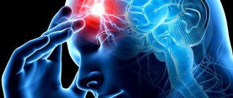
In the brain, ischemia underlies discirculatory encephalopathy with corresponding neurological and psychiatric symptoms, and acute ischemia, turning into necrosis, is the basis of cerebral infarction (stroke) with paresis and paralysis.
Ischemia of the renal cortex, as well as thrombus formation in the microvasculature of the organ, contribute to epithelial necrosis and the development of acute renal failure. Chronic venous stasis or prolonged ischemia provoke sclerotic and atrophic changes with a possible outcome in chronic insufficiency.
Skin microcirculation in normal and pathological conditions
Intravital studies of microcirculation in the middle layers of the skin of the rat's back allowed us to clarify the anatomy of the vascular bed. This opportunity appeared thanks to the new research method we developed [1]. It was previously believed that the dermis contained a large number of arteriovenular anastomoses. It turned out that there is not a single such anastomosis, but there are many venulo-venular anastomoses. This anatomical feature is not accidental. The skin has many functions, among which two main ones can be distinguished: protective and storage (the skin is a blood storage organ). Low metabolism in the skin compared to other organs and the function of depositing large volumes of blood do not imply the presence of arteriolo-venular anastomoses.
Another feature of the skin is that it ranks first among other organs and tissues in terms of the density of lymphatic microvessels. In fact, the human body is protected from pathogenic environmental influences by a powerful lymphatic membrane. Studies have shown that stimulation of lymph flow in the body with the help of direct lymphostimulants of a peptide nature in the event of ultraviolet damage to blood microvessels with the development of stasis can restore microhemocirculation in the dermis and subcutaneous fatty tissue [1]. Subsequent testing of the effectiveness of lymphostimulants applied as an ointment to people's facial skin showed a rejuvenating effect. Thus, it can be assumed that any harmless methods of stimulating lymph flow will restore not only skin turgor, which is especially important in cosmetology, but also all its functions, which deteriorate with age, with dermatological diseases and various extreme exposures.
Since ancient times, massage has been an effective cosmetic remedy, the effect of which is largely determined by the stimulation of lymph flow. Nowadays, bilateral limb massage for neurogenic pathology works wonders. Moreover, stimulation of lymph flow, as experiments have shown, prevents the death of animals with cerebral ischemia [6] and increases survival in acute pulmonary edema by 2-2.5 times.
Consequently, by improving lymph and blood circulation, we relieve the symptoms of chronic stress and prevent the development of “adaptation diseases”. And by stimulating microcirculation in the skin, we not only have a positive effect on this organ, preventing the appearance of skin diseases and aesthetic defects and slowing down the processes of age-related involution, but also normalize the vital processes of the entire organism. In our opinion, in modern medicine this approach has not yet received enough attention. It is necessary to widely use various methods of stimulating the lymph flow of the skin to improve the health of the population. One of the factors that significantly reduces lymph and blood circulation in the dermis and hypodermis, without a doubt, is stress. Microcirculation in these areas is disrupted by both acute and chronic stress; we can observe these changes even externally. Pay attention to the face of a person who has been stressed or suffering from a serious illness. A sallow complexion indicates poor condition of internal organs and is a reflection of prolonged hypoxia of the body. When very frightened, a person turns pale. This is due to reflex vascular spasm, which under conditions of acute stress can be aggravated by the redistribution of blood flow between organs and further increase skin ischemia.
Pharmacological principles of stress treatment
Drug treatment for stress involves the use of the following drugs:
- various tranquilizers that have, along with therapeutic side effects, unfavorable properties;
- adrenoblockers and adrenolytics;
- activators of central and peripheral anti-stress mechanisms (GABA, antioxidants, prostaglandins, adenosine);
- blockers of mechanisms of damage to cells and non-cellular structures (impaired energy supply, damage to membranes, enzymes, genetic apparatus of cells, imbalance of ions and water, disorder of local regulatory mechanisms).
Non-drug ways to relieve chronic stress
The task of a cosmetologist, unlike doctors of other specialties, is not to treat the patient, but to improve his appearance. However, within the framework of existing methods, by influencing the microcirculation of the skin, it is possible to improve the condition of the entire organism. After all, the main target of any cosmetic effect is the skin, a huge organ in area, which is also reflexively connected with all internal organs. In addition, we are able to provide patients with simple but effective recommendations that will help prevent the development of chronic stress or reduce its consequences.
Music has the strongest influence on the human psyche. The appearance of the first sounds forces a person to listen, and therefore, to escape from his experiences. It is no coincidence that lovers of classical music often become people who have suffered severe shocks.
- Nature has a positive influence on humans by creating a sense of permanence. We look at the trees that were there yesterday, are there today and will be there tomorrow. This confidence calms a person who lives in an extremely rapidly changing world: a new job, a new family, new furniture, a car, one-time acquaintances, dishes, dresses. Our brain is not able to change and adapt to environmental changes so quickly. We need to create a stable living environment.
- Eating your favorite food is also calming, since food contains enkephalins and endorphins, which have analgesic, calming and mild hypnotic effects. Pierre Bezukhov, the hero of the novel by L. N. Tolstoy, who suffered stress during the battle on the Borodino field and in occupied Moscow, did not notice how much food he absorbed. It was a subconscious release of stress.
- Sports, physical activity, water treatments.
- Hobbies, interests: reading, collecting, sewing, knitting, etc.
- Sex.
- One of the effective ways to relieve chronic stress is work, and especially favorite work, where a person is forced to be distracted from his experiences.
- Communication with a psychoanalyst: although in our country this form is not very popular. Communication with close and friendly family and friends is more widespread. Don't lose them, but increase them. It is good for health and orientation in life. They say that every man must have two friends ten years older than himself in order to know what happened before; two contemporaries to correctly navigate today, and two friends ten years younger than him to know his future. One friend is a man, the other is a woman.
- Take your time and don’t worry if you are faced with a problem that cannot be solved today. Do not torment yourself with heavy thoughts, but leave the solution to the problem until the last moment. You will be very surprised that doubts have left you and you are confident in your decision. The brain worked on the problem around the clock.
- Don't rush to express your dissatisfaction. Set it aside for 24 hours. The reaction will be significantly reduced. Time cures.
- Never worry about what happened in the past. Think about not repeating mistakes in the future.
Treatment of pathology of peripheral circulation
The nature of treatment for peripheral circulatory disorders depends on the cause of the pathology and the changes that accompany it. In case of obstruction of microcirculatory vessels, it is important to restore blood flow as quickly as possible by:
- Fibrinolytic therapy (alteplase, streptokinase);
- Thrombolysis (heparin);
- Administration of antihypoxants (ascorbic acid), protease inhibitors (contrical, trasylol), antiplatelet agents (aspirin), anticoagulants (heparin, warfarin, fraxiparin), antispasmodics.
In the case of systemic disorders caused by heart failure, the underlying disease is treated, and additional drugs are prescribed to improve microcirculation in the tissues. Shock with blood shunting requires intensive care in an intensive care unit.
Drugs to improve peripheral circulation include:
- Angioprotectors and agents that improve blood rheology - dipyridamole, pentoxifylline, flexital (dipyridamole is often prescribed even to pregnant women with circulatory pathology in the placenta), ascorutin;
- Low molecular weight dextrans - rheopolyglucin, rheomacrodex - reduce blood viscosity by increasing plasma volume;
- Prostaglandins - increase the speed of blood flow and the intensity of microcirculation, have an angioprotective effect, somewhat expand the vascular lumen, while reducing the total peripheral resistance (vasaprostan);
- Calcium channel blockers - improve microcirculation, have a neuroprotective effect, regulate blood pressure - cinnarizine, stugeron, norvasc, nimotop, etc.;
- Vasodilators - promote vasodilation, facilitating blood flow in small vessels, have an antiplatelet, neuroprotective effect, increase tissue resistance to hypoxia - drotaverine, halidor, cavinton, aminophylline;
- Ganglioblockers - cause vasodilation and reduce blood pressure - dimecoline, pachycarpine, pentamine;
- Bioflavonoids - improve rheological parameters and elasticity of red blood cells - troxevasin, venoruton;
- α-adrenergic blockers - dilate the blood vessels of internal organs, reduce vascular resistance and improve blood flow - sermion, prazosin, pyrroxan and others;
- Herbal preparations - obtained from plant extracts, they act more slowly than synthetic drugs, are used for impaired blood flow in the brain, legs - ginkgo biloba, tanakan, bilobil.
Treatment of microcirculation disorders requires an integrated approach and the participation of a specialist; self-medication in this case is unacceptable. In case of serious disorders of peripheral blood flow, you should not rely on traditional methods, but it is better to consult a doctor - a therapist, cardiologist, hemostasiologist, phlebologist, neurologist who deals with vascular pathology of various organs.