© Author: A. Olesya Valerievna, candidate of medical sciences, practicing physician, teacher at a medical university, especially for SosudInfo.ru (about the authors)
The brachiocephalic arteries (BCA) are large vascular trunks that supply blood to one of the most important human organs - the brain. Since the main volume of blood flows to the brain and head tissues through these vessels, their damage not only causes unpleasant symptoms, but is very dangerous due to severe complications.
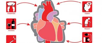
The main pathological process unfolding on the walls of the brachiocephalic arteries is considered to be atherosclerosis, which is so common among modern people. Narrowing of the artery by plaque inevitably leads to obstruction of blood flow, and in this case the brain will suffer.
A variety of diagnostic methods are used to study the brachiocephalic arteries, and the presence of pathology can be determined not only through expensive procedures, but also through conventional ultrasound - a cheap, accessible and safe way.
Causes of the disease
The main cause of non-stenotic atherosclerosis of the extracranial brachiocephalic arteries is high cholesterol levels in the blood plasma.
Factors in the development of pathology include:
- Patient's age. The disease usually develops after 40 years of age.
- Excess weight. Obesity signals a violation of metabolic processes.
- Bad habits (smoking and excessive alcohol consumption).
- High blood pressure. Hypertension itself is a consequence of high cholesterol. In addition, it causes an increase in blood viscosity and slows down its circulation.
- A sedentary lifestyle leads to slower blood circulation and a reduction in the nutrition of organs with valuable substances.
- An unhealthy diet with a large amount of animal fats, which increases the level of low-density lipoproteins in the blood.
Order of conduct
Let's look at how an ultrasound scan of the brachiocephalic arteries (BCA) is done. To undergo Doppler ultrasound of the BCA, the patient needs to undress to the waist and lie down on the couch. The doctor will apply a special thick gel to the skin, which will ensure tight contact of the ultrasound sensor with the skin. During the study, the patient will need to change position - turn over on his side, on his stomach. Also, the doctor may suggest performing some functional tests: holding your breath or changing a horizontal body position to a vertical one.
Classification
There are several stages of non-stenotic atherosclerosis of the BCA.
Among them:
- First stage (fat stain). The vascular wall is prone to swelling and loosening. Special enzymes aim to dissolve lipids and provide protection. But over time, the body’s protective properties decrease. As a result, complex compounds consisting of proteins and lipids are formed on the walls.
- Second stage (liposclerosis). At this stage, the disease is characterized by the proliferation of fatty deposits. A plaque gradually forms, which consists of connective tissue and fats. At first the deposit is liquid and can be dissolved.
- Third stage (atherocalcinosis). At this stage, calcium salts are deposited in the plaques. The formation grows and behaves unstable, narrowing the lumen and causing the situation to worsen. The risk of lumen blockage increases.
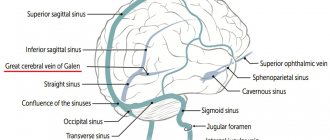
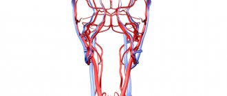
Anatomy of the brachiocephalic arteries
Brachiocephalic vessels are represented by:
- Brachiocephalic trunk and its branches;
- Left subclavian artery;
- Left common carotid artery (CCA).
All of these vessels originate from the aortic arch. The brachiocephalic trunk is a short vessel up to five centimeters long, which at the junction of the clavicle with the sternum on the right gives two large branches - the right subclavian and the right CCA. The left CCA is directed from the aorta upward, to the left sternoclavicular joint.
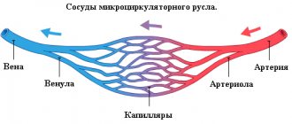
The common carotid arteries have a lumen width of about 6-8 mm, but not less than 4 mm. Having reached the upper edge of the thyroid cartilage, they branch into the right and left internal and external carotid arteries. The bifurcation can also be located at the level of the hyoid bone or the angle of the lower jaw. To this place, the CCA goes with one trunk, “without sending” a single arterial branch to the tissues.
The external carotid artery (ECA), almost immediately after its origin at the common artery, gives rise to nine arterial vessels supplying blood to the soft tissues and structures of the head.
The internal carotid artery (ICA) goes to the cranial cavity, and there, in the suprasphenoid part, it participates in the formation of the circle of Willis and gives off large cerebral arteries - the anterior and middle cerebral.
The first branch of the ICA is the orbital branch, which supplies blood to the eyes and anastomoses with vessels - branches of the ECA. Blood flow occurs through these communication pathways when the ICA is damaged.
The left subclavian artery arises from the aortic arch and leaves the chest cavity at the level of the middle third of the clavicle, then both subclavian arteries run parallel to this bone and go to the axillary region, where the vessels supplying the upper limbs begin. The diameter of the subclavian arteries reaches 9 mm.
The important arterial branches starting from the subclavian are the vertebral ones, going into the cranial cavity and, connecting, forming the main (basilar) artery, which gives off the posterior cerebral arteries, which are included in the circle of Willis.
Thus, going up and entering the cranium, blood flows from the ICA, ECA and subclavian arteries are connected into a large anastomosis - the Circle of Willis, which redirects blood in conditions of impaired patency of one or another arterial system.
In contrast to the variant anatomy of the Circle of Willis, which is important for brain nutrition, the BCAs have a fairly constant structure. Therefore, branching anomalies of the brachiocephalic arteries are diagnosed quite rarely. Among them:
- Complete absence of the brachiocephalic trunk, when the CCA and subclavian arteries begin directly from the aorta, like similar vessels on the left;
- The origin of the left vertebral artery is from the aorta, the right - not from the subclavian, but from the CCA;
- Asymmetry of the lumens of the vertebral arteries - often the left one is larger, their minimum diameter is 2 mm, maximum - 5.5 mm.
Video: anatomy of the brachiocephalic arteries
Symptoms
With atherosclerosis of the extracranial region, patients complain of:
- dizziness;
- blood pressure surges;
- pain in the heart and other organs.
Also, with non-stenotic atherosclerosis of the brachiocephalic arteries, visual and speech disturbances, attacks of nausea, itching in the arms and legs, and angina may occur. When there is a significant disruption of blood flow to the brain, brain failure occurs.
It manifests itself:
- constant headaches caused by oxygen deprivation;
- memory disorders;
- decreased level of intelligence.
During examination, the doctor may detect a change in the shape of the face.
The development of atherosclerosis of the carotid arteries can provoke encephalopathy and stroke.
USDG BCA
Hello. The neurologist prescribed an ultrasound examination of the BCA. What is this procedure, how is it carried out and is special preparation required? Olga M.
Answered by Nadezhda Ivanovna, ultrasound diagnostics doctor at the MedMix Plus clinic.
Doppler ultrasound of the BCA is an ultrasound examination of the brachiocephalic vessels, that is, in other words, ultrasound of the vessels of the neck. This examination is very important in assessing the condition, anatomy and patency of arteries and veins. Using this ultrasound technology, it is possible to judge the state of the blood supply to the brain, determine the degree of stenosis, thereby enabling the attending physician to determine treatment tactics.
In medical practice, there are several methods for studying the vessels of the neck:
1.USDG (ultrasound Dopplerography ) is a method aimed at assessing the patency of blood vessels and their condition. This method gives a general idea of the vessels. Allows you to identify the internal cause of obstruction, for example, a blood clot or atherosclerotic plaque.
2. Duplex/triplex scanning , which is used by ultrasound diagnostic doctors at our MedMix Plus clinic. On the monitor screen, a specialist sees a vessel in color against a gray background and evaluates its blood flow. This method allows you to assess the cause of the disturbance in the blood supply to the vessels, whether it is external or internal.
Using BCA ultrasound, you can diagnose diseases such as:
- vascular atherosclerosis;
- abnormalities in the structure of blood vessels;
- delamination of the vessel wall;
- arterial aneurysms;
- vasculitis, etc.
Before the symptoms of the disease appear, BCA should be performed when:
- hypertension;
- suffered a stroke;
- lupus;
- diabetes mellitus;
- smoking.
Research is also necessary in the presence of conditions such as:
- dizziness;
- decreased visual function;
- memory impairment;
- headache;
- fainting, etc.
Preparing for the study.
Before performing an ultrasound examination, you need to stop taking foods that can affect vascular tone: tea, coffee, energy drinks, alcohol. You should not visit stuffy or smoky rooms, and you should also stop taking medications that improve memory and attention. It is better to discuss the discontinuation of vascular and cardiac medications with your doctor.
Conducting research.
Doppler ultrasound of the BCA is a standard ultrasound examination procedure. The patient lies on the couch, a cushion is placed under the head. A gel is applied to the neck area, and a specialist conducts a study with a special sensor. The procedure lasts on average 30–35 minutes.
Doppler ultrasound of the brachiocephalic vessels is a painless and accurate ultrasound examination method, which in less than an hour will give an answer about the condition of the neck vessels and the causes of discomfort. Performed routinely, ultrasound of the neck vessels will prevent stroke by 80%. Be healthy!
Diagnostics
Diagnosis of pathology includes:
- Inspection. At the appointment, the specialist finds out the patient’s complaints and risk factors for his disease. On a general examination, various atherosclerotic lesions are identified.
- Laboratory research. Before treatment, the patient must take a blood test, which allows you to study the level of cholesterol, triglycerides and lipoproteins.
- Angiography. This study evaluates blood flow disorders.
- Aortography. This examination is carried out to detect elongation of the aorta and its thickening, calcification, expansion in the thoracic or abdominal regions, and aneurysms.
- Coronary angiography. This examination is effective for assessing the condition of the coronary system.
Other modern techniques are also used for diagnosis. They allow not only to make an accurate diagnosis, but also to predict the development of pathology, assess the patient’s condition, select the right medications, and adjust their dosage during therapy.
Treatment
A number of techniques are used to treat non-stenotic atherosclerosis. The patient must always adhere to all doctor's recommendations. Only in this case will he be able to prolong the period of active life and avoid negative consequences (complete blockage of blood vessels, strokes and death).
To prescribe optimal therapy for non-stenotic atherosclerosis, the doctor:
- Determines the stage of the disease.
- Records general symptoms.
All medications are prescribed individually and take into account the patient’s condition.
As a rule, patients take:
- Anticoagulants. This reduces the risk of blood clots and strokes.
- Medicines to lower blood pressure (if it increases). Such remedies can reduce the load on the arteries, dilate blood vessels, improve blood flow and relieve spasms.
- Medicines to lower cholesterol levels. Typically, such drugs are taken throughout life after atherosclerosis of the brachiocephalic arteries is detected.
It is also very important to constantly monitor the patient's lipid balance. If necessary, sedatives are prescribed. They allow you to normalize the emotional background, avoid depression, improve the quality of sleep, and get rid of headaches. Patients are often prescribed physiotherapy.
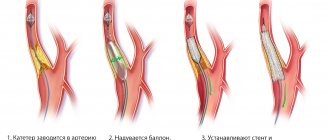
The decision to undergo surgical intervention is usually made if the passages are significantly narrowed and the disease has reached a high stage of development.
The following types of operations are carried out today:
- Removing part of the artery and replacing it with a prosthesis.
- Stitching a vessel to reduce its size.
Endovascular operations are increasingly being performed. They are as safe as possible, and recovery from them takes a short period of time.
Are you interested in all the features of therapy? Contact our specialists. You will not have to overpay for a consultation. Prices are indicated on the website. Note! The cost is approximate. The exact price of therapy will be announced by your attending physician. In any case, you will not overpay. Our private clinic in Moscow adheres to a loyal pricing policy.
Classification of diseases
The main classification of diseases of the vascular bed of the brain depends on the cause of the development of the pathology. This is important for choosing treatment tactics that should affect the etiology. The following are the main groups of diseases of the brachiocephalic vessels:
- congenital pathology - arteries develop with anatomical anomalies, pathological tortuosity, impaired development of connective tissue areas or the muscular layer of arteries may be observed;
- atherosclerotic changes - this type of disease is the most common;
- narrowing of the lumen of the artery due to external pathology - a volumetric process or pathology of the spinal column at the site of the artery;
- inflammation of the vascular wall - vasculitis;
- alternate decreases and increases in blood pressure;
- compression of blood vessels due to pathology of the spinal column;
- aneurysm - expansion of the vessel and protrusion of its wall in the form of a “pocket”;
- changes in blood vessels due to the underlying disease (diabetes, endocrine pathology);
- consequences of injuries and surgical interventions.
If we talk about the frequency of occurrence, atherosclerosis accounts for about 90% of all cases of pathology of the brachiocephalic arteries. Atherosclerosis develops as a result of exposure to risk factors - hereditary predisposition, smoking, age-related changes, disorders of fat metabolism (chronic pathologies).
Chronic cerebrovascular diseases can be compared to a time-delayed explosive device - you never know when the mechanism will go off. However, there is always a risk to health and even life. Timely consultation with a doctor and initiation of treatment minimizes the risk of disability and death.
Prevention
The prognosis for the development of the disease is determined by the patient’s lifestyle. The progression of the pathology can be stopped by eliminating existing risk factors and active drug therapy.
Prevention of vascular problems should be carried out comprehensively.
If there is any risk of developing pathology, you must:
- switch to proper nutrition;
- stop smoking;
- reduce stress factors;
- normalize weight;
- devote time to physical activity.
Prevention is impossible without regular examinations and visits to the doctor. We recommend seeing a cardiologist for anyone who has a history of problems or has negative heredity.