Both arteries and veins are types of blood vessels in the cardiovascular system
An artery carries blood away from the heart, and a vein brings blood back to the heart.
Blood vessels are necessary to transport blood throughout the body. Blood carries oxygen and nutrients to various tissues in the body, allowing them to function.
The heart and blood vessels make up the cardiovascular system. This system contains a complex network of vessels with different structures and functions.
In this article, we discuss the differences between arteries and veins. We also highlight the different types of blood vessels and how they work as part of the cardiovascular system.
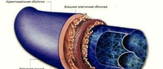
The structure of the walls of veins and their difference from arteries
- The inner wall is lined with epithelial cells
- Average MMC
- External RVST, fixed to surrounding tissues.
Veins have a thinner wall: they expand easily and collapse easily.
There are valves - folds of the inner membrane - distributed unequally: less in the arms than in the legs, the rest above the confluence of the venous tributaries. Designed to prevent reverse blood flow and prevent stagnation in venous inflows. If the valve is not developed, then varicose veins, if stagnation, then a blood clot, and possible damage to the vessel.
The peculiarity of the hemodynamic conditions of the veins is low pressure (15-20 mm Hg) and low blood flow rate, which causes a lower content of elastic fibers in these vessels.
The number of muscle elements in the wall of these vessels depends on whether the blood moves with or against gravity.
Veins of the muscleless type are found in the dura mater, bones, retina, placenta, and red bone marrow. The wall of muscleless veins is lined internally with endothelial cells on the basement membrane, followed by a layer of fibrous SDTK; there are no smooth muscle cells.
Veins of the muscular type with weakly expressed muscular elements are located in the upper half of the body - in the system of the superior vena cava. These veins are usually in a collapsed state. The tunica media contains a small number of myocytes.
Veins with highly developed muscular elements make up the vein system of the lower half of the body. A feature of these veins is well-defined valves and the presence of myocytes in all three membranes - in the outer and inner membrane in the longitudinal direction, in the middle - circular direction.
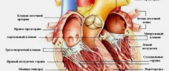
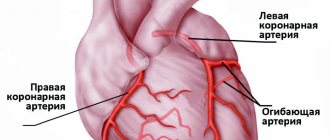
Blood supply to the heart
The blood supply to the heart is carried out by two arteries: the right coronary artery, a. coronaria dextra, and the left coronary artery, a. coronaria sinistra, which are the first branches of the aorta. Each of the coronary arteries emerges from the corresponding right and left aortic sinuses.
- Right coronary artery, a. coronaria dextra, originates from the aorta at the level of the right sinus, follows down the wall of the aorta between the conus arteriosus of the right ventricle and the right appendage into the coronary sulcus; being covered here, in its initial sections, by the right ear, the artery reaches the right edge of the heart and gives off the so-called branch of the right edge to the wall of the ventricle. Having further given a series of branches to the wall of the aorta, appendage and conus arteriosus, the right coronary artery passes to the diaphragmatic surface of the heart, where it also lies in the depths of the coronary sulcus. Here it sends branches to the posterior wall of the right atrium and right ventricle, as well as thin branches accompanying the atrioventricular bundle. On the diaphragmatic surface it reaches the posterior interventricular groove of the heart, in which it descends in the form of the posterior interventricular branch, ramus interventricularis posterior. The latter, approximately at the border of the middle and lower third of this groove, plunges into the thickness of the myocardium. It supplies the posterior part of the interventricular septum and the posterior walls of both the right and left ventricles.
- Left coronary artery, a. coronaria sinistra, larger than the right one; starting at the level of the left aortic sinus, it follows to the left behind the root of the pulmonary trunk, and then between it and the left ear. Heading to the left part of the coronary sulcus, still behind the pulmonary trunk it is most often divided into two branches: the anterior interventricular branch, interventricularis anterior, and the circumflex branch, circumflexus.
At the point where the main trunk passes into the interventricular groove, a large branch branches off from it, passing along the coronary groove to the left half of the heart and feeding the posterior walls of the left atrium and left ventricle with its branches. .
The anterior interventricular branch, interventricularis anterior, is a continuation of the main trunk and descends along the anterior interventricular groove to the apex of the heart, rounding which it enters the terminal section of the posterior interventricular groove; not reaching the posterior interventricular branch, interventricularis posterior, it plunges into the thickness of the myocardium. Along the way, it sends branches to the arterial cone, to nearby sections of the walls of the left and right ventricles, a larger branch to the anterior part of the interventricular septum, anastomotic branches to the stems from the right coronary artery and completely supplies the apex of the heart. Near its origin, the anterior interventricular branch gives off the so-called diagonal artery, which sometimes begins from the main trunk of the left coronary artery. In both cases, it branches in the region of the anterior wall of the left ventricle. The circumflex branch, g. circumflexus, emerging from under the left ear, follows the coronary groove to the left edge of the heart and then along the back of the coronary groove to the diaphragmatic surface of the heart, upon transition to which it sends a large branch that feeds the anterior and posterior walls of the left ventricle. Before reaching the posterior interventricular groove, it descends along the diaphragmatic surface of the left ventricle, but does not reach the apex of the heart. On its way, it sends branches to the walls of the left ear, left atrium and left ventricle. Thus, the right coronary artery supplies the walls of the pulmonary trunk, aorta, right and left atria, right ventricle, posterior wall of the left ventricle, interatrial and interventricular septa. The left coronary artery supplies the walls of the pulmonary trunk, aorta, right and left atria, the anterior walls of the right and left ventricles, the posterior wall of the left ventricle, the interatrial and interventricular septa. The coronary arteries of the heart anastomose with each other in all its parts, with the exception of the edges of the heart, which are supplied only by the corresponding arteries. In addition, there are extracoronary anastomoses formed by vessels supplying the wall of the pulmonary trunk, aorta and vena cava, as well as vessels of the posterior wall of the atria. All these vessels anastomose with the arteries of the bronchi, diaphragm and pericardium. In addition to intercoronary anastomoses (intercoronary), anastomoses of branches of the same artery (intracoronary) are very well developed in the heart. The arteries of the heart, especially in the area of the ventricles, follow the course of the muscle bundles. Thus, in the region of the outer and deep layers of the myocardium, as well as the papillary muscles, the arteries are directed along the longitudinal axis of the heart, and in the middle layer of the myocardium they have a transverse direction.
Most of the veins of the heart (except for the small and anterior) bring blood to a special reservoir - the coronary sinus, sinus coronarius, which opens into the posterior part of the cavity of the right atrium, between the opening of the inferior vena cava and the right atrioventricular opening.
- The coronary sinus, sinus coronarius, seems to be a continuation of its great vein onto the diaphragmatic surface of the heart (see below). It is located in the left part of the posterior coronary sulcus, extending from the place where the oblique vein of the left atrium flows into it from above to its mouth; its length is 2-3 cm. Above the coronary sinus, a thin layer of muscle bundles of the myocardium is thrown, due to which its middle shell, tunica media, is also formed.
- Great vein of the heart, v. cordis magna, begins on the anterior surface of the apex of the heart. First, it lies in the anterior interventricular groove next to the descending branch of the left coronary artery. Having reached the top of the coronary groove, it is located in it and runs along the lower border of the left atrium to the left edge of the heart.
- Oblique vein of the left atrium, v. obliaua atrii sinisiri, begins on the lateral wall of the left atrium and goes from left to right down, in the form of a small branch in the fold
- Posterior vein of the left ventricle, v. posterior ventriculi sinistri, originates on the posterolateral wall of the left ventricle, goes upward and flows either into the great vein of the heart or directly into the coronary sinus.
- Middle vein of the heart, v. cordis media, begins on the posterior surface at the apex of the heart, lies in the posterior longitudinal groove next to the interventricular branch of the right coronary artery and flows into the right end of the coronary sinus. Along the way, the middle vein of the heart receives branches from the posterior walls of both ventricles.
- Small vein of the heart, v. cordis parva, begins on the right edge of the right atrium and right ventricle, lies in the posterior part of the coronary sulcus and flows either into the right end of the coronary sinus, or independently opens into the cavity of the right atrium, sometimes into the middle vein of the heart.
- Anterior veins of the heart
- The smallest veins of the heart, vv. cordis minimae, a group of small veins that collect blood from various parts of the heart and open with the openings of the smallest veins, foramina venarum minimarum. directly into the right and partly into the left atrium, as well as into the ventricles.
The opening of the coronary sinus in the cavity of the right atrium is bordered by the valve of the coronary sinus, valvula sinus coronarii. There are two or three small valves in the sinus itself, not far from its opening.
Having rounded the left edge of the heart, the large vein lies in the diaphragmatic part of the coronary sulcus, where it passes without a sharp border into the coronary sinus. Sometimes there is a small valve at the junction of the great cardiac vein and the coronary sinus. The veins of the anterior wall of both ventricles, the interventricular septum and sometimes near the sinus flow into the large vein of the heart - the posterior vein of the left ventricle.
pericardium. Heading down and to the right along the posterior wall of the left atrium, it passes into the coronary sinus. At the mouth of this vein there is sometimes a small valve.
In the area of the cardiac notch, the middle cardiac vein anastomoses with the great cardiac vein.
,vv. cordis anteriores, have different sizes. They originate in the region of the anterior and lateral walls of the right ventricle, go upward and to the right to the coronary sulcus and flow directly into the right atrium; at the mouths of the anterior veins there are sometimes small valves.
Functions of the venous system
Functions of veins:
- Transport of blood to the heart.
- Blood reservoir - deposition is facilitated by thin-walled, numerous veins (many in the abdominal cavity, they hold 80% of the blood in case of shock => GM, heart, liver, kidneys). In shock, blood pressure drops down to 0. The veins of the lungs can accumulate up to 28% of the blood, and swelling is possible due to inflammation.
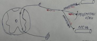
- Regulates hemodynamics: blood flow speed, blood pressure provide blood supply, carried out reflexively. There are many receptors in the walls of the veins: chemo, mechano, baro. When they are irritated, reflexes are triggered:
- venovenous;
- venoarthrial (increased pressure, decreased arterial blood flow);
- venolymphatic.
- Participation in metabolic processes between blood and tissue. The walls of the capillaries are well permeable, metabolic processes are ensured by thin-walled venules, post-capillaries; with inflammation, permeability (swelling) increases, since the walls of the venules react to histamine.
Introduction:
The circulatory system is responsible for moving blood throughout the body. The main components of this system are the heart and blood vessels. The heart is the central organ of the circulatory system. With each heartbeat, blood is pumped through the arteries and delivers oxygen and nutrients to various organs and tissues, after which the blood returns back to the heart through other blood vessels - veins.
There are three types of blood vessels that play different roles. The two main types are arteries and veins. Arteries carry oxygenated and nutrient-rich blood away from the heart, and veins return waste blood back to the heart. Lymphatic vessels are the third component; they filter and “purify” the liquid part of the blood - plasma, before returning it to the general bloodstream.
What connections between extra- and intracranial veins do you know? Their meaning
Extracranial: superficial, deep
Intracranial - GM veins, open in the sinuses of the dura mater, do not collapse, there are no valves. From the sinuses through the internal jugular vein is the main route for the outflow of venous blood from the cranial cavity.
Pathways of communication between intra- and extracranial veins:
- Through the venous outlet: 3 pairs, hole in the bones of the skull, blood from inside to outside
- The ophthalmic vein connects the cavernous sinus with the facial vein
- Through the diploe veins (the spongy substance of the skull bones between the plates)
- Through the foramen magnum into the vertebral venous plexuses
Around which organs are the periorgan venous plexuses more developed? Their meaning
In many places there are well-developed venous plexuses:
- Small pelvis,
- spinal canal,
- Around the bladder
The significance of these plexuses can be seen in the example of the intravertebral plexus. When filled with blood, it occupies those free spaces that are formed when cerebrospinal fluid is displaced when changing body position or during movements. Thus, the structure and location of the veins depends on the physiological conditions of blood flow in them.