B12 anemia or, as it is also called pernicious/megaloblastic, is a pathological condition that is characterized by a disruption in the formation and maturation of blood cells due to a deficiency of vitamin B12 in the body. The latter may be due to its insufficient intake into the human body or problems with absorption. Most often, the disease is diagnosed in patients over the age of fifty of both sexes. According to medical statistics, its prevalence does not exceed 1%, while at least 10% of patients over the age of seventy suffer from B12 deficiency.
The Department of Hematology at CELT offers diagnosis and treatment of B12 deficiency anemia in Moscow. Our multidisciplinary clinic was one of the first in our country to begin offering paid medical services and has been successfully operating for the third decade. Its staff includes leading Russian specialists, who have modern technologies and a diagnostic and treatment base in their arsenal. It allows you to accurately and quickly diagnose and carry out treatment according to international standards. You can find out the cost of services in our price list; check the numbers with our information line operators.
B12 anemia: causes
The daily requirement of the human body for vitamin B12 is from one to five micrograms. It is satisfied through its consumption with food: after entering the stomach, the vitamin is separated from protein under the influence of enzymes. In order for the body to fully absorb it, a process of connection with glycoprotein and other factors must occur. Absorption of the vitamin into the blood occurs in the lower part of the ileum, from where it passes to the tissues. Factors that can initiate the development of anemia are divided into two groups:
- Nutritional – an unbalanced diet that is unable to ensure the required amount of B12 enters the body. This often happens if the patient practices fasting, vegetarianism, or diets that minimize or eliminate the consumption of animal protein;
- Endogenous – disruptions in the process of vitamin absorption due to a deficiency of protein that binds the vitamin and converts it into an absorbable form. A similar phenomenon can be initiated by: a congenital absence of the internal Castle factor;
- inflammatory processes of the gastric mucosa in a chronic form;
- condition after gastric excision surgery;
- condition after partial removal of the ileum or duodenum;
- inflammation of the small intestine (enteritis), pancreas (pancreatitis);
- gluten enteropathy;
- granulomatous inflammation of the digestive tract (Crohn's disease);
- protrusions of the intestinal wall - (diverticula);
- neoplasms of the jejunum of a malignant nature;
- helminthic infestations caused by different types of helminths.
The reasons may lie in physical and mental dependence on alcohol intake, in the use of Colchicine, Neomycin, as well as a number of oral contraceptives.
Reasons for a critical decrease in cobalamin
B12 deficiency anemia occurs when there is insufficient intake of animal protein into the body and in cases where cobalamin is not absorbed and does not enter the blood.
In the first case, people most often suffer who limit themselves in nutrition - losing weight, vegetarians, adherents of methods with an unbalanced diet. B12 is found in meat, beef liver, dairy products, and egg yolks.
The second, endogenous cause requires diagnosis. The absorption of cobalamin is ensured by the enzyme Castle factor. When the synthesis of intrinsic factor Castle decreases or stops, the vitamin that enters the body is not absorbed.
Absorption into the bloodstream is prevented by diseases of the gastrointestinal tract:
- atrophic gastritis,
- chronic pancreatitis,
- Crohn's disease,
- diverticula of the small intestine,
- tumors.
In some hormonal diseases, including hyperthyroidism, B12 consumption increases several times. At the same time, the body experiences a shortage of it even with normal intake and without problems of absorption.
In some cases, the cause of deficiency is helminths and bacteria, taking certain medications and oral contraceptives, and alcoholism. Even frequent intestinal upset can interfere with the absorption of the substance.
B12 anemia: symptoms
The clinical picture of megaloblastic anemia is determined by the severity of four syndromes:
| Syndrome | Clinical manifestations |
| Anemic | Symptoms of B12 deficiency anemia are nonspecific because they arise due to failures in the transport of oxygen by red blood cells. They are:
In the long term, anemia can develop into secondary myocardial damage and heart failure. |
| Gastroenterological | One of the classic clinical manifestations is the so-called “lacquered” raspberry tongue. The rest are as follows:
Diagnosis of B12-deficiency anemia reveals atrophy of the gastric mucosa and decreased secretion production. |
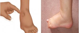
| Neurological | The symptoms of this group are caused by damage to neurons and pathways, which manifests itself:
Neurological examination reveals damage to the peripheral nerves and spinal cord. |
The lack of adequate treatment can cause the development of complications in the form of anemia and degenerative processes of the nerves due to the increased sensitivity of the bone marrow and tissues of the nervous system to the lack of B12 in the body.
B12 anemia: diagnosis
Before starting treatment, CELT hematologists prescribe comprehensive studies to the patient, which allow them to accurately diagnose “B12-deficiency anemia” and identify the causes of its development. In addition to the hematologist, specialists in neurology and gastroenterology take part in the process. Diagnostics is as follows:
- Vitamin deficiency is detected through biochemical analysis;
- A general analysis determines the deficiency of red blood cells, leukocytes and platelets;
- Stool analysis can detect excess fat content, helminth fragments;
- The Schilling test detects failures in vitamin absorption;
- Bone marrow histology and myelogram indicate an increase in the number of megaloblasts.
In order to identify the triggering factors for malabsorption of vitamin B12 deficiency anemia in the gastrointestinal tract, the patient is prescribed gastroscopy and radiography of the stomach, as well as examination of the large intestine using an x-ray contrast agent. Other instrumental methods will also help you obtain the necessary information:
- Ultrasound scanning of the abdominal organs;
- magnetic resonance imaging of the brain;
- encephalography.
A comprehensive study taking into account the quantitative and qualitative composition of cells, as well as biochemical blood parameters, which allows us to identify signs of iron deficiency anemia.
The research results are provided with a free doctor’s commentary.
English synonyms
Iron deficiency anemia screening.
Research method
Colorimetric photometric method, flow cytometry, SLS (sodium lauryl sulfate) method, conductometric method, immunoturbidimetry.
Units
μmol/l (micromoles per liter), *10^9/l (10 in st. 9/l), *10^12/l (10 in st. 12/l), g/l (grams per liter), % (percent), fl (femtoliter), pg (picogram).
What biomaterial can be used for research?
Venous blood.
How to properly prepare for research?
- Eliminate alcohol from your diet 24 hours before the test.
- Do not eat for 12 hours before the test.
- Do not take medications for 24 hours before the test (as agreed with your doctor).
- Avoid physical and emotional stress 30 minutes before the test.
- Do not smoke for 30 minutes before the test.
General information about the study
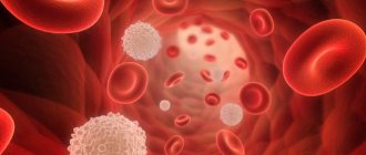
Anemia is a pathological condition characterized by a decrease in the number of red blood cells and/or hemoglobin in the blood. The causes of anemia are varied and can be closely related to chronic somatic diseases (kidney and liver pathologies, autoimmune, infectious and inflammatory diseases, diabetes, tumors), acute and chronic blood loss, hereditary diseases and malnutrition, lack of vitamins and microelements. They occur when there is insufficient production of red blood cells in the bone marrow (for example, with deficiency of iron, vitamins B12 and B6, bone marrow depletion) or premature and excessive destruction of red blood cells in the bloodstream (for example, with hemolysis).
The most common form of anemia, iron deficiency, is detected in 80-90% of patients who have a reduced number of red blood cells and/or hemoglobin in the blood.
Iron deficiency develops gradually. Initially, a negative iron balance is created, in which the body's needs for it and its losses exceed the amount it receives from food. This may be due to blood loss, pregnancy, breastfeeding, growth spurts during puberty, malabsorption of micronutrients in the gastrointestinal tract, or insufficient intake of foods containing iron, as with vegetarianism.
Normally, iron is absorbed in the small intestine, but in some chronic diseases of the stomach and intestines, malabsorption syndrome occurs with impaired absorption of nutrients and microelements from food. In the blood, iron is transported by the protein transferrin to sites of use or accumulation. With iron deficiency, the amount of free transferrin in the blood increases, and the level of serum iron decreases. Hemoglobin synthesis in the bone marrow occurs less actively, and iron deficiency anemia develops with clinical manifestations of anemia. In a general blood test, small pale colored red blood cells are detected, MHC (average hemoglobin content in an erythrocyte), MCV (average erythrocyte volume), MCHC (average hemoglobin concentration in an erythrocyte), and hemoglobin level and hematocrit decrease. Without treatment, the amount of hemoglobin decreases more and more, the shape of red blood cells changes, and the intensity of cell division in the bone marrow decreases. Due to a decrease in the amount of hemoglobin and red blood cells, the transport of oxygen and carbon dioxide in the tissues is disrupted, symptoms of anemia appear: pale skin, headache, memory impairment, drowsiness, spots before the eyes, dizziness, fainting, rapid heartbeat, decreased blood pressure, pain in the area heart, muscle weakness.
In addition to well-being, negative changes occur in appearance: dry skin (as a result, early wrinkles), brittle nails, hair loss.
Children susceptible to anemia are more likely than healthy peers to suffer from colds.
In addition, with iron deficiency, the patient may experience a disturbance of taste and smell: there is a desire to eat chalk, clay, raw cereals and inhale the smells of acetone, gasoline, and turpentine.
Often people do not pay attention to the main signs of anemia - constant fatigue, paleness, dry skin - considering them an insufficient reason to see a doctor. Of course, such symptoms do not always indicate a disease; it cannot be diagnosed only by their presence. Moreover, you should not start taking iron-containing medications on your own - this can lead to serious complications.
It is important to know what exactly causes anemia. For example, in older people it often results from internal bleeding due to a tumor in the gastrointestinal tract. There is a type of anemia associated with poor absorption of vitamin B12. But in order to identify it, you first need to make sure that there is enough iron in the body and that the red blood cells are saturated with it.
At the same time, simply donating blood for hemoglobin is not enough to fully diagnose anemia. For example, in smokers, the level of hemoglobin and red blood cells is often normal: carbon monoxide contained in cigarettes, combining with hemoglobin, forms the substance carboxyhemoglobin, this form of hemoglobin does not have the ability to carry oxygen, and to compensate for oxygen starvation, the level of hemoglobin increases. It turns out that a comprehensive analysis is needed, reflecting the true reserves of this microelement in the body: determination of the levels of serum iron, transferrin, reticulocytes, general blood test, leukocyte formula.
In addition, it is important that timely diagnosis of anemia makes it possible to identify diseases of which it is the first symptom: some malignant tumors, chronic inflammatory processes, helminthic infestations, etc.
What is the research used for?
- For the diagnosis of iron deficiency anemia.
- For differential diagnosis of anemia.
- For the evaluation of patients at high risk of developing anemia.
- To evaluate the effectiveness of treatment for iron deficiency anemia.
When is the study scheduled?
- For symptoms of iron deficiency in the body (pallor and/or yellowness of the skin, general weakness, fatigue, drowsiness, dizziness, tinnitus, flashing spots before the eyes, fainting, atrophy of the tongue mucosa, changes in the structure of the nails, abnormal taste preferences).
- When the number of red blood cells and/or hemoglobin decreases according to a clinical blood test.
- In the presence of risk factors for the development of anemia (pregnancy, breastfeeding, menstrual irregularities, malnutrition, vegetarianism, diseases of the stomach and intestines with impaired absorption of nutrients, chronic stress).
- When examining children during periods of intensive growth.
- When examining patients with asthenia of unknown origin and severe fatigue.
- When monitoring the effectiveness of the use of drugs containing iron.
What do the results mean?
Reference values
- Serum iron
| Floor | Age | Reference values |
| Female | Less than 24 days | 17.9 - 44.8 µmol/l |
| 24 days – 1 year | 7.2 - 17.9 µmol/l | |
| 1-14 years | 9 - 21.5 µmol/l | |
| More than 14 years | 10.7 - 32.2 µmol/l | |
| Male | Less than 24 days | 17.9 - 44.8 µmol/l |
| 24 days – 1 year | 7.2 - 17.9 µmol/l | |
| 1-14 years | 9 - 21.5 µmol/l | |
| More than 14 years | 12.5 - 32.2 µmol/l |
- Leukocyte formula
- Leukocytes
| Age | Reference values |
| Less than 1 year | 6 - 17.5 *10^9/l |
| 1-2 years | 6 - 17 *10^9/l |
| 2-4 years | 5.5 - 15.5 *10^9/l |
| 4-6 years | 5 - 14.5 *10^9/l |
| 6-10 years | 4.5 - 13.5 *10^9/l |
| 10-16 years | 4.5 - 13 *10^9/l |
| More than 16 years | 4 - 10 *10^9/l |
- Neutrophils
| Age | Reference values |
| Less than 1 year | 1.5 - 8.5 *10^9/l |
| 1-2 years | 1.5 - 8.5 *10^9/l |
| 2-4 years | 1.5 - 8.5 *10^9/l |
| 4-6 years | 1.5 - 8 *10^9/l |
| 6-8 years | 1.5 - 8 *10^9/l |
| 8-10 years | 1.8 - 8 *10^9/l |
| 10-16 years | 1.8 - 8 *10^9/l |
| More than 16 years | 1.8 - 7.7 *10^9/l |
- Neutrophils, %
| Age | Reference values |
| Less than 1 year | 16 — 45 % |
| 1-2 years | 28 — 48 % |
| 2-4 years | 32 — 55 % |
| 4-6 years | 32 — 58 % |
| 6-8 years | 38 — 60 % |
| 8-10 years | 41 — 60 % |
| 10-16 years | 43 — 60 % |
| More than 16 years | 47 — 72 % |
- Complete blood count (without leukocyte formula and ESR)
- Leukocytes
| Age | Reference values |
| Less than 1 year | 6 - 17.5 *10^9/l |
| 1-2 years | 6.6 - 11.2 *10^9/l |
| 2-4 years | 5.5 - 15.5 *10^9/l |
| 4-6 years | 5 - 14.5 *10^9/l |
| 6-10 years | 4.5 - 13.5 *10^9/l |
| 10-16 years | 4.5 - 13 *10^9/l |
| More than 16 years | 4 - 10 *10^9/l |
- Red blood cells
| Floor | Age | Reference values |
| Less than 1 year | 4.1 - 5.3 *10^12/l | |
| 1-5 years | 4 - 4.4 *10^12/l | |
| 5-6 years | 4.1 - 4.5 *10^12/l | |
| 6-7 years | 4 - 4.4 *10^12/l | |
| 7-8 years | 4.2 - 4.6 *10^12/l | |
| 8-9 years | 4.1 - 4.5 *10^12/l | |
| 9-14 years | 4.2 - 4.6 *10^12/l | |
| 14-15 years old | 4.4 - 4.8 *10^12/l | |
| Female | 15-19 years old | 3.5 - 5 *10^12/l |
| More than 19 years | 3.5 - 5.2 *10^12/l | |
| Male | 15-19 years old | 3.9 - 5.6 *10^12/l |
| More than 19 years | 4.2 - 5.3 *10^12/l |
- Hemoglobin
| Age | Floor | Hemoglobin, g / l |
| 134-198 | ||
| 14 days – 1 month. | 107-171 | |
| 1-2 months | 94-130 | |
| 2-4 months | 103-141 | |
| 4-6 months | 111-141 | |
| 6-9 months | 110-140 | |
| 9-12 months | 113-141 | |
| 1-5 years | 110-140 | |
| 5-10 years | 115-145 | |
| 10-12 years | 120-150 | |
| 12-15 years | male | 120-160 |
| female | 115-150 | |
| 15-18 years old | male | 117-166 |
| female | 117-153 | |
| 18-45 years old | male | 132-173 |
| female | 117-155 | |
| 45-65 years | male | 131-172 |
| female | 117-160 | |
| > 65 years old | male | 126-174 |
| female | 117-161 |
- Hematocrit
| Age | Floor | Hematocrit, % |
| 41-65 | ||
| 14 days – 1 month. | 33-55 | |
| 1-2 months | 28-42 | |
| 2-4 months | 32-44 | |
| 4-6 months | 31-41 | |
| 6-9 months | 32-40 | |
| 9-12 months | 33-41 | |
| 1-3 years | 32-40 | |
| 3-6 years | 32-42 | |
| 6-9 years | 33-41 | |
| 9-12 years | 34-43 | |
| 12-15 years | male | 35-45 |
| female | 34-44 | |
| 15-18 years old | male | 37-48 |
| female | 34-44 | |
| 18-45 years old | male | 39-49 |
| female | 35-45 | |
| 45-65 years | male | 39-50 |
| female | 35-47 | |
| > 65 years old | male | 37-51 |
| female | 35-47 |
- Mean erythrocyte volume (MCV)
| Floor | Age | Reference values |
| Less than 1 year | 71 – 112 fl | |
| 1-5 years | 73 – 85 fl | |
| 5-10 years | 75 – 87 fl | |
| 10-12 years | 76 – 94 fl | |
| Female | 12-15 years | 73 – 95 fl |
| 15-18 years old | 78 – 98 fl | |
| 18-45 years old | 81 – 100 fl | |
| 45-65 years | 81 – 101 fl | |
| More than 65 years | 81 – 102 fl | |
| Male | 12-15 years | 77 – 94 fl |
| 15-18 years old | 79 – 95 fl | |
| 18-45 years old | 80 – 99 fl | |
| 45-65 years | 81 – 101 fl | |
| More than 65 years | 81 – 102 fl |
- Average hemoglobin content in erythrocytes (MCH)
| Age | Floor | Reference values |
| 30 - 37 pg | ||
| 14 days - 1 month. | 29 - 36 pg | |
| 1 - 2 months | 27 - 34 pg | |
| 2 - 4 months | 25 - 32 pg | |
| 4 - 6 months | 24 - 30 pg | |
| 6 - 9 months | 25 - 30 pg | |
| 9 - 12 months | 24 - 30 pg | |
| 1 – 3 years | 22 - 30 pg | |
| 36 years | 25 - 31 pg | |
| 6 – 9 years | 25 - 31 pg | |
| 9-15 years | 26 - 32 pg | |
| 15-18 years old | 26 - 34 pg | |
| 18-45 years old | 27 - 34 pg | |
| 45-65 years | 27 - 34 pg | |
| > 65 years old | female | 27 - 35 pg |
| > 65 years old | male | 27 - 34 pg |
- Mean erythrocyte hemoglobin concentration (MCHC)
| Age | Reference values |
| Less than 1 year | 290 – 370 g/l |
| 1-3 years | 280 – 380 g/l |
| 3-12 years | 280 – 360 g/l |
| 12-19 years old | 330 - 340 g/l |
| More than 19 years | 300 – 380 g/l |
- Platelets
| Floor | Age | Reference values |
| Less than 1 year | 214 - 362 *10^9/l | |
| 1-2 years | 208 - 352 *10^9/l | |
| 2-3 years | 209 - 351 *10^9/l | |
| 3-4 years | 196 - 344 *10^9/l | |
| 4-5 years | 208 - 332 *10^9/l | |
| 5-6 years | 220 - 360 *10^9/l | |
| 6-7 years | 205 - 355 *10^9/l | |
| 7-8 years | 205 - 375 *10^9/l | |
| 9-10 years | 177 - 343 *10^9/l | |
| 10-11 years | 211 - 349 *10^9/l | |
| 11-12 years old | 198 - 342 *10^9/l | |
| 12-13 years old | 202 - 338 *10^9/l | |
| 13-14 years old | 192 - 328 *10^9/l | |
| 14-15 years old | 198 - 342 *10^9/l | |
| 15-18 years old | 165 - 396 *10^9/l | |
| 18-45 years old | 159 - 376 *10^9/l | |
| Male | 45-65 years | 156 - 300 *10^9/l |
| Female | 45-65 years | 156 - 351 *10^9/l |
| More than 65 years | 139 - 363 *10^9/l |
- Mean platelet volume (MPV)
| Age | Reference values |
| Less than 10 years | 8.3 - 10.9 fl |
| 10 – 18 years | 8.6 - 12.1 fl |
| 18 – 45 years old | 8.5 - 12.8 fl |
| 45 – 65 years | 8.6 - 12 fl |
| 45 – 65 years | 9 - 12.9 fl |
| More than 65 years | 8.8 - 12.4 fl |
- Reticulocytes
- Reticulocytes,% (RET%)
| Floor | Age | Reference values |
| Female | Less than 2 weeks | 0,15 — 1,5 % |
| 2 weeks – 1 month | 0,45 — 1,4 % | |
| 1-2 months | 0,45 — 2,1 % | |
| 2-6 months | 0,25 — 0,9 % | |
| 6 months – 2 years | 0,2 — 1 % | |
| 2-6 years | 0,2 — 0,7 % | |
| 6-12 years | 0,2 — 1,3 % | |
| 12-18 years old | 0,12 — 2,05 % | |
| Over 18 years old | 0,59 — 2,07 % | |
| Male | Less than 2 weeks | 0,15 — 1,5 % |
| 2 weeks – 1 month | 0,45 — 1,4 % | |
| 1-2 months | 0,45 — 2,1 % | |
| 2-6 months | 0,25 — 0,9 % | |
| 6 months – 2 years | 0,2 — 1 % | |
| 2-6 years | 0,2 — 0,7 % | |
| 6-12 years | 0,2 — 1,3 % | |
| 12-18 years old | 0,24 — 1,7 % | |
| Over 18 years old | 0,67 — 1,92 % |
- Reticulocytes (absolute count, RET#)
| Floor | Reference values |
| Female | 17 - 63.8 *109/l |
| Male | 23 - 70 *109/l |
- Immature reticulocyte fraction (IRF)
| Floor | Reference values |
| Female | 3 — 15,9 % |
| Male | 2,3 — 13,4 % |
- Transferrin
| Age | Reference values |
| Less than 11 years old | 2.03 - 3 g/l |
| More than 11 years | 2 - 3.6 g/l |
Serum iron
Reasons for the increase:
- anemia (aplastic, hemolytic, pernicious), thalassemia;
- polycythemia;
- acute hepatitis, liver damage;
- hemochromatosis;
- hemosiderosis;
- nephritis;
- lead poisoning;
- excessive intake of iron supplements.
Reasons for the decline:
- chronic inflammation;
- Iron-deficiency anemia;
- blood loss;
- burns;
- malignant neoplasms;
- rheumatoid arthritis;
- myocardial infarction;
- nephrosis;
- malabsorption;
- pregnancy;
- uremia.
General blood analysis
1) Red blood cell count
Reasons for the increase:
- erythremia;
- chronic heart and pulmonary failure;
- stress, physical activity, staying at high altitudes;
- Itsenko-Cushing syndrome.
Reasons for the decline:
- anemia of various origins;
- acute and chronic leukemia;
- lymphomas;
- myelodysplastic syndrome;
- radiation sickness;
- renal failure;
- hypothyroidism;
- taking cytostatics.
2) Hemoglobin
Reasons for the increase:
- erythrocytosis;
- polycythemia;
- dehydration;
- burns;
- Cushing's syndrome;
- kidney cysts;
- liver cancer;
- heart failure;
- chronic lung diseases with respiratory failure.
Reasons for the decline:
- anemia of various origins;
- blood loss;
- myelodysplastic syndrome;
- Addison's disease;
- overhydration;
- acute and chronic leukemia, lymphoma;
- multiple myeloma;
- cirrhosis;
- chronic infection;
- hypothyroidism;
- kidney disease;
- pregnancy;
- autoimmune diseases;
- deficiency of vitamins B6, B12.
3) Mean erythrocyte volume (MCV)
Causes of increased (macrocytosis):
- megaloblastic anemia;
- myelodysplastic syndrome;
- liver diseases;
- pregnancy;
- hypothyroidism;
- alcoholism;
- treatment with estrogens, barbiturates.
Reasons for decrease (microcytosis):
- anemia (microspherocetary, iron deficiency, sideroblastic, thalassemia);
- anemia in chronic diseases;
- hypohydration;
- aluminum intoxication.
4) Dispersion of red blood cell distribution by volume (RDW)
Reasons for the increase (anisocytosis):
- anemia (hemolytic, iron deficiency, megaloblastic);
- osteomyelofibrosis.
5) Average hemoglobin content in erythrocytes (MCH)
Causes of increase (hyperchromia):
- megaloblastic anemia;
- cirrhosis of the liver;
- neonatal period.
Reasons for decrease (hypochromia):
- Iron-deficiency anemia;
- thalassemia;
- sideroblastic anemia.
6) Mean erythrocyte hemoglobin concentration (MCHC)
Reasons for the increase:
- megaloblastic anemia;
- microspherocytosis;
- prolonged hypohydration;
- neonatal period.
Reasons for the decrease (absolute hypochromia):
- Iron-deficiency anemia;
- thalassemia;
- sideroblastic anemia;
- hydremia.
7) Hematocrit usually decreases with anemia of various etiologies.
 The number of platelets can be increased in iron deficiency anemia and hemolytic crisis, decreased in aplastic or megaloblastic anemia.
9) The number of leukocytes decreases with megaloblastic or aplastic anemia, severe forms of iron deficiency anemia.
Reticulocytes
Reasons for increase (hyperregeneration):
- anemia (hemolytic, acute post-hemorrhagic);
- effective treatment of anemia caused by deficiency of iron, folic acid, vitamins B12 and B6 (on the 6-10th day);
- recovery from the state of bone marrow hypoplasia after cytostatic therapy;
- splenectomy;
- malaria.
Reasons for decrease (hyporegeneration):
- anemia (hypo- or aplastic, megaloblastic);
- anemia of chronic disease;
- myelodysplastic syndrome;
- acute leukemia;
- radiation sickness;
- regeneration crisis in hemolytic anemia;
- kidney pathology;
- taking cytostatics.
Transferrin
Reasons for the increase:
- Iron-deficiency anemia;
- pregnancy;
- taking estrogen.
Reasons for the decline:
- acute inflammation;
- anemia in chronic diseases;
- hypoproteinemia (with chronic infections, chronic kidney diseases, liver diseases, neoplasms, burns and malnutrition);
- genetic defects;
- hemochromatosis;
- oversaturation of the body with iron.
What can influence the result?
- Transfusion of blood and its components.
- Use of intravenous radiopaque agents shortly before the examination.
- Hemodialysis.
- Alcoholic liver disease, acute and chronic inflammatory diseases, neoplasms.
- Taking medications containing iron.
- The use of certain hormonal drugs, as well as other drugs that affect certain test indicators.
B12 anemia: treatment
Treatment tactics for B12-deficiency anemia are developed by CELT specialists according to diagnostic results and individual patient indications. If the diagnosis is confirmed, treatment will be carried out for life, and every five years an endoscopic examination of the gastric mucosa will be required, eliminating the risk of developing malignant neoplasms.
In order to compensate for the deficiency of the vitamin, the patient is prescribed intramuscular administration. Treatment of diseases and disorders that initiated the development of anemia is carried out:
- elimination of helminths;
- taking digestive enzymes;
- operations for diverticula and malignant tumors;
- food with a high content of animal protein;
- taking glucocorticoids for disorders of the production of internal factor.
If you start treating anemia in a timely manner, your blood counts will return to normal in one and a half to two months. Neurological symptoms persist for up to six months; if treatment was started at an advanced stage, they are irreversible and cannot be cured.
The Department of Hematology at CELT welcomes candidates, doctors and professors of medical sciences with over twenty-five years of practical and scientific experience. You can make an appointment with them online or by contacting our operators. No less experienced and qualified specialists perform septoplasty in the otolaryngology department.
At CELT you can consult a hematologist.
- Initial consultation – 3,500
- Repeated consultation – 2,300
Make an appointment
By making an appointment with a hematologist, you can get a comprehensive consultation. The doctor is competent to treat various blood diseases, most of which can be identified in the early stages and prescribe timely treatment to cope with the disease quickly and easily.