Circulation is the movement of blood in the human body. It consists of three main parts: blood, blood vessels (arteries, veins, capillaries) and the heart.
We decided to prepare educational material so that each of you is aware of all the nuances of the cardiovascular system. This is important so that you can understand in time what problem you could or may encounter in the future, and also so that the terminological expressions of our specialist during a face-to-face consultation do not seem like a foreign language to you.
The heart is the basis of the circulatory system
The heart is a muscular organ about the size of a human fist that is located on the left side of the chest, just in front of the lungs. This organ is actually a powerful double pump with four chambers that pumps blood and keeps it moving throughout the body.
The right side of the heart consists of an upper (atrium) and lower (ventricle) chamber. The atrium receives processed venous blood, saturated with carbon dioxide, and then sends it to the ventricle. From there it enters the pulmonary arteries, where it is again saturated with oxygen. “Fresh” blood circulates to the left upper chamber (atrium), from where it enters the aorta and begins renewed transportation throughout the body.
The heart muscle beats more than 3 billion times during a lifetime.
Veins of the lower extremities
Vienna
- these are vessels that ensure the outflow of blood from organs and tissues to the heart. The wall of the veins consists of three layers: internal (endothelium), middle (muscular) and external (adventitia). Unlike arteries, the walls of veins are thinner and contain few elastic fibers. Therefore, the veins are less elastic and collapse easily. In this case, the diameter of the veins is larger than that of the arteries. The peculiarity of the veins is that their diameter depends on many factors: body position, blood pressure, blood flow speed, condition of the valves and breathing phase.
The peculiarity of the veins in the legs is that they have valves. Venous valves are folds of the inner lining; they allow blood to flow towards the heart and prevent it from flowing back.
The flow of blood is carried out due to respiratory movements, the existence of constant muscle tone of the venous wall, constant support of blood from the arterial end of the capillary bed, and the suction action of the right parts of the heart. The main role in moving blood is played by the so-called “muscular-venous pump”. The deep veins that run in the legs are surrounded on all sides by muscles. When walking and physical activity on the legs, the muscles contract and squeeze blood upward.
If the valves are malfunctioning, during the operation of the muscle pump, there is no drop in pressure in the deep veins during muscle contraction. Venous blood is retained in the sinuses and venules, which leads to changes in capillary exchange parameters and the development of edema, pigmentation, itching and other symptoms of venous insufficiency.
The outflow of blood from the lower extremities is provided by three interconnected and clearly interacting systems: superficial veins, deep veins and communicating veins (perforators) connecting them.
Superficial veins
and their tributaries form venous networks under the skin. They can be felt and are quite visible. This network is especially clearly visible on the dorsum of the foot. From the superficial veins of the leg, it is customary to distinguish the large and small saphenous veins of the leg. Both saphenous veins receive other superficial veins along their path. Varicose veins on the legs concern the superficial veins.
Deep veins
. These veins are located between the muscles and connective tissue. The main outflow of blood (85-90%) is through the deep veins. These veins have valves that prevent blood from flowing back.
Superficial and deep veins are connected to each other by communicating veins
. The reason for the existence of these veins is to equalize the pressure between them. Damage to the valves of the communicating (perforating) veins leads to blood flowing from the deep veins to the superficial ones. Normally, the valves of these veins allow blood to flow in only one direction - from superficial to deep.
Blood vessels
Blood vessels have different shapes, structures and volumes, depending on their role in the body.
1. Arteries are the strongest vessels in the human body. Their walls are dense and elastic, consisting of three layers - endothelium, smooth muscle fibers and fibrous tissue. The task of the arteries is to saturate all organs and tissues with blood enriched with oxygen and nutrients. An exception is the arteries of the pulmonary circulation, through which venous blood flows from the heart to the lungs. The largest arterial vessel is the aorta.
2. Veins perform the function of transporting waste blood, saturated with carbon dioxide, back to the heart. This vein fluid is obtained from capillaries. Like an artery, a vein consists of several layers - endothelial, soft connective, dense connective and muscle. Venous walls are several times thinner and more vulnerable than arterial walls. For this reason, as you move away from the heart, the movement of venous blood may be disrupted - the pressure in the capillaries is almost equal to atmospheric pressure, and a normal flow is not created. Therefore, in hemodynamics, the vessels are assisted by the venous valves and the venous pulse.
3. Capillaries are the thinnest vessels, similar in volume to human hair. They are branches of large peripheral arteries. It is through them that tissues and organs are supplied with oxygen and nutrients. They also communicate with the veins to carry cellular waste. Consequently, these tiny vessels are at the same time the breadwinners and caretakers of our body.
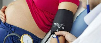
Normal blood circulation within the vascular system is ensured by blood pressure.
Vessels: what you need to know?
22.10.2019
There are three main types of blood vessels in the human body: arteries , veins , and lymphatic vessels . They all look like a rubber pipe with many branches and different passages. pink arteries the veins are bluish and soft. Blood vessels are yellowish.
Story
Ancient anatomists connected arteries and veins with various organs. In the Middle Ages, scientists misunderstood the system of arteries ; they thought that they were not all connected to each other. This theory was refuted by the Italian physician Jacopo Berengario da Carpi at the end of the 15th century. He noticed that an artery is connected to every artery . In the 16th century, anatomists tried to answer the question of how blood gets from veins to arteries . This was clarified in the second half of the 17th century by William Harvey, who discovered blood circulation.
Under a magnifying glass
The wall of the vessel consists of three layers. Its inner part consists of a lining of flat cells (the so-called endothelium; the entire inner layer has other parts and is called intima). The middle layer consists of round and spirally oriented smooth muscle cells. The outer layer of the vessel is a compound that forms a flexible membrane (adventitia). The adventitia connects blood vessels and nerves to nourish and control vascular . Arteries have a thicker layer of muscle than veins . The capillary wall consists of a single layer of cells - the endothelium. In the veins, the endothelium forms a small pocket, a flap, in certain places, preventing blood flow. Blood vessels have a similar structure to veins .
Comparative anatomy of blood vessels
As is known, humans have a closed blood circulation. For example, mollusks, snails, arthropods and jellyfish have open blood circulation. Their “blood” or hemolymph, if present, is located directly between the organs of the body. Insects are less developed, they have only a simple tubular heart .
Anatomy
Blood vessels extend from the heart to all organs and cells. The aorta originates in the left ventricle, descends into the abdomen and flows into the pelvis, where it divides into two arteries that carry blood to the lower extremities. arteries (emanating from the aorta) also function blood to the head, upper limbs, all internal organs and skin. The arteries branch into smaller ones and eventually become capillaries. Arterial capillaries pass into venous capillaries, which converge further into veins . The superior vena cava carries blood to the heart from the lower extremities, abdomen and torso, and it also carries blood from the head and upper extremities. Blood vessels form blindly between cells flowing into stronger trunks and entering veins .
Functions
Blood vessels are used to transport blood in the body. A person has a so-called closed circulation. This means that blood flows only in blood vessels and does not accidentally “wash” certain organs. Arteries carry blood from the heart . Most of this blood is oxygenated, except for the blood that passes through the pulmonary artery from the right ventricle to the lungs . Veins carry blood to the heart . Except for the blood in the pulmonary veins , which is oxidized and flows into the left ventricle.
In the human body there are two circles of blood connected by the heart . The "small" or pulmonary circulation is where oxygenated blood is drawn through the pulmonary artery from the right ventricle into the lungs , where the pulmonary veins lead to the left ventricle. And the “large” circulation, where oxygenated blood from the left ventricle moves to the right atrium, and from there to other arteries and organs. There the blood loses oxygen and the veins return it to the heart . Blood vessels collect saliva in the intercellular spaces and transport it to the veins .
Published in Cardiology Premium Clinic
Cellular structure of blood
Blood consists of two components: plasma (50-60%) and suspended formed elements (40-50%).
The second category includes:
· Erythrocytes (red blood cells) are the most numerous of the formed elements. According to official studies, one drop of blood contains about 5 million red blood cells. Red blood cells are responsible for transporting gases - oxygen and carbon dioxide. They contain the protein hemoglobin, which binds oxygen molecules in the lungs. Red blood cells deliver oxygen to all tissues and organs, after which they absorb carbon dioxide and carry it to the lungs. It is removed from the body during respiration.
· Leukocytes (white blood cells) - elements that protect our body from foreign bodies and compounds, are part of the immune system. White blood cells recognize and attack pathogens through the production of antibodies and macrophages. When an infection enters the body, the production of leukocytes increases significantly. Normally, their quantity is inferior to the concentration in the blood of other formed elements.
· Platelets (blood platelets) are cells that provide coagulation (clotting) of blood flowing from a damaged vessel and protect the body from heavy blood loss. They stick to the hole in the damaged vessel, forming a “sealing” plug to stop bleeding. It is the platelets that can stick together and form pathological blood clots inside the vessels, called thrombi.
All formed elements are synthesized by the bone marrow and distributed through plasma, the liquid part of the blood.