Heart defects can be congenital or acquired.
When planning a pregnancy, a woman should identify (or rule out) a congenital heart defect. This will make it possible to take a conscious and balanced approach to family planning and, if pregnancy and childbirth are possible, to prepare for them in advance.
90% of acquired heart defects develop against the background of rheumatism; they can also occur during pregnancy (exacerbation of rheumatism in pregnant women is most often observed in the first three and last two months of pregnancy). Fortunately, there is now a wide range of methods for diagnosing and treating this disease. It is especially important for women suffering from rheumatism to plan a pregnancy. A favorable prognosis for pregnancy is possible if it occurs against the background of an inactive rheumatic process.
Thanks to the improvement of methods for diagnosing and treating heart diseases, many patients with similar diseases, previously doomed to infertility, have the opportunity to carry and give birth to a child.
Congenital heart defects in the expectant mother
Among congenital heart defects there are three groups:
- Defects with blood discharge from right to left. These include defects of the interatrial and interventricular septum - their non-closure (through the holes, improper discharge of blood occurs), as well as a patent ductus arteriosus *.
- Defects with blood discharge from left to right - transposition (movement) of the main (main) vessels.
- Defects in which there is an obstruction to blood flow (narrowing of large vessels).
*The ductus arteriosus is a short, thin-walled vessel connecting the pulmonary artery and the aorta. Through it, normal blood circulation of the fetus is ensured in the prenatal period - bypassing the lungs, since oxygen enters through the placenta, and normally this duct closes up after birth.
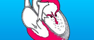
As you know, the heart is a hollow muscular organ responsible for pumping blood in the body. It consists of four departments. These are the right atrium and right ventricle, which make up the right heart, and the left atrium and left ventricle, which make up the left heart. Oxygen-rich blood coming from the lungs enters the left atrium through the pulmonary veins, from there into the left ventricle and then into the aorta. Venous blood enters the right atrium through the superior and inferior vena cava, from there into the right ventricle and further through the pulmonary artery into the lungs, where it is enriched with oxygen and again enters the left atrium.
A heart defect is a persistent pathological change in the structure of the heart that impairs its function. Heart defects can be congenital or acquired. Congenital heart defects are much less common than acquired ones. They arise as a result of disruption of the normal development of the heart and great vessels in fetal development and account for 1-2% of all heart diseases.
The most common defects in pregnant women are the first group. If the disease is accompanied by circulatory failure, i.e. the heart, already working at the limit, can no longer cope with the load, then termination of pregnancy is necessary. Circulatory failure is manifested by fatigue, palpitations, and in more severe cases, shortness of breath and heaviness in the chest. Lack of medical care with the rapid development of heart failure can lead to serious consequences - pulmonary edema (that is, actually turning them off from breathing), thromboembolism (clogging of blood clots) of the pulmonary artery or thrombosis of other large vessels. But usually women with this disease tolerate pregnancy and childbirth well.
Often, after timely surgical correction of a heart defect (if the hole was narrow, it is widened, and vice versa), expectant mothers cope well with the load and the pregnancy ends happily. Recently, more and more women have undergone heart surgery before pregnancy and even during pregnancy.
Defects of the second group are the most severe, and usually the pregnancy has to be terminated, since with these defects circulatory disorders are quite serious and the pregnant woman’s heart often cannot cope with the resulting load.
Obstruction of blood flow in patients of the third group in the absence of circulatory failure usually does not serve as an indication for termination of pregnancy, however, childbirth often ends with a cesarean section, which is associated with a significant increase in the load on the heart during childbirth.
Tactics for managing a pregnant woman
Obstetric tactics for managing a pregnant woman who is carrying a fetus with congenital heart disease involves raising the question of amnio- or cordocentesis after a thorough echocardiographic examination. Their goal is to obtain material for chromosome analysis. If a defect is detected in a non-viable embryo, the pregnant woman will be offered an abortion. The period does not matter at all, especially if the heart defect is combined with genetic abnormalities.
If congenital heart disease is compatible with life, the pregnant woman will be under intensive observation by obstetricians until the end of her term. After forty weeks, she will be asked to be hospitalized for delivery in a specialized perinatal center. As a rule, delivery of such a fetus by cesarean section is indicated.
After birth, the baby will be transferred for examination, treatment and possible surgery to the cardiology department of the children's hospital.
Acquired heart defects
Acquired heart defects are associated with inflammation of the endocardium (the inner lining of the heart) and the myocardium - the heart muscle.
These inflammations can occur with rheumatism - a disease of the heart and connective tissue, sepsis - a generalized infectious lesion, atherosclerosis, syphilis. Under the influence of the inflammatory process, scar tissue develops in the valve, which causes deformation and shortening of the valve leaflets or narrowing of the opening. As a result, the valve cannot completely close the hole and valve insufficiency occurs. If the valves are insufficient during systole (contraction of the ventricles), a reverse unnatural flow of blood occurs from the ventricles into the atria. With stenosis (narrowing) of the left atrioventricular orifice during diastole (relaxation of the ventricles), blood does not have time to move from the atrium to the ventricle. Pathological overflow of the left atrium occurs, and the load on it increases. Thus, heart defects lead to poor circulation. Most often (in 75-90% of cases), acquired heart defects develop against the background of rheumatism. (This disease is caused by streptococcus, the same microbe that usually causes tonsillitis and often affects young women.) The predominant lesions of the heart valves are the mitral valve, located between the left atrium and the left ventricle, and the aortic valve, located between the left ventricle of the heart and the aorta. These lesions lead to disruption of the normal functioning of the valves, overload of the heart muscle and circulatory failure.
Often stenosis and insufficiency develop on the same valve (the so-called combined defect). In addition, there are cases when the defects affect two or more valves - this is called combined heart disease.
How will pregnancy proceed if the mother has a heart defect?
The prognosis of pregnancy depends on the degree and combination of damage, as well as on the activity of the rheumatic process (in other words, on whether it is currently exacerbating) and on the severity of circulatory disorders.
The issue of maintaining or terminating a pregnancy is decided collectively by a cardiologist and an obstetrician-gynecologist in each case individually. If pregnancy occurs after heart surgery, you need to consult a cardiac surgeon. You should know that corrective heart surgery does not always lead to the elimination of organic changes in the valve apparatus or the elimination of congenital developmental anomalies. Often, after surgical treatment, there is a relapse of the underlying disease, for example, in the form of restenosis (re-narrowing) after some operations.
It is extremely difficult to decide whether pregnancy is permissible in women with prosthetic heart valves. They are at high risk of developing blood clots, so pregnant women with mechanical valves constantly receive anticoagulant (anti-clotting) therapy.
Of course, the issue of continuing pregnancy in women with cardiovascular diseases is best decided in advance, before its onset. The basis for proper management and treatment of such pregnant women is an accurate diagnosis that takes into account the cause of the disease.
Defects with blood discharge to the right serve as a contraindication to pregnancy, as well as any type of defect with decompensation, in which circulatory failure has already formed.
Features of pregnancy management with heart defects
Pregnancy management in women with heart defects, as mentioned above, is carried out with the participation of several specialists. It requires not only the coordinated activities of specialists at the antenatal clinic, but also the disciplined behavior of the woman herself: early registration with the antenatal clinic, timely visits to doctors and tests, a full examination, and timely comprehensive treatment.
If possible, then, of course, it is better to entrust your health to a large medical center specializing in this problem. This could be a department for women with cardiovascular pathology at an institute or a specialized department at a large maternity hospital, where competent specialists with experience in managing patients with such pathology can effectively help.
The course of pregnancy in women with heart defects has its own characteristics. Often there are complications such as gestosis (complications of pregnancy, manifested by the appearance of edema, protein in the urine, increased blood pressure), which are characterized by a latent course and are difficult to treat. Pregnancy in such patients is often complicated by the threat of termination - the number of spontaneous abortions and premature births significantly exceeds the average. In addition, the course of pregnancy can be complicated by impaired uteroplacental blood flow, which leads to hypoxia (oxygen starvation) or intrauterine growth retardation. The risk of placental abruption is also high. The accumulation of blood clots in the placenta leads to the exclusion of part of the placenta from the bloodstream and increased oxygen starvation of the fetus.
For all the above reasons, women with heart defects and other pathologies of the cardiovascular system must be hospitalized at least three times during pregnancy:
The first hospitalization is at the 8-10th week of pregnancy to clarify the diagnosis and decide on the possibility of continuing the pregnancy. The issue of termination of pregnancy before 12 weeks is decided depending on the severity of the defect, the functional state of the circulatory system and the degree of activity of the rheumatic process.
The second hospitalization is at the 28-29th week of pregnancy to monitor the state of the cardiovascular system and, if necessary, to maintain heart function during the period of maximum physiological stress. This is due to the fact that it is during this period that the load on the heart normally increases significantly (one of the periods of maximum physiological stress) - the so-called cardiac output increases by almost a third, mainly due to an increase in heart rate.
The third hospitalization is at the 37-38th week to prepare for childbirth and choose a method of delivery, drawing up a birth plan.
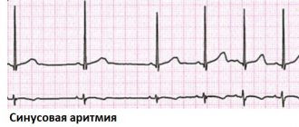
If signs of circulatory failure, exacerbation of rheumatism, the occurrence of atrial fibrillation (frequent irregular heartbeat), gestosis or severe anemia (decreased hemoglobin) and other complications occur, hospitalization is necessary, regardless of the stage of pregnancy.
The issue of termination of pregnancy at a later stage is quite complex. Doctors often have to decide what is less dangerous for the patient: to terminate the pregnancy or allow it to develop further. In any case, if signs of circulatory failure or any concomitant diseases appear, the patient should be hospitalized, subjected to a thorough examination and treatment.
If the situation does not require such drastic measures, the pregnant woman should exercise maximum caution. First of all, you need to take care of sufficient rest and long, 10-12-hour sleep. A 1-2 hour nap during the day is beneficial. Quite effective means of treatment and prevention are physical therapy, morning exercises, and walks in the fresh air. The set of morning exercises should be the simplest, not leading to excessive strain or fatigue.
The diet must be made as varied and nutritious as possible, with a high content of protein products (up to 1.5 g/kg body weight). You need to take multivitamins. In addition, the doctor may prescribe sessions of hyperbaric oxygenation (sessions in a pressure chamber where air with a high oxygen content is supplied under pressure), general ultraviolet irradiation.
What body systems begin to work in a new way?
After conceiving a child, global physiological changes occur in a woman’s body. Great changes are observed in the functioning of the endocrine system. The production of hormones of the anterior pituitary gland, which are responsible for the formation and development of the follicle in the ovary, is suppressed. The corpus luteum is formed - a hormone-producing gland that plays a key role in maintaining pregnancy in the early stages.
A temporary gland forms in the right or left ovary after ovulation and produces progesterone. Functions of progesterone:
- prevents the release of new eggs and the development of menstrual bleeding;
- stimulates the growth of the endometrium, prepares the uterine mucosa for the implantation of the fertilized egg,
- ensures implantation of the embryo and its further normal development;
- promotes the formation of a mucus plug in the cervix, which is necessary to protect the embryo from infections;
- reduces the tone of the muscular layer of the uterus, preventing its contraction and miscarriage.
In non-pregnant women, the corpus luteum dissolves two weeks before menstruation. During the period of bearing a child, it exists until 10-12 weeks of gestation, after which the temporary gland ceases to exist and its functions are performed by the placenta.
The functioning of the corpus luteum is ensured by the hormone hCG, which begins to be produced by chorion tissue 6-8 days after the fusion of male and female gametes. HCG enhances the synthesis of sex hormones and corticosteroids produced in the adrenal cortex.
An important hormone during pregnancy is prolactin, which is produced by the pituitary gland. Starting from the 8th week of pregnancy, prolactin production increases. This is necessary to prepare the mammary glands for breastfeeding, as well as for the normal formation of the lung tissue of the embryo.
In the early stages of pregnancy, the functioning of the thyroid gland changes. The organ increases in size and begins to synthesize more hormones that are important for the development of the embryo.
The immune system
During the period of bearing a child, even in an absolutely healthy woman, the body's defenses decrease. This is due to changes in hormonal levels and the body’s adaptation to new conditions.
The likelihood of acute inflammatory processes and exacerbation of chronic diseases increases. Pathologies of the respiratory, digestive, and genitourinary systems may worsen. The situation is often complicated by the fact that many preventive and therapeutic medications are contraindicated during pregnancy.
What can a pregnant woman do to strengthen her immune system?
- Create a useful menu. Fresh vegetables and fruits, dairy products, fish, nuts, green tea - all this strengthens the immune system.
- Lead an active lifestyle and monitor your body weight. Excess weight and inactivity reduce the body's defenses.
- Avoid stress. Emotional experiences suppress the functioning of the immune system.
In addition, during pregnancy it is important to treat any inflammatory processes in a timely manner. All body systems are interconnected. For example, a common cold can cause acute pyelonephritis or other serious illness.
Psycho-emotional changes
Almost all women during pregnancy are subject to sudden emotional changes, because changes in hormonal levels affect the functioning of the central nervous system. The psycho-emotional state of a pregnant woman often worsens during toxicosis. In this regard, a pregnant woman may experience:
- tearfulness;
- irritability;
- dizziness;
- drowsiness.
As a rule, such manifestations are characteristic only of early pregnancy. Good sleep, moderate physical activity, pleasant activities and hobbies will help you cope with emotional stress in the first trimester.
Online consultation with a gynecologist
Online consultation
During the consultation, you will be able to voice your problem, the doctor will clarify the situation, interpret the tests, answer your questions and give the necessary recommendations.
Management of childbirth with heart disease
The issue of medical tactics during childbirth is of particular importance. The best choice is early hospitalization at 36-37 weeks of pregnancy. The delivery plan is drawn up in consultation with the participation of an obstetrician, cardiologist or therapist and anesthesiologist. The choice of method is strictly individual for each patient, depending on the situation.
Modern medicine has a sufficient arsenal of diagnostic tools to promptly prevent pregnancy complications in women with heart defects. The following methods are usually used:
- Electrocardiography is the recording of electrical phenomena occurring in the heart muscle when it is excited. This study allows you to register changes in the heart muscle by changes in electrical impulses.
- Phonocardiography is a method of recording sounds (tones and noises) resulting from the activity of the heart. It is used to assess heart function and recognize abnormalities, including valve defects.
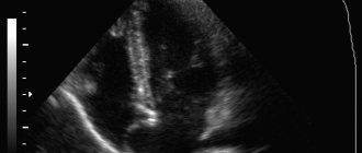
- Echocardiography (ultrasound of the heart). It is used to study blood circulation and cardiodynamics (heart function), determine the size and volume of the cavities of the heart, and assess the functional state of the heart muscle. The method is harmless to the mother and fetus.
- Load testing is used to assess the functional state of the heart muscle. Load tests on a bicycle ergometer are also used when examining pregnant women - during this test, an ECG is taken for the patient at different intensities of physical activity.
- The study of the function of external respiration and the acid-base state of the blood involves studying lung capacity and blood oxygen saturation at rest and during exercise. The study allows you to determine how adequate the blood oxygen saturation is, i.e. How well the heart can cope with the load at the moment.
- Blood tests are usually a standard test that is performed when examining all pregnant women. However, in this case, the doctor pays special attention to the state of the blood coagulation system.
- Fetal ultrasound, cardiotocography , which are regularly performed after 28 weeks to assess the condition of the placenta and fetus. These studies help determine whether the fetus is suffering from a lack of oxygen and nutrients. In addition, fetal ultrasound allows you to identify possible malformations of the child even before birth and take appropriate measures - from urgent surgery after delivery to termination of pregnancy.
The advantage remains with the natural method of delivery. In case of compensated heart disease, therapy is carried out aimed at preventing heart failure and supporting the heart, preventing pulmonary edema, and, if arrhythmia is possible, ECG monitoring. Adequate labor pain relief is provided, as fear and pain lead to additional stress on the heart.
Advertising
As a rule, the most difficult period of labor - pushing (the period of expulsion of the fetus) - is tried to be shortened with the help of an episiotomy - incision of the perineum. Switching off pushing (application of obstetric forceps) is carried out in case of circulatory problems.
Many doctors believe that full-term delivery by cesarean section reduces the burden on the cardiovascular system and reduces mortality among pregnant women with heart defects.
Caesarean section for heart defects is performed in the following cases:
- with an active rheumatic process (fever, pain in the relevant organs, characteristic changes in tests);
- with severe heart disease with severe left ventricular failure and no effect of drug therapy;
- when a heart defect is combined with obstetric pathology requiring surgical delivery.
Successful delivery of patients suffering from severe congenital and acquired heart defects can be facilitated by delivery under conditions of hyperbaric oxygenation.
After the birth of the fetus and the passage of the placenta, there is a rush of blood to the internal organs (and primarily to the abdominal organs) and a decrease in blood circulation in the vessels of the brain and the vessels feeding the heart muscle. Therefore, to prevent deterioration of the condition, immediately after the birth of the child, the woman is given medications to ensure normal heart function.
Clinical manifestations during pregnancy
As a rule, pregnancy with a fetus with congenital heart disease for a woman is not much different from the typical option. The diagnosis becomes obvious only when performing an ultrasound of the fetus and its heart, Doppler sonography or CTG.
The fetus, as a rule, suffers from hypoxia and is characterized by developmental delays. It is possible for a pregnant woman to be prescribed medications that facilitate the functioning of the fetal heart. But, as a rule, we are not talking about full compensation for the condition.
The main task of a mother carrying a child with a heart defect is to maximize the gestational age in the dynamics of the fetal condition.
Postpartum period in women with heart defects
The risk of thrombosis, bleeding and heart failure persists up to 5 days after birth, so during this period the woman is prescribed bed rest. From the 7-11th day, if the defect is of a rheumatic nature, the woman is prescribed a study to determine the activity of rheumatism; The activity of rheumatism is monitored during the year after childbirth.
Postpartum women with heart disease can be discharged from the maternity hospital no earlier than 2 weeks after birth in satisfactory condition under the supervision of a cardiologist at the place of residence.
If decompensation does not occur after childbirth and medications are not required, breastfeeding is preferable.
After the birth of a child, specialists must examine him for the presence of heart defects, since in children whose mothers had such problems, the risk of having them increases many times over.
Thus, timely diagnosis and proper treatment allow many women who were previously unable to have a child to experience this joy. And experts are always ready to help them with this.
Sources
- Pereira SP., Tavares LC., Duarte AI., Baldeiras I., Cunha-Oliveira T., Martins JD., Santos MS., Maloyan A., Moreno AJ., Cox LA., Li C., Nathanielsz PW., Nijland MJ., Oliveira PJ. Sex dependent Vulnerability of Fetal Nonhuman Primate Cardiac Mitochondria to Moderate Maternal Nutrient Reduction. // Clin Sci (Lond) - 2021 - Vol - NNULL - p.; PMID:33899910
- Ben Abdelaziz A., Bchir A., Ben Salah A., Ben Salem K., Mansour N., Hsairi M., Ennigrou S., Nacef T., Dhidah L., Bellaj R., Mehdi F. Professor Mohamed Soussi Soltani : Leader, Innovator and Researcher in Public Health. // Tunis Med - 2021 - Vol99 - N1 - p.5-11; PMID:33899170
- Saavedra LPJ., Prates KV., Gonçalves GD., Piovan S., Matafome P., Mathias PCF. COVID-19 During Development: A Matter of Concern. // Front Cell Dev Biol - 2021 - Vol9 - NNULL - p.659032; PMID:33898461
- Alexanderson-Rosas E., Antonio-Villa NE., Sanchez-Favela M., Carvajal-Juarez I., Oregel-Camacho D., Gopar-Nieto R., Flores-Garcia AN., Keirns C., Hernandez-Sandoval S. ., Espinola-Zavaleta N. Assessment of Atypical Cardiovascular Risk Factors Using Single Photon Emission Computed Tomography in Mexican Women. // Arch Med Res - 2021 - Vol - NNULL - p.; PMID:33896676
- Bhasin D., Arora GK., Isser HS. Young woman with recurrent pregnancy loss. // Heart - 2021 - Vol107 - N10 - p.821-854; PMID:33893215
- Lee K., Laviolette SR., Hardy DB. Exposure to Δ9-tetrahydrocannabinol during rat pregnancy leads to impaired cardiac dysfunction in postnatal life. // Pediatr Res - 2021 - Vol - NNULL - p.; PMID:33879850
- Richards C., Sesperez K., Chhor M., Ghorbanpour S., Rennie C., Ming CLC., Evenhuis C., Nikolic V., Orlic NK., Mikovic Z., Stefanovic M., Cakic Z., McGrath K ., Gentile C., Bubb K., McClements L. Characterization of cardiac health in the reduced uterine perfusion pressure model and a 3D cardiac spheroid model, of preeclampsia. // Biol Sex Differ - 2021 - Vol12 - N1 - p.31; PMID:33879252
- Song Y., Xu J., Li H., Gao J., Wu L., He G., Liu W., Hu Y., Peng Y., Yang F., Jiang X., Wang J. Application of Copy Number Variation Detection to Fetal Diagnosis of Echogenic Intracardiac Focus During Pregnancy. // Front Genet - 2021 - Vol12 - NNULL - p.626044; PMID:33868367
- Khanna R., Chandra D., Yadav S., Sahu A., Singh N., Kumar S., Garg N., Tewari S., Kapoor A., Pradhan M., Goel PK. Maternal and fetal outcomes in pregnant females with rheumatic heart disease. // Indian Heart J - 2021 - Vol73 - N2 - p.185-189; PMID:33865516
- Kirby A., Curtis E., Hlohovsky S., Brown A., O'Donnell C. Pregnancy Outcomes and Risk Evaluation in a Contemporary Adult Congenital Heart Disease Cohort. // Heart Lung Circ - 2021 - Vol - NNULL - p.; PMID:33863666