In order to talk about why a low pulse occurs (bradycardia), it is necessary to recall a little the anatomy and electrophysiology of the heart.
The heart has two atria and two ventricles. First, the atria contract, driving blood into the ventricles of the heart, then the ventricles contract, throwing blood into the largest vessel, the aorta, and through it the blood is distributed to all organs and tissues.
What makes the heart beat?
Like any organ, the heart has a conducting system, in other words, wires through which “current” flows to its various parts. The “electric current” originates spontaneously in the sinus node (a small point in the wall of the right atrium). This node is necessary for generating an impulse and adjusting the frequency of contractions depending on the needs of the body, for example, when we run it speeds up the heart, and when we sleep it slows it down.
From the sinus node, the impulse first goes to the atria, causing them to contract. Then the “wires” merge into one so-called “switch” or AV node (it is located in the middle of the heart between the atria and ventricles, in this node there is a slight delay in the passing “current” for the full contraction of the atria), then the impulse passes to the ventricles. In the ventricles, the “wires” branch and are called the bundle branches (left and right; the left, in turn, is further divided into the anterior branch and the posterior branch) with their help, the impulse causes contraction of the ventricles of the heart.
How to measure correctly?
The main rule is to do this at the same time of day in the same body position. It is best in the morning, immediately after waking up, without getting out of bed. You need to count the beats within a minute: the “economical” approach to measurement, when counting is carried out for 10, 15, 20 or 30 seconds, and then the result is multiplied, does not give an accurate picture. The reason for this is the unequal heart rate even within 1 minute.
There are different opinions about which area of the body is best to take measurements. Experts agree that the radial artery gives the most accurate readings. Place your thumb just below the first crease on your wrist and listen. To count, use three fingers: hold your hand motionless with your middle and index fingers, and measure with your thumb.
Why might your heart rate drop?
Unfortunately, this entire system can break down at different stages and lead either to a decrease and/or cessation of the flow of “current” (blockade) or to incorrect conduction of the impulse in various parts of the heart:
- The first “breakage” may be at the level of the sinus node itself. For various reasons, organic or functional, it loses the ability to produce the required number of impulses. For example, no matter how much a person runs or climbs the stairs, the sinus node cannot rise above a hundred beats, which may result in increased shortness of breath or weakness.
- The second “breakage” may be at the level of the atrioventricular (AV) node. Let me remind you that it is located between the atria and ventricles, an impulse passes through it from the atria to the ventricles; it is needed in order to create a short physiological pause for a more complete contraction of the atria. At this level, all kinds of blockades (AV blocks) can occur. They come in different degrees, 1st, 2nd or 3rd degree depending on the severity of the condition. From the name it is clear that they block the impulse at this stage, as a result the ventricles of the heart cannot contract, this leads to pauses, sometimes significant ones (3 or more seconds); for example, depending on the body’s reserves and concomitant pathology, starting from a 3-second delay in heart contraction, a person may begin to briefly lose consciousness; as these blockades progress, consciousness, sometimes, may not return. Let us reassure you a little: nature has come up with life-saving “(emergency) stations” for generating backup impulses; they are located at all stages of the conduction system, but the “lower” they are located, the less “current” they are able to reproduce. So, in case of this breakdown, the “backup generator” is already in the ventricles and is capable of delivering a maximum of 40 beats or less, so that this can allow one to “hold out” to the resuscitation ambulance team.
- The third “breakdown” can occur below the atrioventricular (AV) node at the level of the bundle branches (these are “wires” that extend from the atrioventricular node and excite the ventricles; we will talk about them in more detail in another article).
Unfortunately, these legs can also be partially or completely blocked due to various reasons, thereby preventing the full contraction of the ventricles of the heart.
Under what circumstances is it not recommended to measure your pulse?
There are a number of circumstances where the results will be biased. Sometimes this happens during a consultation with a cardiologist if the patient is too excited, but there are other situations.
- You should not check your pulse within an hour after eating, taking medications, or especially drinking alcohol. A lot of processes take place in the body, and the heart works at an accelerated pace.
- Indicators will be biased when a person is thirsty or hungry.
- The heart rate is always elevated after physical activity or training.
- Another reason to refuse the procedure is a massage, visiting a sauna or going to the beach: under the influence of high temperatures you should not expect objective information.
- Doctors do not recommend women to do calculations during menstruation, when hormonal changes occur in the body.
The ability to measure your pulse will come in handy sooner or later, so take into account all the nuances and practice on yourself and your loved ones. It is advisable to develop the skill in yourself so that in a critical situation you do not remember the equipment, but quickly come to the rescue.
What causes these “breakdowns” in the conduction of impulses?
- Diseases leading to overload and hypertrophy of the ventricles such as cor pulmonale, congenital and/or acquired heart defects with increased load on the right and/or ventricles (for example, mitral and aortic stenosis/insufficiency or septal defects), arterial hypertension, myocardial infarction various localizations, cardiomyoptia, myocardial dystrophy, etc.
- Various congenital anomalies of the development of the cardiac conduction system (Brugada syndrome)
- Age-related changes (cardiosclerosis)
- Idiopathic (acting as a separate disease, causeless) calcification or sclerosis of the cardiac conduction system
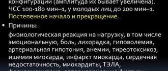
- Also, a low heart rate in itself can be normal in trained athletes (it can reach up to 35 beats per minute)
- changes in the electrolyte composition of the heart (hyperkalemia or hypercalcemia)
- Non-cardiac causes (increased intracranial pressure, decreased body temperature, hypothyroidism, etc.)
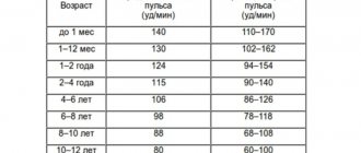
What factors are taken into account when measuring?
- Times of Day. The heart rate, even in a completely healthy person, can change throughout the day, which is confirmed by daily ECG monitoring. The lowest readings occur at night or in the morning; in the evening, on the contrary, the heart rate increases, and the difference can be up to 10 beats. Such fluctuations in the absence of other signs are considered a physiological norm.
- Body position. When sitting, the frequency is always higher than when a person is lying down, but as soon as he gets up, the indicator goes off scale. This is due to the increased workload on the heart associated with physical activity.
Why do different types of bradycardia develop?
- Diseases of the central nervous system. A decrease in heart rate often occurs in patients with VSD (vegetative-vascular dystonia), increased intracranial pressure, neoplasms and brain contusions. People with such diseases suffer from chronic neurogenic bradycardia. It also occurs against the background of neuroses with autonomic disorders. If a person wears clothing that compresses the neck, the carotid sinus is disrupted and the pulse slows down.
- Endocrine pathologies, gastrointestinal diseases. People with stomach and duodenal ulcers often suffer from a slow pulse. When the production of thyroid hormones decreases, metabolic processes are disrupted and the heart rate decreases. With the development of myxedema, which is expressed by an acute lack of thyroid hormones, bradycardia worsens and begins to threaten human life. The same thing happens with diabetic ketoacidosis, a decrease in adrenaline production by the adrenal glands.
- Cardiological pathologies. Bradycardia is a type of arrhythmia and is most often associated with heart disease. Low pulse is one of the symptoms of myocardial infarction or inflammation, cardiosclerosis, and a number of other pathologies. The reason is changes in the tissues of the sinus node and myocardium. The process of generating electrical impulses is disrupted, and tissue conductivity deteriorates. The most severe complication is dysfunction of automatism, in which the production of impulses stops. If the pathology affects the myocardium, the generation of impulses is preserved, but there are difficulties with their transmission to the ventricles. Only a certain proportion of the signals reach the target, so the heart rate becomes slow.
- Incorrect medication intake. Some types of drugs cause drug-induced bradycardia. It is caused by incorrectly selected dosage or hypersensitivity to the components of cardiac glycosides, anti-arrhythmic drugs, adrenergic blockers, and morphine-based drugs.
- Poisoning, intoxication in diseases. Slowing of the pulse occurs in people with impaired water and electrolyte balance, as well as with severe infectious lesions of the body. In severe forms of hepatitis, liver failure develops and, as a result, intoxication. The same happens with sepsis and typhoid fever. Heart rate slows down with excessive intake of calcium and potassium into the body, and poisoning with organophosphorus substances.
Manifestations
With a slight reduction in heart rate, the disease is asymptomatic and is detected by measuring the pulse. More pronounced bradycardia manifests itself as symptoms of circulatory disorders:
- weakness, fatigue, dizziness;
- pallor and bluish tint of the skin;
- slowdown in intellectual and physical development in childhood;
- irritability;
- excessive sweating;
- fainting;
- decrease in blood pressure;
- chest pain.
If the heart rate drops critically, the patient requires emergency medical care.
Surgical treatment
The reason for surgical intervention for bradycardia is the patient’s serious condition, which has developed in connection with heart defects or acquired diseases. Operations are performed for children and adults. For children with congenital pathologies, epicardial stimulators or endocardial electrodes are installed.
Young and older people are advised to have a pacemaker installed. It takes over the production of electrical impulses that are generated at a physiological frequency. This allows you to ensure a normal heart rate and prevent negative consequences.
What affects a sleeper's heart rate?
Pulse is one of the types of biomarkers, relying on its indicators to determine the general condition of the human body. At the time of sleep, jerky oscillations in the artery decrease, the functions of the cardiovascular system slow down, and metabolic processes weaken. Cells require less oxygen.
This process regulates the activity between organs, some are stimulated, others, on the contrary, arrive in complete relaxation. Heart rate and intensity are affected by:
- gender identity;
- age;
- physical state.
The more trained a person is, the lower the heart rate reduction. For men, the difference is considered to be in the range of minus 5-10 beats. In children, compared to the older generation, pulsation increases by 25-30 beats/minute.
Treatment for low heart rate
To cure a decreased heart rate, you need to know exactly its cause. If the condition is caused by hormonal pathology or water-electrolyte imbalance, therapy is selected after conducting the necessary tests for hormones and electrolytes. Taking medications replenishes the lack of necessary substances and thereby normalizes heart rhythm.
If bradycardia is due to drug overdose, other drugs should be selected or daily doses reduced.
A low pulse during intoxication is a side effect caused by the underlying disease, so it is this that needs to be treated.
If the decrease in heart rate is due to cardiac pathology, the cardiologist selects the appropriate treatment for the given clinical case. Even in the absence of symptoms, people with bradycardia need to undergo regular medical examinations to monitor changes in heart function.
For a chronic decrease in heart rate, the following is prescribed:
- cardioprotectors;
- drugs to improve metabolic processes in myocardial tissues;
- antioxidants;
- drugs with anabolic effects.
Duration of treatment is 3-6 months. The doctor selects a combination of medications and monitors the progress of treatment. Complex therapy may include herbal preparations that have a tonic effect. Basically, extracts of ginseng and eleutherococcus are prescribed - natural stimulants.
In severe conditions, it is important to normalize the pulse rate as soon as possible to avoid tissue hypoxia. The patient is hospitalized and parenteral drugs are used to improve myocardial function.
Symptoms of low heart rate
When the number of heartbeats decreases to less than forty, the following are observed: general weakness, increased fatigue, dizziness, loss of consciousness, chest pain (angina), changes in blood pressure, forgetfulness, decreased concentration, blurred vision.
symptoms of low heart rate
These symptoms are caused by hemodynamic disturbances. Due to the slowing of the heartbeat, the brain receives an insufficient amount of oxygen, which is why confusion and convulsions may occur, which disappear in a short period of time. This symptomatology is dangerous and can cause respiratory arrest. Urgent specialized medical care is required.
What to do with bradycardia
? To identify the cause of deviations, a complete comprehensive diagnosis will be required. Initially, the doctor determines the patient’s complaints, collects anamnesis, conducts auscultation of the heart, and measures pulse and blood pressure. In case of bradycardia, an additional examination by a cardiologist is required. The scope of diagnostic measures includes: ECG, 24-hour Holter monitoring, 24-hour blood pressure study, ultrasound of the heart, EchoCG and other methods.
If necessary, the cardiologist may prescribe an additional consultation with an arrhythmologist or cardiac surgeon.