Vasorenal hypertension is one of the symptomatic forms of hypertension that occurs as a result of a disorder of the main renal circulation without signs of primary damage to the renal parenchyma and urinary tract. The development of pathology is based on a narrowing of the arterial lumen of both kidneys or several of its vascular branches. As a result, blood circulation in the affected area is impaired, which leads to the formation of ischemic zones, the severity of which depends on the degree of arterial stenosis. What are the causes of this type of hypertension? What clinical signs does it manifest?
Reasons for formation
In most cases, the main cause of renovascular hypertension is atherosclerotic damage to the renal vascular system. Atherosclerotic formations are localized at the mouth of the renal arteries, causing unilateral deformation. With bilateral damage, a more severe clinical picture appears, which can be complicated by arterial thrombosis.
To date, experts have identified 40 etiological factors against which renal hypertension is formed. Depending on the mechanism and time of development, they can be divided into congenital, they are diagnosed mainly in patients under 40 years of age, and acquired causes.
Congenital factors include:
- fibromuscular dysplasia of the renal arteries;
- hypoplasia of both blood vessels and the kidney itself;
- aneurysm of the renal vessels;
- arteriovenous fistulas.
The main causes causing the formation of the disease
In the development of pathology, the following acquired causes are distinguished:
- atherosclerotic process;
- aortoarteritis of a nonspecific nature;
- anatomical displacement of the paired organ;
- renal infarction;
- frequent injuries;
- aortic aneurysm dissection.
The disease can also develop as a result of arterial embolism and thrombosis, nephroptosis, neoplasms, and congenital anomalies in the development of paired organs.
Development mechanism
The pathogenesis of the development of vasorenal hypertension is associated with stenosis or obstruction of the renal arteries, which leads to a decrease in the circulating blood volume in the organ and decreased perfusion. A circulatory disorder is detected by insufficient stretching of the arterioles located in the cortex of the kidney.
The mechanism of development of renal hypertension due to atherosclerotic process
Granule cells that perform the function of receptors are localized in the medial layer. They respond to the slightest changes in hemodynamic parameters by releasing renal incretrenin into the bloodstream. The formation of foci of ischemia in the tissues of paired organs leads to an increase in the number of structural cells of the JGA, as a result of which excessive production of renin is observed.
An increased concentration of renin provokes the formation of angiotensin 2, a vasoconstrictor hormone that causes vascular spasm and an increase in peripheral resistance. In addition to this property, angiotensin 2 activates the production of aldosterone by the adrenal cortex, which causes secondary retention of fluid and sodium ions in the body, causing additional swelling of the arterioles and increased arterial resistance. The accumulation of sodium ions in cells causes an increase in circulating blood volume and an increase in cardiac output. The amount of extracellular fluid also increases, sodium accumulates in the smooth muscle layer of the arteries, which provokes their swelling, increased sensitivity to catecholamines, vasopressin, and vasoconstrictor enzymes.
Organic and functional changes in renal tissue lead to activation of the renin-angiotensin-aldosterone system, and then gradually provoke inhibition of autonomic mechanisms, which causes the formation of persistent renal hypertension.
Causes of renovascular hypertension
Narrowing of the renal artery leads to insufficient blood supply to the renal tissue. Blood supply to the kidney is critically important for the body, otherwise urine is not produced and kidney failure develops. When there is a lack of blood flow in the kidney, the special kidney hormone renin is released, which promotes the conversion of angiotensin I to angiotensin II, which causes a strong narrowing of the arteries and the release of the hormone aldosterone, which promotes sodium retention and increases circulating blood volume and water retention.
If the second kidney functions normally, then this effect can be neutralized and there will be no stagnation of fluid in the systemic circulation, however, with bilateral damage, the volume of fluid in the body and the level of blood pressure increase.
Main diseases leading to renovascular hypertension
- Obliterating atherosclerosis of the aorta and renal arteries. Most often, atherosclerosis is the cause of renovascular hypertension in elderly men. The examination reveals multifocal atherosclerotic lesions in the coronary arteries and vessels of the lower extremities.
- Fibromuscular dysplasia is the development of narrowing of the arteries due to connective tissue, without atherosclerotic plaque. A uniform narrowing develops in the affected segment, which can then turn into expansion. Young women are most often affected.
- Renal artery aneurysm. Leads to constant embolization of renal tissue by microthrombi, which leads to the development of renovascular hypertension.
- Aortic coarctation is a congenital narrowing in the thoracic aorta leading to decreased perfusion of organs below the narrowing. Most often, arterial hypertension with coarctation of the aorta is of mixed origin.
Characteristic signs
Clinical symptoms diagnosed during examination of the patient make it less likely to suspect renovascular hypertension. The clinical picture has low specificity of symptoms, so the only manifestation of narrowing of the lumen of the renal vessels may be arterial hypertension syndrome.
The distinctive signs of pathology are the following symptoms:
- A persistent increase in diastolic and, to a lesser extent, systolic pressure.
- The body's resistance to drug treatment.
- Malignant course of the disease.
- Rapid development of complications associated with target organ damage.
The occurrence of pathology in childhood should alert specialists, since fibromuscular dysplasia of the renal arteries can form during puberty.
A particularly severe course of renal hypertension against the background of essential hypertension, when aggravation of its course may be observed:
- stabilization of blood pressure indicators at high values;
- increased diastolic pressure;
- decrease in the therapeutic effect of previously used antihypertensive drugs;
- formation of signs of chronic renal failure.
Important! A characteristic sign of renovascular hypertension is the formation of chronic renal failure, which is observed with bilateral damage to the renal vessels or with unilateral damage in the presence of a concomitant pathological process in the organ (pyelonephritis, chronic glomerulonephritis, nephrosclerosis). The appearance of this syndrome indicates the presence of a stenotic process in paired organs.
Arterial hypertension
Symptomatic arterial hypertension is a secondary hypertensive condition that develops as a result of pathology of the organs that regulate blood pressure. Symptomatic arterial hypertension is distinguished by its persistent course and resistance to antihypertensive therapy, the development of pronounced changes in target organs (heart and kidney failure, hypertensive encephalopathy, etc.). Determining the causes of arterial hypertension requires ultrasound, angiography, CT, MRI (kidneys, adrenal glands, heart, brain), studies of biochemical parameters and blood hormones, and blood pressure monitoring. Treatment consists of medication or surgery to address the underlying cause.
Symptoms of the disease
As a rule, renovascular hypertension is characterized by a severe malignant course. One of the most frequently identified complaints among patients is a persistent increase in blood pressure, especially in the “lower” blood pressure.
The main symptoms of the disease include:
- pain in the head area;
- frequent attacks of dizziness;
- a feeling of “tide” in the brain;
- periodic tinnitus;
- pain in the area of the eyeballs and the appearance of “spots” before the eyes;
- memory impairment;
- sleep disorder
Simultaneous damage to the renal and main arteries can provoke ischemia of other organs
As the pathology progresses, an increase in negative symptoms is observed with the involvement of other organs and systems:
- Excessive load on the left side of the heart and the development of coronary insufficiency (pain in the heart area, increased heart rate, rhythm disturbance, discomfort behind the sternum).
- Kidney infarction (heaviness and pain in the lumbar region, the appearance of blood in the urine).
- Secondary hyperaldosteronism (increased diuresis, bedwetting, muscle weakness, parasthesia).
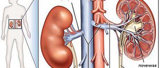
Vasorenal hypertension
What is renovascular hypertension? Vasorenal hypertension is a secondary hypertensive syndrome that occurs due to various disorders of the main blood flow in the kidneys. Normally, blood flowing through the kidneys is filtered there and cleansed of toxins. The kidneys filter waste into urine, which is collected in the bladder, and from where the urine is released through the urethra. The kidneys also regulate blood pressure by secreting the enzyme renin into the bloodstream. Renin in the blood combines with hypertensinogen, and thereby regulates blood pressure. With stenosis (narrowing) of the renal arteries, the kidneys are poorly supplied with blood, which leads to decreased filtration in the kidneys and increased secretion of renin, which in turn leads to increased blood pressure. Renal artery stenosis is a narrowing of the renal arteries. This condition leads to hypertension and can eventually lead to kidney failure.
CAUSES Congenital • Fibromuscular dysplasia • Hypoplasia • Atresia • Renal artery aneurysms Acquired • Atherosclerosis • Nonspecific aortoarteritis • Thrombosis and embolism • Extravasal compression by tumors and scars • Traumatic injuries COMPLAINTS Complaints may be absent for a long time. The main complaint is persistently high blood pressure that is resistant to medications, and in 20-40% of patients it takes a malignant course. Systolic pressure always exceeds 160 mm Hg, and diastolic pressure always exceeds 100 mm Hg. There are 4 groups of complaints: • Complaints characteristic of cerebral hypertension - headache, heaviness in the occipital region, a feeling of rushing in the head, tinnitus, flashing “flies” before the eyes, poor sleep, decreased performance; • Complaints associated with increased stress on the heart - pain and discomfort in the heart area, palpitations, shortness of breath; • Complaints caused by damage to the kidneys themselves - dull pain, a feeling of heaviness in the lumbar region, hematuria (blood in the urine), protein in the urine; • Complaints associated with damage and ischemia in other arterial basins, pain and intermittent claudication in the lower extremities and weakness in the upper extremities, manifestations of carotid and vertebrobasilar insufficiency. This indicates the systemic nature of the disease. What examinations are needed? • Ultrasound duplex scanning of the renal arteries is the most accessible method and allows to identify disturbances in the main blood flow on an outpatient basis. • Spiral computed tomoangiography, along with visualization of the renal arteries, allows you to see the condition of the renal parenchyma itself. • X-ray contrast angiography allows you to visually determine the nature, location and extent of the lesion, assess the degree of their severity and hemodynamic significance. • Magnetic resonance imaging provides two- or three-dimensional reconstructions of blood vessels. • Radionuclide testing is a mandatory test for suspected cerebral hepatitis; it allows one to evaluate renal function. How to treat renovascular hypertension? In the natural course of the disease and the absence of appropriate treatment, about 70% of patients die over the next 5 years from complications of arterial hypertension (cerebral stroke, myocardial infarction, renal failure). Conservative treatment is ineffective or produces an inconsistent and short-lived effect. The main treatment is endovascular. Angioplasty and stenting (endovascular treatment) Angioplasty is a method of expanding the internal lumen of blood vessels using a special balloon. In this case, a specialist - interventional radiologist inserts a thin tube (catheter) through a small puncture and brings a balloon to the narrowed area of the vessel, under the control of X-rays. The balloon expands, pushing apart atherosclerotic plaques, and blood flow through the vessel is restored. After this, a metal frame is installed in this place - a stent, which, when implanted, allows you to maintain the achieved result.
How to behave after surgery? Remember that even after surgery you need to continue to treat the underlying disease (atherosclerosis) and prevent its progression. Some recommendations that must be followed, regardless of his age, gender, financial status, etc.: • Stop smoking. At the same time, passive smoking is just as harmful as active smoking. It has been proven that quitting smoking for a year reduces the risk of developing atherosclerosis of the arteries by 50%. • Take so-called antiplatelet drugs for life, the most common of which is regular aspirin in small doses (50-100 mg per day). This thins your blood and reduces the risk of blood clots in blood vessels and prostheses. • Reduce blood pressure, that is, treat arterial hypertension. • Reduce blood cholesterol levels. Excessive intake of fats from food leads to the deposition of cholesterol in the artery wall. By reducing the level of cholesterol in the blood as a result of a strict diet and taking medications, you promote the release of cholesterol from the so-called “depot”, which includes atherosclerotic plaques. By losing cholesterol, the plaque becomes smoother, the possibility of its rupture and the formation of blood clots at the rupture site decreases, that is, it becomes safer. • Fight excess weight. Losing even 4-5 kilograms can significantly reduce blood pressure, cholesterol and blood sugar levels, and will also significantly reduce the dosage of medications you take. • Fight physical inactivity. Increasing physical activity is not only an opportunity to burn excess calories, but also an excellent way to control blood pressure, cholesterol and blood sugar levels. Don’t be afraid of physical activity, the implanted prosthesis will not come off! You need daily walking, at least 1 hour a day.
Diagnostic principles
Diagnosis of pathology consists of collecting anamnestic data, examination, palpation and conducting instrumental and laboratory studies, on the basis of which a specialist can establish a diagnosis and select the optimal type of treatment.
At the preliminary diagnostic stage, non-invasive research methods are used, with the help of which it is possible to localize the area of the lesion, its nature and the functional state of paired organs. These methods are not absolutely informative, but should precede X-ray contrast angiography.
During the examination, it is necessary to measure blood pressure in the upper and lower extremities, which makes it possible to exclude differences in the blood flow above and below the site of aortic stenosis, as well as to determine the degree of vascular damage. Measurements are taken in both horizontal and vertical positions of the patient. If, when changing the position of the body in space, blood pressure readings are higher than normal, then nephroptosis can be suspected in the patient.
Diagnostic signs that can help identify pathology
Laboratory data:
- Radioimmune method (determines the activity of renin in the bloodstream).
- Blood biochemistry (increased concentration of creatinine, residual nitrogen).
- Clinical analysis of urine (increased amount of albumin, red blood cells).
The ultrasound research method is the most informative screening technique that allows identifying disorders of the main renal circulation at the initial diagnostic stage.
Special research methods:
- Duplex scanning. Allows you to visualize the functional state of the renal vessels and assess the speed of blood flow using contrast agents.
- Magnetic resonance angiography. When using the contrast agent Gadolinium, high-quality three-dimensional image visualization can be achieved.
- Spiral computed tomography angiography. The non-invasive technique is carried out by administering contrast agents intravenously. Allows you to assess the state of renal circulation, as well as obtain an image of the arteries in three dimensions.
- Angiography of the kidneys. Determines the degree of narrowing of the vascular lumen and its location, and also excludes other types of hypertension.
A comprehensive study makes it possible to establish an accurate diagnosis, as well as exclude diseases with similar clinical presentations.
Conservative treatment methods
In most cases, conservative therapy does not bring the desired effect, since the therapeutic effect of the drugs is short-lived. In cases where it was possible to achieve a hypotensive effect in case of arterial stenosis, then in the future there is a deterioration in the blood supply to the affected organ, due to which it decreases in size.
Long-term drug treatment is prescribed when there is no possibility of surgical intervention, and in the postoperative period to stabilize the patient.
The following groups of drugs are used in the treatment of renovascular hypertension:
- Antiplatelet agents (Aspirin, Clopidogrel, Ticlopidine).
- ACE inhibitors (Captopril, Lisinopril, Losartan).
- Diuretics (Furosemide, Hypothiazide).
- Calcium antagonists (Diltiazem, Amlodipine, Lercandipine).
- Beta blockers (Atenolol, Nebivolol, Bisoprolol).
When symptoms of chronic renal failure develop, depending on the severity of the pathology, hypoazotemic drugs are prescribed and the dosage of antiplatelet agents is increased.
Important! Patients are advised to follow a low protein diet combined with a sharp restriction of sodium salts. This diet will help remove toxic substances from the body, restore blood circulation and reduce blood pressure.
Forms of symptomatic arterial hypertension
Nephrogenic parenchymal arterial hypertension
Most often, symptomatic arterial hypertension is of nephrogenic (renal) origin and is observed in acute and chronic glomerulonephritis, chronic pyelonephritis, polycystic and hypoplastic kidneys, gouty and diabetic nephropathy, trauma and kidney tuberculosis, amyloidosis, SLE, tumors, nephrolithiasis.
The initial stages of these diseases usually occur without arterial hypertension. Hypertension develops with severe damage to the tissue or apparatus of the kidneys. The features of renal arterial hypertension are predominantly the young age of patients, the absence of cerebral and coronary complications, the development of chronic renal failure, and the malignant nature of the course (in chronic pyelonephritis - in 12.2%, chronic glomerulonephritis - in 11.5% of cases).
In the diagnosis of parenchymal renal hypertension, ultrasound of the kidneys, urine examination are used (proteinuria, hematuria, cylindruria, pyuria, hyposthenuria - low specific gravity of urine), determination of creatinine and urea in the blood (azotemia is detected). To study the secretory-excretory function of the kidneys, isotope renography and urography are performed; additionally - angiography, ultrasound of renal vessels, MRI and CT of the kidneys, kidney biopsy.
Nephrogenic renovascular (vasorenal) arterial hypertension
Renovascular or vasorenal arterial hypertension develops as a result of unilateral or bilateral disturbances of arterial renal blood flow. In 2/3 of patients, the cause of renovascular arterial hypertension is atherosclerotic damage to the renal arteries. Hypertension develops when the lumen of the renal artery narrows by 70% or more. Systolic blood pressure is always above 160 mm Hg, diastolic blood pressure is always above 100 mm Hg.
Renovascular arterial hypertension is characterized by a sudden onset or sharp worsening of the course, insensitivity to drug therapy, and a high proportion of malignant course (in 25% of patients).
Diagnostic signs of vasorenal arterial hypertension are: systolic murmurs over the projection of the renal artery, determined by ultrasonography and urography - a decrease in one kidney, a slowdown in the removal of contrast. Ultrasound shows echoscopic signs of asymmetry in the shape and size of the kidneys, exceeding 1.5 cm. Angiography reveals a concentric narrowing of the affected renal artery. Duplex ultrasound scanning of the renal arteries determines a violation of the main renal blood flow.
In the absence of treatment for renovascular arterial hypertension, the 5-year survival rate of patients is about 30%. The most common causes of death in patients: cerebral strokes, myocardial infarction, acute renal failure. In the treatment of vasorenal arterial hypertension, both drug therapy and surgical techniques are used: angioplasty, stenting, traditional operations.
With significant stenosis, long-term use of drug therapy is unjustified. Drug therapy produces a short-term and inconsistent effect. The main treatment is surgical or endovascular. For vasorenal arterial hypertension, an intravascular stent is installed to expand the lumen of the renal artery and prevent its narrowing; balloon dilatation of a narrowed section of a vessel; reconstructive interventions on the renal artery: resection with anastomosis, prosthetics, vascular bypass anastomoses.
Pheochromocytoma
Pheochromocytoma, a hormone-producing tumor that develops from chromaffin cells of the adrenal medulla, accounts for 0.2% to 0.4% of all occurring forms of symptomatic arterial hypertension. Pheochromocytomas secrete catecholamines: norepinephrine, adrenaline, dopamine. Their course is accompanied by arterial hypertension, with periodically developing hypertensive crises. In addition to hypertension, pheochromocytomas cause severe headaches, increased sweating and palpitations.
Pheochromocytoma is diagnosed when an increased content of catecholamines is detected in the urine by performing diagnostic pharmacological tests (tests with histamine, tyramine, glucagon, clonidine, etc.). Ultrasound, MRI or CT scan of the adrenal glands allows you to clarify the location of the tumor. By performing a radioisotope scan of the adrenal glands, it is possible to determine the hormonal activity of pheochromocytoma, identify tumors of extra-adrenal localization, and metastases.
Pheochromocytomas are treated exclusively with surgery; before surgery, arterial hypertension is corrected with α- or β-adrenergic blockers.
Primary aldosteronism
Arterial hypertension in Conn's syndrome or primary hyperaldosteronism is caused by an aldosterone-producing adenoma of the adrenal cortex. Aldosterone promotes the redistribution of K and Na ions in cells, fluid retention in the body and the development of hypokalemia and arterial hypertension.
Hypertension is practically not amenable to drug correction; attacks of myasthenia gravis, convulsions, paresthesia, thirst, and nictruria are noted. Hypertensive crises with the development of acute left ventricular failure (cardiac asthma, pulmonary edema), stroke, and hypokalemic cardiac paralysis are possible.
Diagnosis of primary aldosteronism is based on determining plasma levels of aldosterone and electrolytes (potassium, chlorine, sodium). There is a high concentration of aldosterone in the blood and high excretion in the urine, metabolic alkalosis (blood pH - 7.46-7.60), hypokalemia (<3 mmol/l), hypochloremia, hypernatremia. When examining blood from the adrenal veins, a 2-3-fold increase in the level of aldosterone on the affected side is detected. Radioisotope studies and ultrasound scanning of the adrenal glands reveal an enlargement of the adrenal gland affected by aldosteroma or bilateral hyperplasia of the adrenal cortex.
For malignant arterial hypertension caused by aldosteroma, surgical treatment is performed to normalize or significantly reduce blood pressure in 50-70% of patients. Before surgery, a hyposodium diet is prescribed and treatment with an aldosterone antagonist, spironolactone, which relieves hypokalemia and arterial hypertension (25-100 mg every 8 hours).
Itsenko-Cushing's syndrome and disease
Endocrine arterial hypertension develops in 80% of patients with the disease and Cushing's syndrome. Hypertension is caused by hypersecretion of glucocorticoid hormones by the adrenal cortex (hypercortisolism) and is characterized by a stable, crisis-free course, resistance to antihypertensive therapy, and a proportional increase in systolic and diastolic blood pressure. Another characteristic manifestation of the disease is Cushingoid obesity.
With Itsenko-Cushing's syndrome/disease, the level of 11 and 17-OCS, corticotropin, and hydrocortisone increases in the blood. Excretion of 17-KS and 17-OX is increased in urine. For differential diagnosis between corticosteroma and pituitary adenoma, MRI and CT scanning of the adrenal glands, pituitary gland, ultrasound and radioisotope scanning of the adrenal glands, and a craniogram are performed. Treatment of hypercortisolism and the arterial hypertension caused by it can be medication, surgery or radiation.
Coarctation of the aorta
Coarctation of the aorta is a congenital malformation of the aorta, manifested by its segmental narrowing, which impedes systemic blood flow. Coarctation of the aorta is a rare form of hypertension.
With secondary arterial hypertension caused by coarctation of the aorta, there is a difference in blood pressure measured in the arms (high) and legs (normal or low), an increase in blood pressure at the age of 1-5 years and its stabilization after 15 years, weakening or absence of pulsation in the femoral arteries , increased cardiac impulse, systolic murmurs over the apex, base of the heart, and on the carotid arteries. Diagnosis of coarctation of the aorta is based on radiography of the lungs and chest organs, aortography, and echocardiography. If the degree of stenosis is severe, surgical treatment is performed.
Dosage forms of arterial hypertension
The development of medicinal forms of arterial hypertension can cause vascular spasm, increased blood viscosity, sodium and water retention, the effect of drugs on the renin-angiotensin system, etc. Intranasal drops and cold remedies containing adrenomimetics and sympathomimetics in their composition (pseudoephedrine, ephedrine , phenylephrine) may cause hypertension.
Taking non-steroidal anti-inflammatory drugs causes the development of arterial hypertension due to fluid retention and suppression of the synthesis of prostaglandins, which have a vasodilating effect. Oral contraceptives containing estrogens have a stimulating effect on the renin-angiotensin system and cause fluid retention. Secondary arterial hypertension develops in 5% of women using oral contraception.
The stimulating effect of tricyclic antidepressants on the sympathetic nervous system can cause the development of arterial hypertension. The use of glucocorticoids increases blood pressure due to increased vascular reactivity to angiotensin II.
To establish the cause and form of secondary arterial hypertension, the cardiologist needs a detailed collection of the patient’s medical history, analysis of the coagulogram, and determination of blood renin.
Neurogenic arterial hypertension
Arterial hypertension of the neurogenic type is caused by lesions of the brain or spinal cord due to encephalitis, tumors, ischemia, traumatic brain injury, etc. In addition to increased blood pressure, they are typically characterized by severe headaches and dizziness, tachycardia, sweating, salivation, vasomotor skin reactions, abdominal pain, nystagmus, convulsive seizures.
Diagnostics use angiography of cerebral vessels, CT and MRI of the brain, and EEG. Treatment of arterial hypertension of the neurogenic type is aimed at eliminating brain pathology.
Minimally invasive techniques
The goal of the minimally invasive technique is to correct vascular lesions using special devices located at the end of the inserted catheter. The technique can be performed not only by surgeons, but also by an angiography specialist.
Indications for stenting of the renal arteries are:
- atherosclerotic narrowing in critical form;
- complication after endovascular balloon dilatation;
- recurrence of stenosis.
Basic minimally invasive techniques:
- X-ray endovascular balloon dilatation. It is based on the insertion of a catheter into the lumen of the artery, at the end of which an elastic balloon is attached. The balloon must be placed in the area of narrowing, after which it is inflated with liquid under pressure, thus achieving the optimal size of the vessel.
- Superselective embolization of the segmental renal artery. Allows you to reduce local blood flow, which makes it possible to reduce the manifestations of hematuria and hypertension.
Dear patients!
All organs and tissues of the body are penetrated by small arterial vessels - arterioles. Sometimes, for a number of reasons, these vessels begin to narrow. In this case, the heart, in order to push the same amount of blood from the arteries into the veins, needs to apply more force, creating more pressure in the arteries. It is this increase in pressure in the arteries that is called arterial hypertension (simply hypertension or hypertension).
Hypertension occurs in 10-20% of people aged 25-45 years and in 30-40% over 45 years of age.
There are many causes of hypertension; chronic stress, poor nutrition, smoking, age and hereditary factors have been proven to influence the development of this disease. But these are only factors predisposing to the development of the disease. In most cases, the exact cause cannot be determined and a systematic increase in blood pressure is called primary hypertension or essential hypertension. Treatment in this case is aimed at eliminating these negative factors and prescribing medications to lower blood pressure.
However, in some cases, hypertension occurs due to impaired blood supply to the kidneys - a cause that can be radically eliminated. The cause in such situations is most often a narrowing of the renal artery by an atherosclerotic plaque, or due to a congenital defect in the development of the artery walls (fibromuscular dysplasia). This disease is called renovascular (renovascular) hypertension or renal hypertension.
Vasorenal hypertension (hypertension) and its causes
If the kidney, due to narrowing of the artery, does not receive enough blood, it begins to release substances into the blood that contribute to the occurrence of arterial spasm and increased blood pressure. Thus, the kidney provides itself with greater blood flow through the narrowed artery. In other words, renovascular hypertension is a protective reaction of the kidney to decreased blood supply. In this case, drug-induced reduction of blood pressure can lead to disruption of the blood supply to the kidney and a deterioration in its function, up to and including renal failure.
The most common cause of renovascular hypertension is narrowing of the renal artery by atherosclerotic plaque. In addition, there are cases of congenital disorders of the development of the wall of the renal arteries - fibromuscular dysplasia, which also significantly narrow the vascular lumen. Impaired blood supply to the kidney, leading to vasorenal hypertension, can also be caused by kidney prolapse, aortoarteritis, aneurysm of the aorta and renal artery, or external compression, but these cases are quite rare.
Difference between renovascular hypertension and hypertension
The assumption of a renovascular cause of hypertension should arise in cases of unresponsiveness or poor responsiveness to therapy, especially if hypertension was previously well responsive to drug correction; with the sudden development of severe hypertension in patients with initially normal blood pressure. You should also be wary of hypertension at a young age, especially in young women, the presence of concomitant kidney diseases, and changes in the retina.
If a vasorenal cause of hypertension is suspected, it is necessary to perform a duplex scan (Doppler ultrasound) of the renal arteries, which can reveal clinically significant (more than 50%) narrowing of the renal artery. Similar results are obtained using angiographic examination (angiography) of the renal vessels. In doubtful cases, laboratory determination of the level of specific hormones is performed.
Treatment of renovascular hypertension
Treatment of renovascular hypertension is aimed at eliminating the narrowing of the renal arteries. Drug antihypertensive therapy, in the case of narrowing of the renal arteries, as a rule, does not bring sufficient effect; moreover, it can contribute to the development of renal failure.
1. Surgical treatment: removal of an atherosclerotic plaque, damaged area of a vessel, or application of a bypass shunt. This is a rather complex and traumatic surgical procedure. Currently, due to the rapid development of endovascular surgery, it is used much less frequently, mainly in cases where endovascular treatment is impossible.
2. Endovascular treatment: balloon dilatation (angioplasty) or stenting of the renal artery.
Procedure for endovascular restoration of the renal artery lumen
The endovascular surgeon performs a puncture of the femoral artery. In some cases, access through the arteries of the arm is possible. A catheter is passed through the puncture hole into the aorta, and an angiographic study is performed to clarify the location of the renal arteries and the degree of their narrowing. The catheter is brought to the renal artery, through which a balloon is installed at the site of narrowing. The balloon is then inflated at the site of the narrowing, which causes the narrowed section of the artery to smooth out. After the balloon is deflated, a control angiogram is performed, which shows changes in the lumen of the artery. In fibromuscular dysplasia, such intervention usually completely eliminates the narrowing. If the narrowing of the artery lumen is caused by an atherosclerotic plaque, a control angiogram often shows residual stenosis of the renal artery. This is caused by the fact that during dilatation the plaque is not physically removed, but is evenly distributed along the artery wall, “smoothed out.” Therefore, in many cases, to completely eliminate the narrowing, a stent is installed in the renal artery - a special device that allows you to support the artery wall from the inside, “reinforce” the narrowing site. Stenting allows you to completely restore the patency of the renal artery without damaging the surrounding tissue. After the intervention is completed, the catheter is removed from the artery, a pressure bandage is applied to the puncture site, and in the case of femoral access, the patient is prescribed bed rest until the next morning.
The intervention is practically painless, is performed under local anesthesia, and the stay in the clinic can last 2-3 days. After the intervention, drugs that reduce thrombus formation are prescribed for several months to prevent thrombosis of the dilated artery.
results
After the narrowing of the renal artery is eliminated, blood pressure begins to decrease. The prospects for its stabilization depend on the duration of the stenosis. If the narrowing exists for a long time (for years), eliminating it may not completely normalize the pressure, but a positive effect is achieved in most cases.
In the first 1-2 days after endovascular intervention, blood pressure may be unstable and may fluctuate. This occurs as a result of changes in blood supply conditions; the kidney “adapts” to the redistribution of blood flow.
Side effects, complications, relapses
The most common complication of any endovascular intervention is puncture site hematoma. This complication does not require additional hospitalization; in the vast majority of cases it is treated conservatively. Complications at the dilation/stenting site are extremely rare. These include renal artery thrombosis, its rupture with a balloon or stent, and stent dislocation. As a rule, such complications may be associated with insufficient experience of the doctor or imperfect instruments. If the operating specialist has many years of experience and high-quality instruments, the risk of such complications can be less than 0.5-1%, in addition, they can be eliminated by endovascular methods, without open surgery.
Is it possible for the renal artery to narrow again? In the case of fibromuscular dysplasia, relapse is unlikely, but in atherosclerosis, repeated narrowing of the artery over time is possible and may be associated with the development of a new plaque or growth of the intima (inner lining of the artery) at the site of the stent. If such a relapse occurs, repeated dilatation is often sufficient to eliminate it (re-stenting is rarely required). In aortoarteritis, repeated narrowing of the renal artery is associated with an inflammatory process in the aorta. A solution to this situation can be the use of stents covered with a thin plastic sheath (grafts). Without eliminating inflammation, this design prevents the pathological process from moving to the mouth of the renal artery and narrowing it again.
And finally...
The currently accumulated experience in the use of endovascular reconstruction of the renal arteries allows us to recommend the method to everyone who faces the problem of treating this disease.
So, angioplasty and stenting of hemodynamically significantly narrowed renal arteries help combat renovascular hypertension. But the technique also has other positive aspects. It is important to note that timely detection and correction of stenotic lesions of the renal arteries significantly increases the effectiveness of treatment and avoids complications of hypertension. In addition, the natural course of stenotic lesions of the renal arteries (especially with critical degrees of narrowing) in the absence of radical treatment over time can lead to complete closure (occlusion) of the renal artery and loss (shrinkage) of the kidney. Such a kidney ceases to perform its function, which in the long term brings the patient closer to the possible occurrence of renal failure. Thus, this intervention also performs an organ-preserving function.
For timely detection of this disease, all patients with suspected vasorenal hypertension must undergo a duplex scan (Doppler ultrasound) of the renal arteries, based on the results of which further treatment tactics are determined.
You can ask questions related to angiographic and X-ray endovascular interventions at Medservice LLC:
Tel., email mail
Ivanov Andrey Gennadievich (head of the department of x-ray surgical methods of diagnosis and treatment, doctor of the highest category in the specialty “endovascular diagnostics and treatment”)
Radical surgical techniques
If it is impossible to use minimally invasive techniques or their ineffectiveness, surgical treatment is prescribed.
The following main techniques of open surgical treatment are distinguished:
- Reconstruction of renal vessels.
- Conditional reconstruction.
- Nephrectomy.
In case of stenosis that disrupts hemodynamics by more than 50%, surgical intervention is prescribed, the purpose of which is to restore the main circulation.
Surgery identifies contraindications for the use of surgery:
- severe heart failure;
- the first few months after a stroke, heart attack;
- anatomical deformation of paired organs (wrinkling).
As a rule, patients have bilateral organ damage, so specialists resort to a phased elimination of the pathological process. Thus, to prepare the patient for the second stage, reconstruction of the right vessel is initially performed with ligation of several lumbar arteries.
Method of aortic anastomosis of the renal arteries
The following methods are widely used:
- Aortic anastomosis. Using a synthetic prosthesis, the damaged area of the aorta is restored.
- Thromboendarterectomy. The deformed area is removed, after which a venous flap is applied to prevent re-narrowing of the arteries.
- Alternative techniques. Using splenorenal anastomosis on the arteries of the left kidney or performing organ autotransplantation for severe atherosclerosis.
One of the techniques for correcting renovascular hypertension is nephrectomy, which eliminates hypertension and reduces the dose of antihypertensive medications. Aggressive treatment makes it possible to increase life expectancy, effectively control hypertension, and restore the functional capacity of the renal system.
What is the outcome after the illness?
The patient’s life expectancy depends on the severity of the manifestations, as well as the correctness of the doctor’s recommendations for correcting blood pressure. A favorable prognosis is observed in persons who have undergone surgery, since they can completely abandon blood pressure-lowering medications.
In most cases, positive dynamics were recorded after angioplasty, since patients managed to eliminate all manifestations of the disease within 6 months.
Vasorenal hypertension is a dangerous pathology, since it can lead to serious complications, ending in complete inhibition of the activity of paired organs. Therefore, at the first signs of the disease, hypertensive patients need to consult a specialist for a full diagnosis of the condition. The choice of treatment method is based on the degree of organ damage, the degree of narrowing of the arteries, the level of blood pressure, as well as health conditions.