Daily catheter care.
If hygiene requirements are not observed, the catheter can allow bacteria to enter the bladder.
- Always wash your hands before and after handling the catheter
- Take a bath or shower daily. Use only “mild” soap that does not irritate the skin and mucous membranes. Do not disconnect the foot urinal!
- Rinse the catheter using downward, outward movements, and also rinse the area around the catheter. This procedure must be performed at least once a day or more often if there is urine leakage. The area around the epicystomal catheter should be washed with water and mild soap once or twice daily. The use of a bandage is not necessary except in cases of urine leakage.
- Men should be sure to wash the area under the lower flesh, women should perform hygienic treatment of the perineal area from front to back.
- To minimize the risk of irritation, do not use strongly scented soaps, talc-based creams, or antiseptic/bubble baths.
If your catheter is handled by a healthcare professional, ensure that they wash their hands before and after the procedure and use gloves.
Materials for production
The first medical catheters were made of durable materials - latex, elastomer or silicone rubber. They were inflexible and intended to be used over and over again. Gradually, other substances were replaced by silicone. The main advantage of this material is that it does not react with biological fluids. But there are also significant drawbacks - it is quite fragile, and in some cases small particles of the tube can remain inside the body, causing various complications.
Depending on the characteristics of the material used, all modern catheters are divided into soft and hard. The first ones are made of a special composition of rubber or polyvinyl chloride and can be used for both medicinal and surgical purposes.
The material for the manufacture of rigid catheters is metal. These tools are intended for the implementation of diagnostic measures.
For example, if a microcamera needs to be inserted into the stomach to visualize the state of its walls and mucous membrane, then the sensor is inserted using a metal tube. Modern catheters, made of high-quality polymer material that is safe and hypoallergenic, can be disposable or reusable.
What is a urinal?
There are two main types of urinals that can be connected to a catheter and used with it as a single system.
A foot urinal is a small, almost invisible bag that is worn on the thigh or lower leg. Urinals vary in volume and length of the drive tube. Your choice depends on the volume of urine produced and how you plan to wear the urine bag.
- Urinals with a short tube are worn on the hip under shorts or a skirt.
- Urinals with a long drive tube are placed under trousers.
The drain valve is of the cross-shaped type and easily slides in the transverse direction and, when the hole in the valve coincides with the lumen of the outlet tube of the urinal, ensures emptying of the urinal. There are special notches on one side of the tap, which makes it easy to determine by touch whether the tap is closed or open.
Features of catheterization
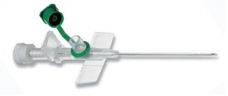
Regardless of the type and reason for use, all catheters require mandatory fixation. The tube is secured to the skin using medical tape or suture material. Modern models are initially equipped with special clamps, which greatly facilitates the catheterization process. Additionally, it is necessary to set the position of the tube inside the cavity; most often, the instrument has a device that allows you to quickly change its shape after insertion into the hollow organ.
The most widely used system is the Pigtail system - the tip of the catheter made of polyvinyl material looks very similar to a pig's tail, hence the name. During production, this device is placed in a special stylet or conductor, and after installation it is released and, twisting, prevents the tube from falling out of the organ. This type of fixation system is recognized as the safest and easiest to implement.
For a more rigid fixation, a loop is used, which is tightened with a fishing line previously placed in the catheter cavity.
How to attach a night urine bag?
When using a night urine bag simultaneously with a foot bag, do not disconnect the foot bag from the catheter.
Place the urine bag below the level of the bladder.
- Select a night bag.
- Attach the ribbed connector of the night bag to the silicone tube in the area of the drain valve of the foot bag.
- Open the drain valve, ensuring the flow of urine from the foot bag into the night bag.
- Loosen the fastening straps so that the maximum comfort is achieved while minimizing the risk of components coming apart.
- Fix the night urine bag with a hook, ensuring the optimal position of the product and the unimpeded outflow of urine.
Classification
Depending on the area of use, the following types of catheters are distinguished:
- aspiration – effective cleaning of the nasal and oral cavities in order to restore respiratory function;
- epidural - injected into the epidural space to administer anesthesia;
- urological – used in the absence of natural urination or urinary incontinence;
- umbilical - used in neonatology for cord blood transfusion;
- gastric - inserted into the stomach;
- trocar catheters – designed for the rapid drainage of fluid from the pleural cavity.
Despite the fact that many types have a similar structure, it is highly recommended not to replace them interchangeably. Such actions can cause complications.
How to empty and replace a foot urine bag?
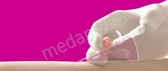
The leg bag should be emptied before it reaches its maximum filling level. If the foot urine bag is full, it will become uncomfortable to use and will begin to put pressure on the catheter. Always wash and dry your hands thoroughly before and after emptying or changing a urine bag and avoid direct contact with the bag connector.
- Release the urine using the drain tap
- Pinch the end of the catheter before disconnecting the urine bag to avoid splashing urine.
- Remove the new urine bag from the packaging
- Immediately connect the ribbed inlet connector of the urine bag to the catheter.
To reduce the risk of infection, do not disconnect the catheter from the leg bag unless you are replacing it.
Publications in the media
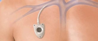
Resuscitation or intensive care is impossible without constant transfusion therapy and taking blood samples for laboratory tests, often emergency. Under these conditions, it is necessary to have a constantly functioning venous access, providing both the infusion of any solution with optimal speed and efficiency, as well as the possibility of laboratory diagnostics and monitoring. Central venous catheterization serves this purpose. In resuscitation practice, the subclavian vein, external and internal jugular veins are most often punctured and catheterized, less often - the femoral and even less often - the ulnar vein (peripheral veins collapse and become empty during hypovolemia, and therefore are poorly contoured).
• Subclavian vein •• Indications: shock of any etiology, because Due to anatomical features, this vein does not collapse during hypovolemia. In addition, when using the subclavian vein, good comfort is provided for the patient and the possibility of careful care even with a long stay of the catheter in the vein (for several months) •• Disadvantages of catheterization of the subclavian vein - a high risk of developing pneumothorax, the possibility of injury to the subclavian artery and difficulties when necessary to stop bleeding (impossibility of compression).
• External jugular vein •• Indications ••• Access to this vein is preferable in the elderly, it is well contoured even in obese patients ••• Less danger of developing pneumothorax ••• Its rapid effective compression is possible in case of hemorrhagic complications due to coagulation system disorders •• Inconveniences - the impossibility of its long-term use due to discomfort for the patient, difficulties in care, and relatively unreliable fixation.
• Internal jugular vein •• Characterized by rare complications, especially pneumothorax, good control in case of hemorrhagic complications, high rate of successful catheterizations even during cardiopulmonary resuscitation •• Disadvantages ••• Possibility of injury to the thoracic lymphatic duct or carotid artery during catheterization on the left •• •• The impossibility of its use in intracranial hypertension and during hemodialysis and tracheostomy ••• This vein can be collapsed with deep shock or hypovolemia.
Execution technique. The patient's position is lying on his back, the head end of the table is lowered, a cushion is placed under the patient's back, the patient's head is turned in the direction opposite to the puncture site. The patient's arm on the puncture side is brought to the body, sharply supinated, the assistant pulls it in the caudal direction. The patient may be positioned according to Trendelenburg. There are two operational approaches for puncture catheterization of the subclavian vein.
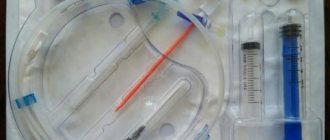
• Subclavian access •• A 0.5% procaine solution is injected intradermally with a syringe with a thin needle to create a “lemon peel” at a point located 1 cm below the clavicle on the line separating the middle and inner third of the clavicle. The needle is advanced medially towards the upper edge of the sternoclavicular joint, continuously applying procaine solution. The needle is passed under the collarbone and the rest of the procaine is injected there. The needle is removed •• Using a thick sharp needle, limiting the depth of its insertion with the index finger, the skin is pierced to a depth of 1–1.5 cm at the location of the “lemon peel”. The needle is removed •• A syringe with a capacity of 20 ml is filled up to half with 0.9% sodium chloride solution, and a not very sharp (to avoid puncture of the artery) needle 7–10 cm long with a bluntly beveled end is put on. The direction of the bevel should be marked on the cannula. When inserting the needle, its bevel should be oriented in the caudal-medial direction. The needle is inserted into a puncture previously made with a sharp needle (see above), and the depth of possible needle insertion should be limited to the index finger (no more than 2 cm). The needle is advanced medially towards the upper edge of the sternoclavicular joint, periodically pulling the plunger back, checking the flow of blood into the syringe. If unsuccessful, the needle is pushed back without removing it completely, and the attempt is repeated, changing the direction of advancement by several degrees. As soon as blood appears in the syringe, part of it is injected back into the vein and again sucked into the syringe, trying to obtain a reliable reverse blood flow. If a positive result is obtained, ask the patient to hold his breath and remove the syringe from the needle, squeezing its hole with a finger. •• A conductor is inserted into the needle with light screwing movements halfway; its length is slightly more than two times the length of the catheter. The patient is again asked to hold his breath, the guide is removed, closing the catheter hole with a finger, then a rubber stopper is put on the latter. After this, the patient is allowed to breathe. If the patient is unconscious, all manipulations associated with depressurization of the lumen of the needle or catheter located in the subclavian vein are performed during exhalation •• The catheter is connected to the infusion system and fixed to the skin with a single silk suture. Apply an aseptic dressing.
• Supraclavicular approach •• Position of the patient - as with subclavian access •• The doctor pierces the skin in the area of the angle formed by the lateral leg of the sternocleidomastoid muscle and the upper edge of the clavicle, moving 0.5 cm upward from the clavicle. After puncturing the skin, the needle is directed at an angle of 45° to the sagittal and 15° to the frontal plane of the clavicle and the vein is pierced. Further actions are the same as for infraclavicular access.
• After each infusion, 0.1 ml of heparin is injected into the catheter (the so-called “heparin lock”), so that the catheter can be used for several weeks without complications.
• If thrombosis, phlebitis or catheter sepsis is suspected, the catheter must be removed, followed by appropriate treatment measures.
Solving emerging problems.
If the catheter is leaking.
A leak can be caused by kinking both the catheter itself and the drive tube of the urinal. Make sure that all components of the drainage system are secure and that there are no kinks throughout. Leakage may be caused by constipation. Eat a balanced diet and drink enough fluids. If the problem cannot be solved, contact your doctor or nurse.
There is no outflow of urine.
Possible reasons:
- Kink in the area of the catheter or urinal tube - fix the problem.
- Insufficient fluid intake into the body - increase the amount of fluid consumed.
- Constipation - Eat a balanced diet and drink plenty of fluids.
- The urinal is fixed too high - make sure that the urinal is below the level of the bladder.
If you develop a catheter blockage or bladder spasm, contact your doctor or nurse.
The urine is cloudy and has an unpleasant odor. There is pain and discomfort in the catheter area. There is bleeding, increased sensitivity, itching...
Possible causes: urinary tract infection, bladder or urethral irritation. Drink enough fluids, contact your doctor or nurse.
The catheter fell out.
The likely cause is premature deflation of the catheter balloon or bladder spasm. Tell your doctor or nurse immediately.
General information about the catheter
When should the catheter be replaced? - In most cases, long-term catheters should be replaced every 1-3 months.
What should you do if your catheter is blocked? — If a catheter blockage or urinary tract infection develops, consult your doctor or nurse, who will decide whether additional treatment or catheter replacement is necessary.
Storing Catheters - Catheters should be stored horizontally in a cool, dark, dry place.