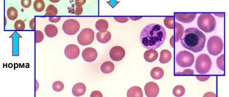
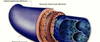
Vascular tone is the tension of blood vessels, supported by smooth muscles, an important factor in the blood supply to internal organs, one of the tools for general blood circulation. Vascular tone is influenced by the endothelium, the inner layer of blood vessels. Very thin (one cell thick), but plays a significant role in regulating vascular and cardiac tone, as well as in angiogenesis (formation of new vessels) and immune defense.
The fact that the endothelium is formed by a single layer of flat mesenchymal cells should seem to mean its insignificance, “microscopicity”. In fact, it is the largest organ in humans, present in all tissues. It affects blood clotting, kidney filtration, brain nutrition, and many other processes.
Theory
Tests
Help the site
Disable advertising
All vessels, with the exception of capillaries, have smooth muscle cells (SMCs) , due to which the lumen of the vessel , therefore the resistance to blood flow and the intensity of blood flow changes in a given region.
Local regulatory mechanisms:
- all vessels with SMCs are characterized by the original - basal tone created by the automaticity of smooth muscles;
- under the influence of various factors, basal tone can increase, while the vessels narrow and less blood enters the region;
- when vascular tone decreases, they dilate and blood flow into the region increases.
A decrease in tone leads to vasodilation, while an increase leads to vasoconstriction.
Vascular tone
Tone is the tension created by the asynchronous contraction of the SMC of the middle layer of the vascular wall, which has automaticity.
Tone components:
- basal tone,
- humoral,
- central (neurogenic).
Mechanisms for regulating vascular tone:
- Local mechanisms that ensure blood flow through individual organs and tissues, that is, controlling the amount of blood flow in individual regions.
- The central mechanisms regulating systemic blood circulation are the constancy of blood pressure, blood flow, blood volume, etc.
Local regulatory mechanisms
The principle of local regulation is to ensure the independence of blood flow in organs from changes in systemic hemodynamics, that is, the provision of blood to a given region in its interests.
Local mechanisms for regulating blood vessel tone include :
- myogenic,
- metabolic.
Myogenic mechanism
- myogenic autoregulation is characteristic of the vessels of the brain, kidneys, heart, liver, celiac region, that is, regions where it is necessary to maintain constant blood flow;
- an adequate stimulus to SMCs is their stretching;
- with an increase in blood pressure (BP) -> stretching of the vascular walls -> contraction of the SMC of the vessels -> an increase in vascular tone and maintaining the same lumen -> the blood flow in the vessels does not change;
- a decrease in blood pressure causes a decrease in vascular tone due to relaxation of the SMC: in this case, despite the decrease in blood pressure, the flow of the same volume of blood into the vessels remains,
- Thus, the amount of basal tone is influenced by the level of blood pressure.
Metabolic mechanism
- metabolic products, dilating blood vessels, increase blood flow in working organs;
- as a result of insufficient supply of oxygen and nutrients to the region, metabolites accumulate in the tissues and blood flow increases due to the expansion of precapillaries.
Vascular tone decreases with a decrease in oxygen and carbon dioxide pressure, an increase in H and C3H6O3 ions and temperature - as a result, blood flow in working organs increases in proportion to their activity.
Central regulatory mechanisms
- nervous (reflex),
- humoral.
Nervous mechanisms
Vasomotor - vasomotor nerves:
- vasoconstrictors - vasoconstrictor nerves,
- Vasodilators are vasodilating nerves.
Vasoconstrictors
- All vasoconstrictors are sympathetic adrenergic nerves.
- The vasoconstrictor effect occurs when norepinephrine (NA) acts on α-adrenergic receptors.
- Impulses through sympathetic vasoconstrictors constantly arrive to the vessels from the neurons of the lateral horns of the thoracolumbar segments of the SC with a frequency of 1-3 impulses/s, maintaining resting tone.
- At a frequency of more than 3 pulses/s (from 3 to 15) - increased tone.
Vasodilators
- Parasympathetic cholinergic nerves:
- chorda tympani - drum string - dilates the vessels of the submandibular salivary gland;
- n. lingualis - lingual nerve - dilates the vessels of the tongue;
- n. glossopharyngeus - glossopharyngeal - dilates the vessels of the tonsils, posterior third of the tongue, parotid salivary gland;
- n. pelvicus - pelvic - dilates the vessels of the area of the same name.
- Sympathetic nerves:
- cholinergic, innervating vessels of skeletal muscles;
- adrenergic - a vasoconstrictor effect occurs when NA acts on β-adrenergic receptors in the vessels of the heart, brain and lungs.
- Dorsal root sensory nerves - dilate skin vessels according to the axon reflex (mediator - ACh).
Axon reflex:
- dilation of skin blood vessels is observed when insects bite, under the influence of mustard plasters, rubbing, scratching the skin;
- blood vessels that do not have special vasodilators dilate due to a decrease in the tone of vasoconstrictors (for example: in the abdominal organs).
Impulses along the vasomotor nerves to the vessels constantly come from the vasomotor center (VMC) .
The main localization of the vasomotor center is in the medulla oblongata (Ovsyannikov, 1871).
How does the endothelium affect vascular tone?
Among the products of endothelial activity, nitric oxide (NO) attracts attention. It regulates vascular tone. The production of nitric oxide increases with increasing pressure at a given vascular site. Blood pressure may rise due to increased physical activity or under the influence of certain hormones (for example, acetylcholine).
Increased pressure on the vessel wall activates a special set of enzymes called endothelial nitric oxide synthase (eNOS). These enzymes spur increased production of NO. Nitric oxide molecules are freely released through cell membranes and penetrate into smooth muscle. Under the influence of NO, muscle tissue relaxes - the walls of blood vessels, without encountering resistance from smooth muscles, expand, and the pressure inside these vessels drops.
A decrease in pressure weakens eNOS activity in the endothelium. Less nitric oxide is released - the vascular muscles tense again, maintaining the pressure at the working level.
Vasomotor center (VDC)
SC centers (lateral horns of gray matter) -> bulbar centers: vasoconstrictor, vasodilator -> hypothalamic centers (anterior ( depressor zone ) and posterior ( pressor zone ) parts of the hypothalamus) -> cortical representation of the SDC.
After transection of the brainstem above the quadrigeminal artery, blood pressure does not decrease , but when the brain is transected between the medulla oblongata and the spinal cord, it drops from 120 mm Hg. Art. up to 70-80.
SDC consists of 2 departments:
- pressor department,
- depressor department.
Both of these departments do not have clear boundaries. They are located at the bottom of the 4th ventricle among the neural structures of the reticular formation and mutually overlap each other.
Pressor and depressor neurons of the SDC are in a reciprocal relationship.
There are more pressor neurons than depressor neurons. The state of the SDC is judged by pressor neurons .
The SDC also includes other parts of the central nervous system.
At rest, the hypothalamus does not actively participate in the regulation of blood pressure.
The influence of the cortex on the regulation of blood pressure is conditioned reflex - an increase in blood pressure before the start, with excitement.
Conclusion: a multi-level system for regulating the functions of the cardiovascular system ensures adequate adaptation to the conditions of the external and internal environment.
The tone of the SDC depends on the nerve impulses constantly coming to it from the receptors of various reflexogenic zones.
What is vegetative-vascular dystonia?
Vegetative-vascular dystonia (VSD)
) is a malfunction in the functioning of the whole organism. By and large, this is not a disease, but a malfunction of the autonomic nervous system, a violation of the regulation of the dynamic constancy of the internal environment of the body and the stability of its basic physiological functions: breathing, blood circulation, digestion, thermoregulation, metabolism, excretion, reproduction, etc. This is a functional failure of the body that can turn into disease.
VSD is a very common disease. About half of all women and a fifth of men are susceptible to autonomic dysfunction syndrome. Complaints can be very diverse: patients complain of weakness, pain in the heart area and under the left shoulder blade, headache, pressure surges and neurological disorders. If no measures are taken, the patient only gets worse. In the future, more serious surges in pressure, disruption of brain function and panic attacks are possible - acute stressful conditions characterized by a serious disturbance in a person’s mental state. If left untreated, functional disorders of VSD can cause more serious diseases, such as hypertension.
Quite often, vegetative-vascular dystonia is situational in nature: this means that an exacerbation of symptoms occurs against the background of stress, during weather changes, menstruation or other ordinary situations.
To date, doctors have described about 150 symptoms and 32 syndromes of clinical disorders with the diagnosis of “vegetative-vascular dystonia”. This is too much for an accurate diagnosis of any disease, and this is why diagnosing VSD is very difficult.
Many patients do not understand what serious consequences vegetative-vascular dystonia can lead to. Unfortunately, feeling unwell and having headaches is considered a “habitual” occurrence that can be alleviated with painkillers.
The availability of medications does its job: it’s easier to go to the pharmacy and relieve pain with pills, but medications only eliminate the symptoms of VSD and have nothing to do with treatment.
The mechanism of development of vegetative-vascular dystonia is as follows: in a normal state, the autonomic nervous system regulates the activity of internal organs, ensuring their adequate response to processes occurring in the body. In turn, the activity of the human autonomic nervous system is regulated by two subsystems: parasympathetic and sympathetic.
The parasympathetic subsystem is responsible for sleep and restoration of the body's strength, and the sympathetic subsystem is responsible for the period of wakefulness. Their work “alternates,” allowing a person to expend energy during the day and recover at night, during sleep and rest. The systems are closely connected to the brain and spinal cord and their failure affects the activity of the entire nervous system. The disruption of the relationship between these subsystems and the body’s incorrect response to natural processes is called vegetative-vascular dystonia. Since we are talking about malfunctions of the nervous system, VSD is very difficult to diagnose. The patient visits several specialists without success, but his condition continues to worsen. At the same time, the test results or cardiogram are in perfect order, but the patient feels worse and worse.
Vascular reflexes
Vascular reflexes are divided into:
- own and
- conjugated.
Own reflexes
They are carried out from mechanoreceptors located in the heart and blood vessels ( baroreceptors ).
These receptors stabilize blood pressure.
There are own reflexes:
- pressor - increasing low blood pressure,
- depressor - lowering high blood pressure.
Reflexogenic zones (areas of maximum accumulation of receptors):
- aortic arch,
- carotid sinus (bifurcation of the common carotid artery into external and internal).
Depressor reflex: with an increase in blood pressure -> the baroreceptors of the aortic arch and carotid sinus are irritated -> excitation along the sensory nerves - aortic (depressor) and sinus (Hering's nerve) -> medulla oblongata -> the vagal center is excited and the vasomotor center is inhibited -> heart rate decreases - > blood vessels dilate -> blood pressure decreases (normalizes).
When blood pressure drops, the opposite is true, that is, the pressor reflex .
Own reflexes:
- are also carried out from chemoreceptors located in the aortic and carotid bodies;
- they are excited when CO2 and H ions increase in the blood and when O2 decreases;
- impulses arriving from chemoreceptors in the medulla oblongata increase the tone of the central nervous system, which leads to an increase in pressure.
Chemoreceptors are not in the vessel wall, but in the aortic and carotid bodies or glomeruli under the adventitia of the vessel and are penetrated by a network of capillaries.
From chemoreceptors -> SDC of the medulla oblongata -> SDC is excited -> vasoconstriction -> increase in blood pressure -> rapid blood renewal.
Conjugate reflexes
Carried out from receptors located outside the heart and blood vessels:
- they disrupt the stability of blood pressure, causing pressor reactions;
- distinguish conjugate reflexes: exteroceptive - from skin receptors,
- interoceptive - from internal organs.
How to treat vegetative-vascular dystonia with the ALMAG-01 device
IMPORTANT: the ALMAG-01 device can only be used for the treatment of VSD due to hypertension. Treatment must begin after the acute phase of the vegetative crisis.
THE DESIGN OF ALMAG-01 INCLUDES THE FOLLOWING ELEMENTS:
Physiotherapeutic procedures with the ALMAG-01 device are carried out once a day. The standard course of treatment lasts three weeks: after every six days, a one-day break is required. The course of treatment is 18 procedures. A repeated course can be carried out after 30-40 days, and a maintenance course - 3-4 months after completion of treatment.
The treatment procedure is simple and does not require special knowledge. Inductor coils are sequentially applied to the projection area of the adrenal glands and the collar zone, and the exposure time is set according to a table developed by specialists.
Fortunately, vegetative-vascular dystonia is a curable disease. To do this, it is necessary not only to undergo treatment, but also to change your lifestyle, reconsidering your views on your daily routine, rest and physical activity.
Humoral regulation
- Hormones formed in the endocrine glands: adrenaline, norepinephrine, vasopressin, etc. - constrict blood vessels.
- Vasoactive agents (local hormones) formed in tissues - acetylcholine, bradykinin, histamine, prostaglandins, etc. - dilate blood vessels.
- Dual-acting substances - catecholamines:
- alpha - narrowing
- betta - extension.
The hormones adrenaline and norepinephrine narrow the arteries and arterioles of the skin, skeletal muscles, and abdominal organs. Coronary vessels, vessels of the brain, and lungs expand at the same time, since it all depends on which adrenergic receptors perceive the hormone. When NA interacts with α-adrenergic receptors, the vessels narrow, and when interacting with β-adrenergic receptors , they dilate. β-adrenergic receptors predominate in the vessels of the heart, lungs, and brain.
Vasopressin mainly constricts arterioles and veins.
Angiotensin II is formed from plasma α-globulins under the influence of renin (JA cells of the renal cortex) and also constricts blood vessels.
Vascular tone:
- basal tone - the tone of the SMC and the influence of sympathetic vasoconstrictors;
- resting tone - tone of the SMC and the influence of sympathetic nerves with a frequency of 1-3 impulses/s;
- increased tone - impulses along sympathetic vasoconstrictors with a frequency of 3-15 impulses/s.
Symptoms
An increase in cerebral vascular tone refers to cerebral hypertonicity and the following symptoms develop:
- Headache in diffuse localization. The area of pain depends on the place where hypertonicity is most pronounced. Also, unpleasant sensations can spread throughout the entire head at once. The pain can spread to the neck, eyes, ears.
- Due to the deterioration of blood supply, intellectual and mnestic activity deteriorates: the pace of thinking slows down, the volume and concentration of attention decreases.
- Changes in physiological state: fatigue, exhaustion. Even simple work requires mental and physical effort.
- Emotional disorders: mood lability (sudden transitions from a bad mood to a good one and vice versa), irritability. Low threshold of excitability: quiet sounds and dim light can make a person angry.
- Nausea and vomiting.
- Hypertonicity, combined with difficulty in venous outflow, is manifested by a feeling of fullness in the head.
- Syncope is rarely observed in the clinical picture. However, especially sentimental people may faint from happiness.