General information
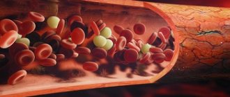
As part of the coagulogram, a study of thrombin time is carried out, which characterizes the last stage of blood coagulation - the conversion of fibrinogen (a protein in a blood clot) into fibrin (the basis of a blood clot) as a result of the reaction of blood with thrombin and calcium.
The analysis is prescribed to determine hereditary disorders, blood diseases, oncological processes, liver pathologies, as well as during pregnancy (to prevent the development of critical conditions during pregnancy and childbirth).
Thrombin time is an indicator of the rate of conversion of fibrinogen to fibrin under the influence of the coagulation factor thrombin. Based on the results of this study, pathologies of blood clotting are identified in the process of secondary hemostasis (formation of a blood clot), which occurs in 3 stages.
- The coagulant thrombin acts on the fibrinogen protein in such a way that it turns into fibrin monomer (a clot with a jelly-like consistency).
- From several fibrin monomers, fibrin polymer is obtained (the trace element calcium is involved in the reaction).
- Fibrin polymers combine with red blood cells and platelets to form fibrin, which is insoluble in the blood. As a result, bleeding is stopped by a blood clot - a thrombus.
All stages of secondary hemostasis occur within a limited time (with the exception of dysfunctional disorders of the hematopoietic organs). The rate of clot formation depends on the biological usefulness of fibrinogen and the level of coagulants and anticoagulants.
One of the most dangerous conditions is hypofibrinogenemia - when the walls of blood vessels or organ tissues are damaged, prolonged bleeding occurs. Also, prolongation of thrombin time occurs when taking heparin or other anticoagulants that neutralize thrombin. Sometimes the cause of slow blood clotting is the secretion of autoimmune antibodies to thrombin or the presence of paraproteins in the plasma that interfere with the second stage of fibrin conversion.
In the case of hyperfibrinogenemia, the blood clots faster than necessary, which can lead to thrombosis (blockage of the lumen of blood vessels).
The thrombin time test is most indicative in the diagnosis and treatment of DIC syndrome (disseminated intravascular coagulation) associated with fibrin degradation.
Liver diseases can cause deviations from the normal thrombin time.
Diseases that affect the duration of clotting can be acquired, hereditary or congenital. Acquired ones are associated with lifestyle, nutrition, bad habits, and a history of diseases (arthritis, cardiovascular pathologies, oncology). Also, thrombin time may deviate from the norm during a pathological pregnancy and/or after a difficult birth. Most often, hypofibrinogenemia is observed, which increases the likelihood of bleeding.
Congenital or hereditary bleeding disorders are quite rare, as they are formed as a result of genetic mutations.
Thrombin time
This is a laboratory test that detects quantitative and qualitative changes in fibrinogen.
Synonyms Russian
TV, antithrombin I.
English synonyms
Thrombin time, TT, Thrombin Clotting Time, TCT, antithrombin I, AT I.
Research method
Side scatter detection method, end point percentage determination.
Units
Sec. (second).
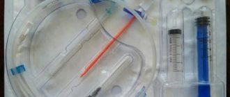
What biomaterial can be used for research?
Venous blood.
How to properly prepare for research?
- Do not eat for 12 hours before the test.
- Avoid physical and emotional stress and do not smoke for 30 minutes before the test.
General information about the study
Thrombin time is the time required for the formation of a fibrin clot when thrombin is added to the plasma - an enzyme (factor IIa), which appears as a result of the interaction of blood coagulation factors when a vessel is damaged. Thrombin is necessary for the final stage of the coagulation cascade - the transformation of the fibrinogen molecule into insoluble fibrin, which is able to polymerize and form a stable fibrin clot, which stops bleeding when small and medium-sized vessels are damaged and contains, in addition to fibrin polymers, cellular elements - platelets and erythrocytes. Qualitative or quantitative changes in fibrinogen lead to insufficient/excessive fibrin production, which is manifested by increased bleeding or a tendency to thrombosis. In laboratory conditions, the final stage of the coagulation cascade is reproduced by adding ready-made thrombin to plasma, and changes in the structure or concentration of fibrinogen are reflected in an increase or decrease in thrombin time.
Fibrinogen is a glycoprotein secreted by hepatocytes into the blood. In addition to being a blood clotting factor (factor I), it also provides some reactions of fibrinolysis, the process of dissolving blood clots, binds excess thrombin (therefore fibrinogen is also called antithrombin I) and activates plasminogen. In this regard, diseases with changes in the structure or concentration of fibrinogen can manifest themselves as bleeding, thrombosis, and in some cases a combination of both.
Pathologies that are accompanied by fibrinogen deficiency are called hypo- and afibrinogenemia, and those in which the structure and function of fibrinogen are impaired are called disfibrinogenemia. They can be hereditary or acquired. Acquired diseases include liver disease, disseminated intravascular coagulation (DIC), primary fibrinolysis, and drug reactions (eg, thrombolytic agents and L-asparaginase).
The most common cause of acquired dysfibrinogenemia is liver disease. In cirrhosis, chronic active hepatitis, acute liver failure, obstructive liver disease and hepatoma, too much sialic acid is added to the secreted fibrinogen. This modified fibrinogen has a greater negative charge, which prevents the polymerization of fibrin molecules. Patients with liver disease have multiple coagulation disorders that manifest as increased bleeding. Determination of thrombin time is the main screening test to suspect dysfibrinogenemia as one of the causes of bleeding. Abnormal fibrinogen is also produced by some tumors (some forms of squamous cell carcinoma of the cervix, adenocarcinoma of the breast, hypernephroma, hepatoma). In certain diseases accompanied by the synthesis of antibodies that interact with fibrinogen (systemic lupus erythematosus, multiple myeloma), its activity decreases, which is manifested by an increase in thrombin time.
The most common cause of acquired hypofibrinogenemia is DIC syndrome, a systemic thrombohemorrhagic disorder in which excessive formation of fibrin microthrombi and consumption of blood coagulation factors and platelets occurs. DIC always develops secondarily as a complication of any disease. There are acute and chronic DIC syndrome. The causes of acute DIC are infectious diseases (E. coli - sepsis, HIV, cytomegalovirus infection, malaria), acute myeloblastic leukemia, complications of pregnancy and childbirth (premature placental abruption, eclampsia, amniotic fluid embolism), extensive burns, massive blood transfusions and others . Causes of chronic DIC syndrome: solid tumors, chronic leukemia, pregnancy complications (intrauterine fetal death), myeloproliferative diseases, rheumatoid arthritis and Raynaud's disease, myocardial infarction, nonspecific ulcerative colitis and Crohn's disease, etc. “Triggers” the development of DIC syndrome in all In the listed conditions, a large amount of thromboplastin (tissue factor, factor III) enters the blood. At the same time, multiple microthrombi are formed in the vessels of the kidneys, brain, liver, and lungs, which determine the clinical picture of the syndrome in the form of multiple organ failure. As a result of massive consumption of fibrinogen and other blood coagulation factors, their secondary insufficiency develops and hypercoagulation is replaced by hypocoagulation and disseminated bleeding. It should be noted that hypercoagulation and hypocoagulation are often present simultaneously in the same patient, so the identification of successive periods of DIC is very arbitrary. The peculiarities of the clinical picture of DIC syndrome lead to the fact that the diagnosis of this condition remains one of the most difficult tasks of modern medicine. In this situation, monitoring thrombin time in combination with other laboratory tests is necessary to assess the risk and early diagnosis of DIC, as well as at the stage of monitoring its treatment.
Inherited fibrinogen disorders are quite rare. About 80 mutations are known to cause fibrinogen deficiency or absence. As a rule, such mutations lead to a complete stop in fibrinogen synthesis or the formation of altered fibrinogen, which cannot be secreted from hepatocytes and accumulates in them. With afibrinogenemia, there is no fibrinogen in the plasma; this is the most severe form of its deficiency. The disease is inherited in an autosomal recessive manner and occurs with a frequency of 1:1,000,000. As a rule, afibrinogenemia manifests itself already in the neonatal period (the first 28 days from birth). In this case, bleeding is observed when crossing the umbilical cord, hemorrhages in the skin, gastrointestinal and genitourinary tract, and intracranial hemorrhage. However, this disease may first appear later, in childhood and even in adulthood. Women with afibrinogenemia are characterized by metro-, menorrhagia, habitual spontaneous miscarriage, and bleeding in the postpartum period. In some patients with afibrinogenemia, along with increased bleeding, symptoms of thrombosis (more often venous than arterial) are observed.
Hypofibrinogenemia is much more common than afibrinogenemia (1:500). As a rule, it is asymptomatic, since the amount of fibrinogen that is still present is enough to stop minor bleeding from small and medium-sized vessels and carry the pregnancy to term. However, with extensive injuries, operations, as well as with concomitant pathology of blood coagulation, hypofibrinogenemia can be accompanied by life-threatening bleeding.
Hereditary dysfibrinogenemia is a heterogeneous group of diseases in which the structure of fibrinogen is disrupted. However, its plasma concentration remains normal or slightly reduced (hypodysfibrinogenemia). To date, about 400 mutations are known that lead to amino acid substitutions in the fibrinogen sequence. Such a replacement leads to a decrease in its activity, disruption of its interaction with thrombin, or the absence of further polymerization of fibrin. The majority of patients with dysfibrinogenemia (55%) do not have clinical manifestations of the disease. The disease is suspected on the basis of incidentally detected abnormalities in laboratory tests or when a diagnosis is made in one of the family members. 25% of patients with dysfibrinogenemia experience episodes of increased bleeding (usually after injury, surgery or in the postpartum period), 20% have a tendency to thrombosis (more often venous than arterial), and 27% have a combination of both conditions. In some cases, abnormal fibrinogen is deposited in the kidney tissue as amyloid. This form of hereditary amyloidosis is characterized by the early development of chronic renal failure.
While the main function of fibrinogen is the formation of fibrin thrombus, it is also involved in several other processes: inflammation, angiogenesis and wound healing. In addition, it is believed to promote the formation of atherosclerotic plaques in blood vessels, the proliferation of smooth muscle cells and the uptake of oxidized lipids by macrophages. It has been established that high concentrations of fibrinogen in the blood are associated with an increased risk of arterial thrombosis.
What is the research used for?
- For the diagnosis of acquired dysfibrinogenemia (in liver diseases, autoimmune diseases).
- For the diagnosis of hereditary dysfibrinogenemia.
- To find out the cause of recurrent miscarriage.
- To assess the risk of developing DIC, diagnose it and monitor its treatment.
- To assess the risk of arterial thrombosis.
When is the study scheduled?
- For liver diseases: cirrhosis, chronic active hepatitis, obstructive liver diseases, hepatomas.
- For symptoms of hypo(a)fibrinogenemia and dysfibrinogenemia: increased bleeding (metro-, menorrhagia, nosebleeds, soft tissue hematomas, bleeding in the postpartum or postoperative period) or a tendency to thrombosis (phlebothrombosis, thromboembolism of the branches of the pulmonary artery, arterial thrombosis), as well as with a combination of both conditions.
- With two or more spontaneous abortions up to 22 weeks.
- For diseases accompanied by a high risk of developing DIC: severe infectious diseases, acute and chronic leukemia, complications of pregnancy and childbirth, severe autoimmune diseases.
- With existing risk factors for arterial thrombosis: high concentrations of homocysteine and C-reactive protein.
What do the results mean?
Reference values: 14 - 21 sec.
Reasons for increasing thrombin time:
- liver cirrhosis, chronic active hepatitis, hepatoma;
- DIC syndrome;
- hereditary hypo(a)fibrinogenemia and dysfibrinogenemia;
- acute leukemia, polycythemia vera;
- amyloidosis;
- complications of pregnancy and childbirth (eclampsia, premature placental abruption);
- drug interactions: L-asparaginase, heparin, streptokinase, urokinase.
Reasons for decreased thrombin time:
- thrombocytosis.
What can influence the result?
- Transfusion of fresh frozen plasma masks qualitative and quantitative fibrinogen abnormalities.
- Therapy with unfractionated heparin leads to an increase in thrombin time.
Important Notes
- Thrombin time is normally longer in newborns than in adults.
- To accurately interpret the result, it is necessary to combine the analysis with other laboratory tests that characterize the blood coagulation system.
- Treatment with fractionated heparin and warfarin does not affect the test result.
Also recommended
- Activated partial thromboplastin time (aPTT)
- Coagulogram No. 1 (prothrombin (according to Quick), INR)
- D-dimer
- Fibrinogen
- Coagulogram No. 2 (prothrombin according to Quick, INR, fibrinogen)
- Coagulogram No. 3 (prothrombin (according to Quick), INR, fibrinogen, ATIII, APTT, D-dimer)
Who orders the study?
General practitioner, hematologist, anesthesiologist-resuscitator, obstetrician-gynecologist.
Literature
- Chernecky CC Laboratory Tests and Diagnostic Procedures / S.S. Chernecky, V.J. Berger; 5th ed. — Saunder Elsevier, 2008.
- Feinbloom D, Bauer KA. Assessment of Hemostatic Risk Factors in Predicting Arterial Thrombotic Events. Arterioscler Thromb Vasc Biol. 2005 Oct; 25(10):2043-53.
- Mark T. Cunningham et al. Laboratory Diagnosis of Dys?brinogenemia. Arch Pathol Lab Med. 2002 Apr; 126(4):499-505.
- Triplett DA. Coagulation and bleeding disorders: review and update. Clin Chem. 2000 Aug; 46(8 Pt 2): 1260-9.
- Kaushansky K. et al. Williams Hematology / K. Kaushansky, M. Lichtman, E. Beutler; 8th ed. — The McGraw-Hill Companies, 2010.
Indications
- Identification of defective forms of fibrinogen or its deficiency (congenital/acquired);
- Diagnosis of hyper- and hypofibrinogenemia;
- The presence of fibrinogen and fibrin breakdown products in the blood;
- Diagnosis and treatment of DIC syndrome;
- Dysfunction or pathology of the liver structure;
- Pain in the liver area of unknown etiology;
- Deviations of pressure inside the vessels in one direction or another;
- Monitoring treatment with anticoagulants (heparin), thrombolytics and fibrinolytics;
- Pregnancy (pathological, spontaneous termination);
- As part of a comprehensive coagulogram.
A coagulogram test is prescribed and its results are interpreted by doctors: hematologist, resuscitator, anesthesiologist, gynecologist, pediatrician, therapist, etc.
Indications for the purpose of the study
- For liver diseases: cirrhosis, chronic active hepatitis, obstructive liver diseases, hepatomas.
- For symptoms of hypo(a)fibrinogenemia and dysfibrinogenemia: increased bleeding (metro-, menorrhagia, nosebleeds, soft tissue hematomas, bleeding in the postpartum or postoperative period) or a tendency to thrombosis (phlebothrombosis, thromboembolism of the branches of the pulmonary artery, arterial thrombosis), as well as with a combination of both conditions.
- With two or more spontaneous abortions up to 22 weeks.
- For diseases accompanied by a high risk of developing DIC: severe infectious diseases, acute and chronic leukemia, complications of pregnancy and childbirth, severe autoimmune diseases.
- With existing risk factors for arterial thrombosis: high concentrations of homocysteine and C-reactive protein.
Thrombin time is normal
To measure thrombin time, the following reference values are accepted:
- 10.3 – 16.6 seconds.
Factors influencing the result
Sometimes the results may be distorted. There are a number of reasons for this:
- Age (in newborns, thrombin time is normally prolonged);
- Violations of the rules for preparing for a coagulogram;
- Violations of the algorithm for blood collection, its transportation and storage;
- Incompetence of laboratory workers;
- Taking interfering drugs;
- Transfusion of frozen plasma shortly before the test (hides quantitative and structural fibrinogen abnormalities);
- Menstruation (reduces clotting rates);
- Acute febrile syndrome in a patient during venous blood sampling;
- Recent injuries, wounds, burns, surgery.
Material under study
Deoxygenated blood
Interpretation of research results contains information for the attending physician and is not a diagnosis. The information in this section should not be used for self-diagnosis or self-treatment. The doctor makes an accurate diagnosis using both the results of this examination and the necessary information from other sources: medical history, results of other examinations, etc.
Units: Sec. (second).
Reference values: 9–12 sec
Increasing values (extending interval)
- Liver pathologies;
- Uremia (intoxication of the body due to kidney disease);
- DIC syndrome;
- Deficiency of protein intake in the body;
- Pathological pregnancy (miscarriage, placental abruption);
- Cardiovascular diseases (heart attack, stroke);
- Pneumonia (pneumonia);
- Hyperbilirubinemia (increased levels of bilirubin in the blood);
- Tuberculosis (infectious disease caused by Koch's bacillus);
- Hypofibrinogenemia (with the level of fibrinogen in the blood less than 0.5 g per 1 l) in acquired or congenital form;
- Dysfibrinogenemia (defect in the structure of fibrinogen at the molecular level);
- Paraproteinemia (presence of defective and abnormal protein compounds in the plasma);
- An increase in fibrin and fibrinogen breakdown products in the blood;
- The presence of anticoagulants in the plasma (antithrombins, hirudin, heparin and others);
- Fibrinolytic therapy (a type of treatment aimed at restoring blood flow by dissolving a blood clot);
- Development of autoimmune antibodies to thrombin;
- Production of lupus anticoagulant (antibodies against enzymes of the circulatory system);
- Myeloma multiforme (cancer of blood plasma cells) and other oncological processes.
DIC - syndrome
DIC syndrome is a systemic thrombohemorrhagic disorder in which excessive formation of fibrin microthrombi and consumption of blood coagulation factors and platelets occurs. DIC - the syndrome always develops secondary, as a complication of any disease.
There are acute and chronic DIC syndrome.
The causes of acute DIC syndrome are:
- infectious diseases (E. coli - sepsis, HIV, cytomegalovirus infection, malaria)
- acute myeloblastic leukemia
- complications of pregnancy and childbirth (premature placental abruption, eclampsia, amniotic fluid embolism)
- extensive burns
- massive blood transfusions and others.
Causes of chronic DIC syndrome:
- solid tumors
- chronic leukemia
- pregnancy complications (intrauterine fetal death)
- myeloproliferative diseases
- rheumatoid arthritis
- Raynaud's disease
- myocardial infarction
- nonspecific ulcerative colitis and Crohn's disease, etc.
The development of disseminated intravascular coagulation (DIC) in all of the above conditions is “triggered” by the entry into the blood of a large amount of thromboplastin (tissue factor, factor III). At the same time, multiple microthrombi are formed in the vessels of the kidneys, brain, liver, and lungs, which determine the clinical picture of the syndrome in the form of multiple organ failure. As a result of massive consumption of fibrinogen and other blood coagulation factors, their secondary insufficiency develops and hypercoagulation is replaced by hypocoagulation and disseminated bleeding. It should be noted that hypercoagulation and hypocoagulation are often present simultaneously in the same patient, so the identification of successive periods of DIC syndrome is very arbitrary.
Features of the clinical picture of DIC syndrome lead to the fact that the diagnosis of this condition remains one of the most difficult tasks of modern medicine. In this situation, monitoring thrombin time in combination with other laboratory tests is necessary to assess the risk and early diagnosis of DIC syndrome, as well as at the stage of monitoring its treatment. Inherited fibrinogen disorders are quite rare. About 80 mutations are known to cause fibrinogen deficiency or absence. As a rule, such mutations lead to a complete stop in fibrinogen synthesis or the formation of altered fibrinogen, which cannot be secreted from hepatocytes and accumulates in them. With afibrinogenemia, there is no fibrinogen in the plasma; this is the most severe form of its deficiency. The disease is inherited in an autosomal recessive manner and occurs with a frequency of 1:1,000,000. As a rule, afibrinogenemia manifests itself already in the neonatal period (the first 28 days from birth). In this case, bleeding is observed when crossing the umbilical cord, hemorrhages in the skin, gastrointestinal and genitourinary tract, and intracranial hemorrhage. However, this disease may first appear later, in childhood and even in adulthood. Women with afibrinogenemia are characterized by metro-, menorrhagia, habitual spontaneous miscarriage, and bleeding in the postpartum period. In some patients with afibrinogenemia, along with increased bleeding, symptoms of thrombosis (more often venous than arterial) are observed.
Hypofibrinogenemia is much more common than afibrinogenemia (1:500). As a rule, it is asymptomatic, since the amount of fibrinogen that is still present is enough to stop minor bleeding from small and medium-sized vessels and carry the pregnancy to term. However, with extensive injuries, operations, as well as with concomitant pathology of blood coagulation, hypofibrinogenemia can be accompanied by life-threatening bleeding.
Hereditary dysfibrinogenemia is a heterogeneous group of diseases in which the structure of fibrinogen is disrupted. However, its plasma concentration remains normal or slightly reduced (hypodysfibrinogenemia). To date, about 400 mutations are known that lead to amino acid substitutions in the fibrinogen sequence. Such a replacement leads to a decrease in its activity, disruption of its interaction with thrombin, or the absence of further polymerization of fibrin.
The majority of patients with dysfibrinogenemia (55%) do not have clinical manifestations of the disease. The disease is suspected on the basis of incidentally detected abnormalities in laboratory tests or when a diagnosis is made in one of the family members.
25% of patients with dysfibrinogenemia experience episodes of increased bleeding (usually after trauma, surgery or in the postpartum period), 20% have a tendency to thrombosis (more often venous than arterial), and 27% have a combination of both conditions. In some cases, abnormal fibrinogen is deposited in the kidney tissue as amyloid. This form of hereditary amyloidosis is characterized by the early development of chronic renal failure.
While the main function of fibrinogen is the formation of fibrin thrombus, it is also involved in several other processes: inflammation, angiogenesis and wound healing. In addition, it is believed to promote the formation of atherosclerotic plaques in blood vessels, the proliferation of smooth muscle cells and the uptake of oxidized lipids by macrophages. It has been established that high concentrations of fibrinogen in the blood are associated with an increased risk of arterial thrombosis.