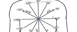
To answer these questions, you need to understand the three basic rules of the ECG (see Figure 4-1).
Rice. 4-1. A - the complex is positive in any lead if the depolarization wave is directed towards its positive pole; B — the complex is negative if the depolarization is directed to the negative pole (from the positive pole) of the lead; B - biphasic complex (partially positive, partially negative), if the average depolarization vector is located perpendicular to the lead; these three principles are true for the P wave (atrial depolarization) and the QRS complex (ventricular depolarization).
The T wave on the ECG is normal in children and adults
The beginning of the T wave coincides with the repolarization phase, that is, with the reverse transition of sodium and potassium ions through the membrane of heart cells, after which the muscle fiber becomes ready for the next contraction. Normally, T has the following characteristics:
- begins on the isoline after the S wave;
- has the same direction as the QRS (positive where R is dominant, negative when S is dominant);
- smooth in shape, the first part is flatter;
- amplitude T up to 8 cells, increases from 1 to 3 chest leads;
- may be negative in V1 and aVL, always negative in aVR.
In newborns, T waves are low in height or even flat, their direction is opposite to the adult ECG. This is due to the fact that the heart turns in direction and takes a physiological position by 2 - 4 weeks. At the same time, the configuration of the teeth on the cardiogram gradually changes. Typical features of a pediatric ECG:
- negative T in V4 persists up to 10 years, V2 and 3 – up to 15 years;
- adolescents and young adults may have negative T waves in the 1st and 2nd chest leads; this type of ECG is called juvenile;
- height T increases from 1 to 5 mm; in schoolchildren it is 3–7 mm (as in adults).
We recommend reading about how an ECG is performed. You will learn about the principles of operation of an electrocardiograph, preparation for an ECG, methods of conducting, and interpretation of indicators. And here is more information about what myocardial ischemia looks like on an ECG.
What it is?
This is where we will begin preparing for the ECG interpretation. The T wave is the most important indicator on the electrocardiogram, which can help the doctor make a conclusion about the recovery process after contraction of the heart ventricles. He is the most changeable in the schedule.
By its shape and location, one can judge the amplitude of heart contractions, the presence of such dangerous diseases, conditions and pathologies as myocardial damage, endocrine diseases, intoxication of the body, taking incorrectly selected medications, etc.
Let's take a closer look at deciphering the ECG and the norm for this indicator.
ECG changes and their meanings
Most often, changes are suspected of coronary heart disease, but such a disorder may be a sign of other diseases:
- thromboembolism,
- myocarditis, pericarditis,
- tumors, infections and injuries,
- ventricular hypertrophy,
- intoxication, including cardiac glycosides, antiarrhythmic drugs, aminazine, nicotine,
- stress, neurocirculatory dystonia,
- diseases of the endocrine system,
- potassium deficiency,
- decreased blood circulation in the brain,
- osteochondrosis.
Therefore, to make a diagnosis, all clinical signs and changes in the cardiogram are taken into account as a whole.
Two-phase
On the cardiogram, T first decreases below the isoline, and then crosses it and becomes positive. This symptom is called the “roller coaster” syndrome. May occur in the following pathologies:
- left ventricular hypertrophy;
- Hiss bundle branch block;
- increased calcium levels in the blood;
- intoxication with cardiac glycosides.
Biphasic T wave with left ventricular hypertrophy
Smoothed
Flattening of the T wave can be caused by:
- taking alcohol, Cordarone or antidepressants;
- diabetes mellitus or eating a lot of sweets;
- fear, excitement;
- cardiopsychoneurosis;
- hypokalemia;
- myocardial infarction in the scarring stage.
Decrease in indicator
A reduced T is indicated by its amplitude, which is less than 10% of the QRS complex. This symptom on the ECG causes:
- coronary insufficiency,
- cardiosclerosis,
- obesity,
- elderly age,
- hypothyroidism,
- dishormonal cardiomyopathy,
- myocardial dystrophy,
- taking corticosteroids,
- anemia,
- tonsillitis.
The T wave on the ECG is smoothed
The T wave can be smoothed in the same conditions as an absent wave, since both definitions characterize low-amplitude oscillations. It should be taken into account that violation of the rules for ECG registration can also cause smoothing of T. It also occurs in metabolic diseases - low function of the thyroid gland (myxedema, hypothyroidism). It can be found in completely healthy people throughout the day in several cardiac cycles (according to Holter monitoring).
Inversion
Inversion (turning over) of the T wave means a change in its position relative to the isoline, that is, in leads with a positive T, it changes its polarity to negative and vice versa. Such deviations can also be normal - in the right chest leads with a juvenile ECG configuration or a sign of early repolarization in athletes.
T wave inversion in leads II, III, aVF, V1-V6 in a 27-year-old athlete
Diseases that are accompanied by T inversion:
- myocardial or cerebral ischemia,
- influence of stress hormones,
- bleeding in the brain,
- attack of tachycardia,
- violation of impulse conduction along the branches of the Hiss bundle.
Negative T wave
For coronary heart disease, a characteristic sign is the appearance of negative T waves on the ECG, and if they are accompanied by changes in the QRS complex, then the diagnosis of a heart attack is considered confirmed. In this case, changes in the cardiogram depend on the stage of myocardial necrosis:
- acute – abnormal Q or QS, ST segment above the line, T positive;
- subacute – ST on the isoline, negative T;
- in the scar stage, weakly negative or positive T.
A negative T wave in leads V5-V6 (highlighted in red) indicates ischemia.
A variant of the norm may be the appearance of negative T wave with frequent breathing, excitement, after a heavy meal, which contains a lot of carbohydrates, as well as with individual characteristics in some healthy people. Therefore, the detection of negative values cannot be considered a serious illness.
Pathological conditions that are accompanied by negative T waves:
- heart disease - angina pectoris, heart attack, cardiomyopathy, inflammation of the myocardium, pericardium, endocarditis, mitral valve prolapse;
- violation of hormonal and nervous regulation of cardiac activity (thyrotoxicosis, diabetes mellitus, diseases of the adrenal glands, pituitary gland);
- pulmonary heart;
- after paroxysmal tachycardia or frequent extrasystoles;
- subarachnoid hemorrhage.
Subarachnoid hemorrhage is accompanied by negative T waves
Absence of T wave on ECG
The absence of T on the ECG means that its amplitude is so low that it merges with the isoelectric line of the heart. This happens when:
- drinking alcohol;
- against the backdrop of excitement, anxiety;
- cardiomyopathy in patients with diabetes mellitus;
- neurocirculatory dystonia (with a sudden change in body position or after rapid breathing);
- insufficient intake of potassium or its loss through sweat, urine, intestinal contents (diarrhea);
- scarring of myocardial infarction;
- use of antidepressants.
High rate
Normally, in those leads where the highest R is recorded, the maximum amplitude is noted; in V3 - V5 it reaches 15 - 17 mm. Very high T can occur when the influence on the heart of the parasympathetic nervous system, hyperkalemia, subendocardial ischemia (first minutes), alcoholic or menopausal cardiomyopathy, left ventricular hypertrophy, and anemia predominate.
Changes in the T wave on the ECG during ischemia: a - normal, b - negative symmetrical "coronary" T wave, c - high positive symmetrical "coronary" T wave, d, e - biphasic T wave, f - reduced T wave, g - smoothed T wave, z - weakly negative T wave.
Flat
A slightly inverted or flattened T may be either a normal variant or a manifestation of ischemic and dystrophic processes in the heart muscle. It occurs with complete blockade of the conduction pathways in the ventricles, myocardial hypertrophy, acute or chronic pancreatitis, taking antiarrhythmic medications, and hormonal and electrolyte imbalance.
Coronary
When the heart muscle is hypoxic, the fibers located under the inner membrane, the endocardium, are most affected. The T wave reflects the ability of the endocardium to maintain a negative electrical potential, therefore, in case of coronary insufficiency, it changes its direction and becomes this shape:
- isosceles;
- negative (negative);
- pointed.
These signs characterize the ischemic wave, or it is also called coronary. Manifestations on the ECG are maximum in those leads where the greatest damage is localized, and in mirror (reciprocal) leads it is sharp and isosceles, but positive. The more pronounced the T wave, the deeper the degree of myocardial necrosis.
Rise of the T wave on the ECG
Moderate physical stress, hyperkalemia, infectious processes in the body, thyrotoxicosis, and anemia lead to an increase in the amplitude of T waves. Elevated T without changes in well-being can occur in healthy people, and can also be a symptom of vegetative-vascular disorders with a predominance of vagal tone.
Depression
A reduced T wave can be a manifestation of cardiomyodystrophy; it occurs with pneumonia, rheumatism, scarlet fever, acute inflammatory process in the kidneys, cor pulmonale and hypertrophic increase in the muscular layer of the myocardium.
The T wave is positive
Normally, T waves in leads should be positive: first, second standard, aVL, aVF, V3-V6. If it appears where in healthy people it is negative or close to the isoelectric line, then this indicates a lack of blood flow through the arteries of the heart (myocardial ischemia), blockade of the branches of the His bundle. Temporary changes are caused by stress, an attack of rapid heartbeat, and intense exercise in athletes.
Reduced rate
This refers to the amplitude of the T wave - it will be less than 10% of the QRS complex. What does this deviation from the norm indicate?
There are several reasons for a reduced T-wave:
- Obesity, excess body weight.
- Cardiosclerosis.
- Hypothyroidism.
- The patient's advanced age.
- Tonsillitis.
- Myocardial dystrophy.
- Anemia.
- Dyshormonal cardiopathy.
Also, the cause of the deviation may be the patient taking corticosteroid drugs.
Nonspecific T wave changes
Nonspecific changes in the T wave include all its deviations from the norm that cannot be associated with any disease. There are such ECG descriptions:
- variant of the norm;
- with strong compression of the limbs with electrode cuffs;
- after taking cardiac glycosides, diuretics, and some drugs to lower blood pressure;
- with frequent and intense breathing;
- due to abdominal pain;
- associated with an imbalance of the main blood electrolytes (sodium, potassium, calcium, magnesium) with vomiting, diarrhea, dehydration, and drinking alcohol on the eve of diagnosis.
In the absence of symptoms (heart pain, shortness of breath, rapid resting pulse, rhythm disturbances, swelling, enlarged liver), such changes are considered minor and do not require treatment. If there are signs of cardiac disease, then daily Holter ECG monitoring is necessary to clarify the diagnosis. It will show whether the restoration of cardiac polarity will be impaired during normal physical activity.
In some cases, nonspecific disturbances in the shape and size of the T wave occur when:
- insufficient nutrition of the myocardium (ischemic disease);
- increased blood pressure, especially with concomitant hypertrophy (thickening of the heart muscle) of the left ventricle;
- violation of intraventricular conduction (branch block).
A synonym for nonspecific changes in the T wave is a doctor’s conclusion: a violation of ventricular repolarization.
Main deviations
Negative T waves are just one type of deviation of this electrocardiogram indicator from the norm. But there is a whole list of them - each name will talk about its own violation.
The main ones will be:
- Negative T waves.
- Two-phase.
- Flat.
- Smoothed.
- Inversion.
- Coronary.
- Depression.
- Decreased performance.
- Raising the tooth.
- High performance.
We will provide an explanation of a number of deviations in the following sections of the article.
Double-humped T wave
Double-humped T waves are their shape, in which instead of one dome-shaped tip, 2 waves appear on the ECG. Such changes most often occur with a lack of potassium. This is manifested by the appearance of a distinct U wave, which is normally indistinguishable. With a pronounced lack of a microelement, this rise is so pronounced that the wave reaches the T level and can even overtake it in amplitude.
Possible causes of double-humped T include:
- the use of diuretics that remove potassium;
- laxative abuse;
- diarrhea, vomiting due to infection;
- long-term use of antibiotics, hormones;
- profuse sweating;
- diseases of the kidneys, adrenal glands, intestines;
- overdose of vitamin B12 and folic acid.
Discordant T wave
A T wave is called discordant if its direction is opposite to the ventricular QRS complex. It happens with bundle branch block, as well as during the period of restoration of blood circulation in the heart muscle after a heart attack.
Discordant T-waves may also appear in cases of severe hypertrophy of the left ventricular myocardium, as well as Wellens syndrome - blockage of the left anterior coronary artery. The latter condition is characterized by attacks of angina-type pain, a high risk of heart attack and the absence of other significant ECG changes, except for the direction of T, and normal blood tests.
Tall T waves in precordial leads
Tall T waves in the chest leads are accompanied by angina pectoris. It can be both stable and progressive, that is, threatening the development of myocardial infarction. In this case, it is important to take into account the clinical picture and other ECG changes. A typical sign of ischemic waves is their symmetry.
High T can also manifest itself as:
- hyperkalemia (excessive intake of potassium, taking drugs that inhibit its excretion);
- anemia;
- circulatory disorders in the brain;
- left ventricular hypertrophy.
What do the changes mean?
Let's take a closer look at the reasons for a negative T wave on an ECG. In general, an electrocardiogram helps diagnose the following diseases:
- Osteochondrosis.
- Poor circulation in certain areas of the brain.
- General potassium deficiency.
- Diseases of endocrine nature.
- Cardiopsychoneurosis.
- Constant stress, severe nervous overload.
- Various types of intoxication of the body. Including nicotine, glycosides, aminazine, antiarrhythmic drugs.
- Hypertrophy of the cardiac ventricles.
- Injuries, infections and tumors of various nature.
- Pericarditis.
- Thromboembolism.
- Myocarditis, etc.
T wave alternation
T wave alternans is understood as any change during exercise: on a treadmill, exercise bike, or administration of medications compared to the ECG at rest. One of the options is to analyze the daily recording (monitoring) of the cardiogram.
The doctor may discover that the shape, direction, duration of T, and its amplitude (height) have changed. But there are also micro-changes that are found when analyzed with special equipment - signal-averaged ECG.
By identifying T wave alternans, the electrical instability of the heart muscle is determined. This means that under the influence of stress or stress, a life-threatening arrhythmia with cardiac arrest can occur. Studying the characteristics of T is necessary if you have:
- changes in the duration of the QT interval;
- cardiomyopathy due to arrhythmia;
- ventricular tachycardia;
- ventricular fibrillation.
For information on changes in the T wave on an ECG, watch this video:
Normal QT interval
Normally, the QT interval does not have a constant value. The distance from the beginning of Q to the end of T depends on:
- gender and age of the subject;
- time of day;
- states of the nervous system;
- use of medications, especially analogues of stress hormones (Adrenaline, Dopamine, Hydrocortisone);
- calcium, magnesium and potassium levels in the blood.
The most significant dependence can be traced to heart rate. Therefore, the calculation formulas that take this indicator into account have been continued. The faster the heart rate, the shorter the QT. When mathematically analyzing ECG data from healthy people, an approximate pattern was derived and is reflected in the table.
| QT characteristic | Men, ms. | Women, ms. |
| Normal | 360-390 | 370-400 |
| Bit longer | 391-450 | 401-460 |
| Lengthened | 451-470 | 461-480 |
| Significantly lengthened | From 470 | From 480 |
| Shortened | 360-330 | 370-340 |
| Significantly shorter than normal | Up to 330 | Up to 340 |
Shortening of the QT interval on the ECG
Shortening the QT interval on the ECG is dangerous, as it provokes complex types of rhythm disturbances. This syndrome can be a congenital feature, and also appears when:
- treatment with cardiac glycosides in the usual dose, progresses with its increase;
- increased concentrations of potassium and calcium in the blood;
- fever;
- a shift in the blood reaction to the acidic side (acidosis).
Short QT syndrome can be constant and repeated from cycle to cycle or paroxysmal due to changes in heart rate. Patients with such disorders are prone to dizziness, lightheadedness, and sudden loss of consciousness. In severe cases, there is a risk of sudden cardiac arrest.
Nonspecific ST-T changes
Nonspecific ST-T changes include all minor violations of ST height, smoothing or the opposite direction of T. They “do not reach” obvious pathologies, but the doctor pays attention to them when deciphering them. This can be important, since if there are complaints of heart pain, further examination is necessary. It is also carried out with risk factors:
- high pressure,
- smoking,
- elderly age,
- high cholesterol,
- sedentary lifestyle.
The main causes of nonspecific symptoms include:
- imbalance of electrolytes (potassium, magnesium, calcium);
- use of medications;
- angina pectoris;
- infectious diseases, pulmonary pathology;
- pain attack;
- consumption of large amounts of food, alcoholic beverages;
- left ventricular hypertrophy;
- cerebrovascular accident.
Since all these factors are diverse, when making a diagnosis, the doctor takes into account the symptoms and, if necessary, prescribes blood tests, an ECG using the Holter method (24-hour monitoring), and stress tests with exercise.
ST segment elevation
ST segment elevation occurs in the following diseases:
| Diseases | Description |
| Myocardial ischemia, infarction | In the leads from the site of the lesion it is increased, passes into T, a pathological Q wave may appear, and in the opposite ST it decreases. Then the T first becomes negative and then the ST returns to normal. |
| Accumulation of fluid in the pericardial sac | It differs from a heart attack in that there are changes in many leads and there is no decrease in the opposite leads, there is no Q. First, ST returns to normal, then T. |
| Aneurysm of the left ventricular wall (bulging due to muscle destruction) | There is a deep Q, all changes in ST and T are persistent. |
Increasing the segment is a variant of the norm. In this case:
- the ST dome is directed downward, turns into a unipolar (concordant) T;
- T extended;
- changes can be traced in all leads and cycles.
Rising (elevation) can be caused by an increased concentration of potassium in the blood, inflammation (myocarditis) and a tumor process in the heart.
We recommend reading about the normal ECG in children. From the article you will learn about why children undergo an ECG, indications for examination, rules of conduct, decoding of indicators in children of different ages. And here is more information about ECG for myocarditis.
Inversion
Inversion - in other words, the inversion of the T wave. What does this look like on an electrocardiogram? The tooth changes its position relative to the isoline. That is, in leads with positive (normal) T, it suddenly reverses its own polarity.
Inversion will not always indicate pathology. It is considered normal with a juvenile configuration (if observed only in the right leads), signs of early repolarization, which is typical for professional athletes.
Inversion of T at the same time will be a sign of a number of diseases and pathologies:
- Bleeding in the brain.
- Recent attack of tachycardia.
- Ischemia of the brain or myocardium.
- Disturbances in the conduction of impulses along the Hiss bundle.
- A state of severe stress.
ST Down Shift
A pronounced downward ST shift is a sign of insufficient myocardial nutrition - coronary heart disease. It is clinically manifested by angina pectoris, heart attack, and post-infarction cardiosclerosis. Similar changes, but without clear localization, are characteristic of:
- overdose of cardiac glycosides;
- use of diuretics;
- tachycardia;
- increased and frequent breathing;
- hypertrophy of the ventricles of the heart;
- intraventricular conduction disorders.
The T wave reflects the process of ventricular repolarization after their contraction. This is the most labile wave on the ECG; its changes may be the first sign of impaired blood supply to the myocardium in coronary heart disease. To make a diagnosis, you need to compare the clinical symptoms and other signs on the cardiogram.
Pathologies indicated by negative T
However, in most cases, this indicator indicates various kinds of pathological conditions. A negative T wave will be observed in the following diseases and disorders:
- Subarachnoid hemorrhage.
- Condition after frequent extrasystoles, paroxysmal tachycardia.
- The so-called “pulmonary heart”.
- Violation of the nervous or hormonal regulation of the heart - diabetes mellitus, thyrotoxicosis, diseases affecting the adrenal glands or pituitary gland.
- A number of cardiac pathologies - cardiomyopathy, heart attack, inflammatory process in the pericardium, myocardium, angina pectoris, mitral valve prolapse, endocarditis.