03 Jun 2021 at 09:50 MRI of the heart in Tushino 8650
Left ventricular hypertrophy is a fairly common heart disorder. The disease in most cases begins to develop in patients suffering from hypertension. Hypertrophy provokes an increase in the size of the wall of the left ventricle. The disease can provoke a change in the size of the septum, which is located between the left and right ventricles. The development of hypertrophy in most cases occurs over several years.
From simple to complex
Structure of the heart
Before we begin to explain why such a phenomenon as the predominance of the potentials of the left ventricle (LV) or right ventricle (RV) is observed, let us dwell on a simple one. But what does this incomprehensible and frighteningly scientific conclusion of a functionalist doctor mean? This conclusion is a purely electrocardiographic recording. Similar conclusions can be drawn during the interpretation of the cardiogram. The entire muscle mass of both the left and right ventricles is individually represented by small cells - cardiomyocytes.
The peculiarity of these cardiomyocytes is that they are able to accept negative and positive charges. The electric charge, like a chain reaction, spreads from one fiber to another, exciting the entire mass of the ventricular myocardium. The greater the mass of the ventricle, the greater the total potential it will have. In an adult, due to the fact that the LV has to work with greater force, its myocardium is much thicker than that of the RV. And since the ECG records the total potential, the potential of the left ventricle should normally prevail.
ANALYSIS OF LATE VENTRICULAR POTENTIALS USING PORTABLE CARDIAC ANALYZERS
Published in 2021, Issue No. 12(66) December 2021, TECHNICAL SCIENCES | No comments yet
Sedov S.S.1, Shcherbakova T.F.2, Galimzyanov E.R.3
1ORCID: 0000-0002-9417-3919, associate professor, candidate of technical sciences, associate professor, 2ORCID: 0000-0002-1733-968X, associate professor, candidate of technical sciences, associate professor, 3ORCID: 0000-0002-6500-4366, senior lecturer,
Kazan National Research Technical University named after. A.N. Tupolev –KNRTU-KAI
ANALYSIS OF LATE VENTRICULAR POTENTIALS USING PORTABLE CARDIAC ANALYZERS
annotation
The article is devoted to the issue of expanding the functionality of a portable cardiac analyzer when analyzing late ventricular potentials of the heart based on the Simson method. A portable cardiac analyzer registers an electrocardiosignal taken only from standard lead I, whereas the Simson method requires recording a signal from three orthogonal leads X, Y, Z. Therefore, the shape of the signal we are studying differs from the signal obtained by the standard Simson method. In this work, we pose and solve the problem of determining new threshold values for the parameters of the Simson method as applied to the electrocardiosignal recorded in standard lead I.
Key words: portable cardiac analyzer, electrocardiographic signal, Simson method, late ventricular potentials, electrocardiographic leads.
Sedov S. _ S.1 , Shcherbakova T. _ F .2, Galimzyanov E . R.3 _
1ORCID: 0000-0002-9417-3919, Associate professor, PhD in Engineering, 2ORCID: 0000-0002-1733-968X, Associate professor, PhD in Engineering, 3ORCID: 0000-0002-6500-4366, Senior Lecturer,
Kazan National Research Technical University named after AN Tupolev (KNRTU-KAI)
ANALYSIS OF LATE POTENTIALS OF HEART VENTRICLES WITH USE OF PORTABLE CARDIO ANALYZERS
Abstract
The article is devoted to the issue of extending the functionality of a portable cardiac analyzer in the analysis of late heart ventricles potentials on the basis of the Simson method. The portable cardiac analyzer registers an electrocardiac signal taken only from the 1st standard lead, whereas by the Simson method it is required to register a signal from three orthogonal leads X, Y, Z. Therefore, the shape of the signal we are testing is different from the signal obtained by the standard Simson method. In this paper, we set and solve the problem of determining new threshold values for the parameters of the Simson method in application to the electrocardiac signal recorded in the 1st standard lead.
Keywords : portable cardio analyzer, electrocardiogram, Simson method, late ventricular potentials, electrocardiographic leads.
Introduction
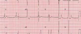
Currently, modern electrocardiography is actively developing in the field of registration and analysis of low-amplitude potentials (LAPs) of the heart. This direction is called high-resolution electrocardiography (HR ECG) [1], [2, pp. 15-21]. Analysis of the NAP allows you to obtain important additional information about the patient’s heart condition. One type of LAP is late ventricular potentials (LVPs). Their occurrence on the electrocardiosignal (ECS) at the end of the QRS complex and the beginning of the ST segment means an increased (up to 50%) risk of developing cardiac arrhythmias in this patient, including life-threatening ones. However, the problem is that PPV cannot be detected on a conventional cardiogram, since their level is below the noise level.
Within the HR ECG, the standard method for recording and analyzing PPV is the Simson method [3, pp. 235-242], [4, pp. 26-29], based on the temporary synchronous accumulation of many cardiac cycles and subsequent analysis of the average signal. As a result of temporary accumulation, we increase the signal-to-noise ratio by the required number of times, since the useful signal is added in phase. According to this method, the ECS is first recorded from the surface of the human body using three bipolar orthogonal leads X, Y, Z [5, pp. 79-87]. Next, the signal is averaged over time and filtered in a certain frequency range. Then the amplitude of the cardiac EMF vector is calculated, called the filtered QRS complex, for which three amplitude-time parameters are calculated [3, pp. 236-237]. If any two of these three parameters exceed certain threshold values, then a conclusion is made about the presence of PPV on the test pacemaker. The specified parameters and their threshold values have the following names and meanings:
– DQRS – duration of the filtered QRS complex (duration of QRS); DQRS>120 ms;
– LAS – duration of the low-amplitude final part (below 40 µV) of the filtered QRS complex (low amplitude signals); LAS>39 ms;
– RMS – root mean square amplitude of the last 40 ms of the filtered QRS complex (root mean square); RMS<25 µV.
Purpose and objectives of the work
The purpose of this work is to modify the Simson method for analyzing PPV in portable cardiac analyzers. Such cardiac analyzers are used in hospitals, commercial clinics, and also by individuals as a personal device for monitoring the condition of the heart. The task of expanding the functionality of portable cardiac analyzers is very important. So, for example, if a person is found to have PPV, then timely measures can be taken to eliminate the risk of arrhythmias. Thus, the purpose of the work is very relevant.
However, cardiac analyzers record the pacemaker, as a rule, only in one – standard lead I [6, pp. 412-414], [7, pp. 1-2]. Therefore, the shape of the filtered QRS complex obtained from one lead will differ from the shape of the filtered QRS complex obtained from three orthogonal leads X, Y, Z. Standard threshold values of the parameters of the Simson method cannot be applied when using one lead. Therefore, it is necessary to obtain new threshold values for the three amplitude-time parameters of the Simson method with a new form of the filtered QRS complex. In [8, p. 253], we obtained preliminary threshold values for a small number of ECS recordings (149) from the www.physionet.org database. Therefore, the task of this work is to clarify the specified threshold values based on statistical analysis of a larger number of signals.
The scientific novelty of the work lies in the use of the well-known method for detecting low-amplitude potentials - the Simson method for the detection and subsequent analysis of NAP on the pacemaker from standard lead I. The new threshold values of the amplitude-time parameters of the Simson method obtained in this way are scientific knowledge of practical interest.
Materials and methods
For the study, 290 pacemaker recordings were taken from the PTB Diagnostic ECG Database [9]. All signals were analyzed using the standard Simson method for the presence or absence of PPV in three bipolar orthogonal leads X, Y, Z. The results of signal analysis - the presence (51 cases) and absence of PPV (239 cases) - will be taken as the basis for further research. That is, we accept the division of the entire data array into 2 classes - the presence of PPV and the absence of PPV based on the results of processing using the standard Simson method for three leads. Next, the same recordings (290 pacemakers) were analyzed for the presence or absence of PPV using the Simson method, but using standard lead I. In this case, in contrast to the standard method, the filtered QRS complex was calculated using the signal of one lead. And the threshold values of the amplitude-time parameters were taken from [8, P. 253].
results
As a result of the analysis, in 16.2% of cases, the conclusion about the presence or absence of PPV on the pacemaker in the first standard lead did not coincide with the conclusion taken as a basis. Therefore, it is necessary to clarify the threshold values of the parameters of the Simson method for analyzing the ECS using standard lead I.
Using the example of the duration parameter of the filtered QRS complex DQRS, we will show the principle of obtaining a new threshold value. First of all, two histograms of the distribution of the DQRS parameter were constructed on one graph for the cases of the presence and absence of PPV.
In this case, the division into classes PPV+ and PPV- was carried out on the basis of a standard analysis of three orthogonal leads X, Y, Z. And the durations of the filtered QRS complexes - DQRS were taken from the statistics we collected (290 cases of ECS) in standard lead I. By testing statistical hypotheses [10, pp. 244-262], it was determined that the mathematical expectations of the two constructed histograms with a confidence probability of p=0.95 are significantly different. After this, using the constructed histograms, the decision threshold was determined using the maximum likelihood criterion. As a result, we obtained a new threshold value for the duration parameter of the filtered QRS complex Dthor, equal to 105 ms.
New threshold values for the other two amplitude-time parameters of the Simson method were determined in a similar way. These parameters are the duration of the low-amplitude part (less than 40 μV) of the filtered QRS complex - LAS and the root-mean-square amplitude of the last 40 ms of the filtered QRS complex - RMS. New threshold values for these parameters, obtained in the same way as the above-described threshold Dthr, were: LASthr – 39 ms; RMSpore – 18 µV.
Taking into account the new refined threshold values of the amplitude-time parameters, a repeated analysis of the ECS implementations for the presence of PPV was carried out. As a result, the percentage of erroneous conclusions dropped to 12%. This value is acceptable from a practical point of view for automated algorithms for analyzing bioelectrical signals of the heart.
Conclusion
As a result of the study, new threshold values of the amplitude-time parameters of the Simson method were determined for the analysis of the ECS recorded using standard lead I in order to determine the presence of late potentials of the ventricles of the heart. The Simson method modified in this way can be used in portable cardiac analyzers. In the future, we plan to implement this method in the portable cardiac analyzer we are developing. This will certainly expand its functionality.
List of literature / References
- Ivanov G.G. High-resolution electrocardiography / G.G. Ivanov, S.V. Goryacheva, A.L. Syrkina – M.: “Triad-X”, 2003. – 304 p.
- Kulambaev B.B. High-resolution electrocardiography: some methodological approaches to the analysis of late ventricular potentials / B.B. Kulambaev, G.G. Ivanov, D.U. Akasheva et al. // Cardiology. – 1994. – T. 34, No. 5-6. – pp. 15-21.
- Simson MB Use of signals in the terminal QRS complex to identify patients with ventricular tachycardia after myocardial infarction // Circulation. – 1981. – Vol. 64. – P. 235-242.
- Galimzyanov E.R. Optimization of the method of processing electrocardiosignals to identify low-amplitude cardiac potentials. // Methods and devices for transmitting and processing information. – 2011. – Issue. 13. – pp. 26-29.
- Dekhtyar G.Ya. Electrocardiographic diagnostics. M.: Medicine, 1966. – 543 p.
- Sherbakova TF Cardiac arrhythmia analysis used in the system of electrocardiosignal transmission to the supervision center" / Sherbakova TF, Sherbakov GI, Sedov SS // 2015 International Conference on Antenna Theory and Techniques (ICATT). – Kharkiv, 2015. – P. 412-414. doi: 10.1109/ICATT.2015.7136904
- Korobkov AA A portable device for registration and transfer of patient's electrocardiogram using microwave technologies / Korobkov AA, Ulanov AV // 2015 International Conference on Antenna Theory and Techniques (ICATT). – Kharkiv, 2015. – P. 1-2. doi: 10.1109/ICATT.2015.7136900.
- Sedov S.S. Expanding the functionality of portable cardiac analyzers / Sedov S.S., Shcherbakova T.F., Zaripov B.A. // XVII International Scientific and Technical Conference “Problems of Engineering and Technologies of Telecommunications” PTiTT-2016. – Samara, 2021. – pp. 252-253.
- PhysioNet/PTB Diagnostic ECG Database. – URL: https://physionet.org/physiobank/database/ptbdb/ (access date: 05/12/2017)
- Smirnov N.V. Course on probability theory and mathematical statistics / N.V. Smirnov, I.V. Dunin-Barkovsky - M.: Nauka, 1969. - 511 p.
List of literature in English / References in English
- Ivanov GG Jelektrokardiografija vysokogo razreshenija [High-resolution electrocardiography] / GG Ivanov, SV Gorjacheva, AL Syrkina – M.: “Triad-X”, 2003. – 304 p. [in English]
- Kulambaev BB Jelektrokardiografija vysokogo razreshenija: nekotorye metodicheskie podhody pri analize pozdnih potencialov zheludochkov [High-resolution electrocardiography: some methodical approaches in the analysis of late potentials of the ventricles.] / BB Kulambaev, GG Ivanov, DU Akasheva and others // Kardiologija [Cardiology ]. – 1994. – Vol. 34, no. 5-6. – P. 15-21.
- Simson MB Use of signals in the terminal QRS complex to identify patients with ventricular tachycardia after myocardial infarction // Circulation. – 1981. – Vol. 64. – P. 235-242. [in English]
- Galimzyanov ER Optimizacija metoda obrabotki jelektrokardiosignalov dlja vyjavlenija nizkoamplitudnyh potencialov serdca // Metody i ustrojstva peredachi i obrabotki informacii [Methods and devices of information transmission and processing]. – 2011. – No. 13. – P. 26-29.
- Dehtjar' G.Ja. Jelektrokardiograficheskaja diagnostika. M.: Medicina, 1966. – 543 p. [in English]
- Sherbakova TF Cardiac arrhythmia analysis used in the system of electrocardiosignal transmission to the supervision center" / Sherbakova TF, Sherbakov GI, Sedov SS // 2015 International Conference on Antenna Theory and Techniques (ICATT). – Kharkiv, 2015. – P. 412-414. doi: 10.1109/ICATT.2015.7136904 [in English]
- Korobkov AA A portable device for registration and transfer of patient's electrocardiogram using microwave technologies / Korobkov AA, Ulanov AV // 2015 International Conference on Antenna Theory and Techniques (ICATT). – Kharkiv, 2015. – P. 1-2. doi: 10.1109/ICATT.2015.7136900
- Sedov SS Rasshirenie funkcional'nyh vozmozhnostej portativnyh kardioanalizatorov / Sedov SS, Shherbakova TF, Zaripov BA // XVII Mezhdunarodnaja scientific-tehnicheskaja konferencija “Problemy tehniki i tehnologij telekommunikacij” PTiTT-2016 [XVII International scientific and technical conference "Problems of telecommunications engineering and technologies "PTEaT-2016]. – Samara, 2021. – P. 252-253.
- PhysioNet/PTB Diagnostic ECG Database. – URL: https://physionet.org/physiobank/database/ptbdb/ (accessed: 05/12/2017)
- Smirnov NV Kurs teorii verojatnostej i mate-maticheskoj statistiki / NV Smirnov, IV Dunin-Barkovskij – M.: Nauka, 1969. – 511 p.
Signs of predominance of ventricular potentials
Predominance of left ventricular potentials
The predominance of ventricular potentials or the predominance of its electrical activity is detected by the electrocardiographic method. ECG signs of the predominance of LV activity are changes in the leads responsible for the left parts of the heart. Since the R wave on the cardiogram is responsible for the excitation of the ventricles, the dominance of RV or LV activity will influence changes in this wave in the corresponding leads. If LV activity predominates in the left precordial leads (I, II, AVL, V5-V6), the R wave will be larger than normal - more than 25 mm.
In the right leads, changes in the negative S wave will be observed. It will be more than 15 mm in depth in III, AVF, V1-V2. If the activity of the pancreas predominates, the opposite picture will occur. In the right leads, the height of the R wave will be greater than 7 mm. In the left chest leads, the negative S wave will be deeper than 7 mm. However, you should not draw conclusions only based on the R and S waves. It is necessary to evaluate other elements of the electrocardiogram, combining everything with clinical data.
When the predominance of potentials is normal
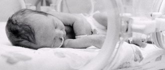
ECG of a newborn
The ECG of a full-term child up to one year old shows signs of predominant activity of the pancreas, since at the moment of the child’s birth the pulmonary circulation begins to function. But anatomically, the LV myocardium has not yet had time to “increase” its mass, so the thickness of the RV and LV are approximately the same at the time of birth. After one year and up to 6-8 years of age, as a rule, a restructuring of the cardiovascular system occurs. Due to the fact that the load on the ventricles changes, not only the anatomical location of the heart in the chest changes, but also its electrical axis.
After the age of six, RV dominance begins to wane and the ECG pattern begins to reflect increased LV activity. Therefore, if an ECG reveals dominance of RV potentials in a six-year-old child, this is not a reason to panic. Another question is if the child has some complaints and deviations in physical development. In an adult, a normal ECG should reflect a predominance of left ventricular potentials. The patient's body type may also have an impact. In people with an asthenic (thin) build, the heart occupies a vertical position in the chest, since the dome of the diaphragm is low.
This position leads to a change in the electrical axis of the heart. The largest R wave will be in lead I, and the smallest in lead III. In persons with a hypersthenic physique, the heart occupies a horizontal position due to the high position of the dome of the diaphragm. Therefore, the maximum R wave will be in III, and the smallest in I. In such situations, you should not look for pathology. Another situation where left ventricular activity dominates is physiological hypertrophy. Physiological hypertrophy can occur in athletes, as well as people whose work involves heavy physical activity.
Symptoms of the disease
Angina is the most common symptom that indicates left ventricular hypertrophy of the heart. As a rule, in the first stages there are no symptoms. With further development of hypertrophy, the patient may develop atrial fibrillation. Often there may be a condition in which the heart freezes and does not beat (this condition provokes loss of consciousness). In some cases, in a person suffering from left ventricular hypertrophy. In this case, severe shortness of breath may occur. Additionally, a number of symptoms are identified:
- unstable blood pressure;
- high blood pressure;
- general weakness, poor health;
- pain in the heart, chest;
- sleep disturbances, arrhythmia, headaches.
The main symptoms of left ventricular hypertrophy are listed above.
Diagnosis of the disease
Only a qualified cardiologist can examine a patient who is suspected of having a disease. The most effective diagnostic method is electrocardiography (ECG). Thanks to this method, a specialist can determine the thickness of the heart muscle. With this disease, the following changes are visible on the ECG:
- increase in teeth SI, V6 and Rv I, III;
- conduction disturbances in the myocardium;
- modifications of the electrical position;
- complete or partial blockade of the His bundle;
- deviation of the average QRS vector to the right or forward.
In addition to the above diagnostic method, the specialist conducts an examination using MRI, chest X-ray or computed tomography. Cardiac MRI in Moscow also allows the specialist to obtain a lot of useful information about the patient’s condition.
Get a free consultation Consultation does not oblige you to anything
Disease prevention
The risk of further development of the disease is reduced by avoiding drinking alcohol, cigarettes, and coffee. With left ventricular hypertrophy, it is recommended to lead a healthy lifestyle, systematically monitor heart function, and also reduce salt intake and reduce the amount of fatty foods in the diet. With this disease, it is necessary to include more vegetables and fresh fruits in the menu.