The ventricles of the brain are a system of interconnected cavities where cerebrospinal fluid circulates. The ventricles provide:
- Mechanical protection of the brain, creating a soft “buffer” upon impact.
- The production of cerebrospinal fluid - a fluid that nourishes brain tissue.
- Circulation of cerebrospinal fluid. The ventricles ensure the removal of toxic metabolic products from the brain.
- Filtration of substances entering the cavities of the nervous system.
Dilatation or enlargement of the ventricles of the brain leads to dysfunction, which develops neurological and psychological symptoms.
Dilation develops in two types:
- Increase in the volume of all ventricles.
- An increase in the volume of individual ventricles, which leads to their asymmetry.
Expansion or asymmetry of the cavities leads to changes in intracranial pressure. As it increases, it compresses areas of the brain, forming a clinical picture that is determined primarily by general cerebral symptoms, since the entire ventricular system is affected. A separate brain area is not affected.
Dilatation is not an independent disease, but is part of a symptom complex of other major leading diseases or defects. For example, expansion can be represented as an anatomical change in infectious and inflammatory diseases.
Dilatation is considered a pathology only when it manifests itself: it worsens a person’s quality of life and gives rise to symptoms. When ventricular expansion occurs latently, asymptomatically, this is considered an anatomical feature of the nervous system.
Dilatation may not manifest itself as a syndrome. The severity of specific symptoms depends on the degree of expansion of the brain cavities.
Expansion of cavities also occurs in newborns. In this case, the pathology is a defect in the intrauterine development of the fetus.
Indicators of normal sizes
In the human body, the ventricular system consists of several cavities that anastomize with each other. They communicate with the subarachnoid space, as well as the spinal cord canal. A special liquid, cerebrospinal fluid, moves directly inside the cavities. With its help, tissues receive nutrients and oxygen molecules.
The largest intracerebral hollow formations are, of course, the lateral ventricles. They are localized below the corpus callosum - on both sides of the midline of the brain, symmetrical relative to each other. In each, it is customary to distinguish several sections - the anterior and lower, as well as the posterior horns and the body itself. The shape resembles the English S.
The normal size of the ventricles is assessed taking into account individual anatomical characteristics - there are no uniform standards. Experts focus on average parameters. It is important to know these sizes for babies under one year old - for the purpose of early diagnosis of hydrocephalus.
Normal values for children:
| Anatomical unit | Newborns, mm | 3 months, mm | 6 months – 9 months, mm | 12 months, mm |
| Lateral ventricle | 23.5-/+ 6.8 | 36.2 -/+3.9 | 60.8-/+6.7 | 64.7-/+12.7 |
For adults, the parameters should be in the range - the anterior horn of the lateral ventricle is less than 12 mm in people under 40 years of age, while its body is 18–21 mm in people under 60 years of age. Exceeding the age-related size of the brain ventricles by more than 10% requires additional research to identify and eliminate the root cause.
Forecast
Every patient with left ventricular dilatation, already knowing what it is, must follow all medical recommendations. With this diagnosis, early diagnosis and initiation of treatment are important. In advanced forms, there is a high probability of developing heart failure. In these same patients, the valve apparatus is deformed, which leads to mitral insufficiency. This diagnosis significantly affects the quality of life and reduces its duration. The prognosis for patients is unfavorable.
The average survival rate for left ventricular dilatation is 10 years. If the course is asymptomatic, then life expectancy is on average 5 years. Patients with chronic heart failure observed in the hospital survive up to 50% of those admitted.
It is important for each patient to remember that the first symptoms are not considered normal and require a set of diagnostic procedures. Timely treatment will reduce the risk of complications, and treatment will prolong life for many years.
Classification
The main criteria for separating dilatations of the lateral ventricles in the brain are the size of the cavities, the etiology of the dilation, the age of the patient, and the localization of pathological changes.
Each neurologist chooses the optimal classification of the disorder for himself. However, most doctors adhere to the average principles of diagnosis:
- According to the time of expected appearance of the lesion in the brain:
- prenatal period;
- detection of enlarged cerebral ventricles in newborns;
- expansion of brain cavities in adults.
- By localization:
- left ventricle enlargement;
- right-sided lesion;
- bilateral defeat.
- By etiology:
- post-infectious ventricular dilatation;
- post-traumatic changes;
- toxic expansion;
- tumor focus in the brain;
- vascular diseases.
- By severity:
- slightly enlarged ventricles of the brain in infants;
- moderate dilatation;
- severe changes in the ventricles.
Additionally, the specialist can indicate in the diagnosis whether complications are present - for example, hydrocephalus or intellectual/neurological problems.
Features and classification
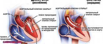
One of the known variants of cardiomyopathy is ventricular dilatation.
Expansion of cavities occurs in many patients for no apparent reason. As a result, the superficial function of the myocardium is disrupted, which leads to a rapid increase in its size. The appearance of dysfunction is associated with a decrease in the strength of contractions of the muscular wall of the ventricles. At the same time, there is a decrease in the release of blood into the aorta. During the examination of some patients, the thickness of the heart wall does not change when the cavities are dilated. The following options for dilatation of the left ventricle of the heart are distinguished:
- tonogenic;
- myogenic.
With tonogenic dilatation, expansion of the heart cavity is noted due to increased blood flow to them and increased pressure. The myogenic form is characterized by an irreversible change in the volume of the chamber. It appears against the background of elongation of fibers and their stretching with a simultaneous lack of contractility.
The latter variant of dilatation is most often combined with a decrease in wall tone. It is divided into primary and secondary. The first form develops with myocarditis in acute or chronic stages, cardiosclerosis caused by atherosclerosis. During primary expansion, the cavity grows uniformly in size. The function of myocardial contraction is significantly reduced. The pulse and heart rate become weak and difficult to feel.
The secondary form occurs against the background of established myocardial hypertrophy. The size of the heart is significantly increased in comparison with the primary one.
There are many factors that have a negative effect on the myocardium, but there are certain conditions that contribute to dilatation of the left ventricular cavity:
- Pathology associated with damage to the myocardium itself.
- Excessive load.
Some patients are characterized by an asymptomatic course of the disease against the background of complete health. Over time, if it is impossible to compensate for the condition, signs of the disease appear. This is typical for dilated cardiomyopathy. Other causes are inflammation, arterial hypertension, which over time make the muscle wall weak. This condition leads to loss of elasticity and excessive extensibility, which leads to dilatation of the cavity.
Overload of the left chamber of the heart occurs when the functioning of the valve that opens into the aorta is disrupted. The narrowing creates an obstruction to the flow of blood, which over time leads to stretching of the heart tissue and dilatation of the cavity.
This condition is observed in people with defects in which a large volume of blood enters the ventricle.
Causes
The stages of development of the central nervous system in humans provide that as the size of the brain increases, the parameters of the ventricles will also change. For each period, the reasons for dilatation of the lateral cavities have their own characteristics.
In general, the main provoking factors will be as follows:
- brain injuries or falls;
- neuroinfections - for example, meningitis or congenital syphilis;
- brain tumors;
- thrombosis of cerebral vessels;
- strokes;
- abnormalities in the development of brain structures - for example, the anterior horns of the ventricles.
The mechanism for the development of dilatation is the overproduction of cerebrospinal fluid, or a violation of its adsorption/outflow from the cavities of the brain.
In some cases, it is not possible to determine the exact cause of the expansion of the cavities - the idiopathic version of the disorder. The doctor will select the treatment regimen taking into account the main clinical signs. Less often, the basis of dilatation is seen as an atypical anlage of brain structures - it is necessary to carefully collect an anamnesis from the child’s mother, what diseases she suffered during pregnancy. Sometimes the pathology is hereditary in nature - genetic abnormalities.
Symptoms
Moderate expansion of one or two chambers in the heart may not manifest itself for a long time. Often, pathology is discovered by chance, during a routine examination or treatment of another disease. Severe dilatation of the cavity leads to a decrease in pumping function, which leads to the appearance of signs of heart failure or arrhythmia. These include the following:
- Palpable palpitations.
- Dyspnea.
- Blueness of the nasolabial triangle, lips, earlobes, fingertips.
- As the severity of the course worsens, cyanosis spreads to the skin.
- Swelling in the arms and legs.
- Memory impairment.
- Fatigue and weakness that persists after rest.
- The appearance of discomfort when lying down.
- Dizziness.
- Headache.
- Feeling of interruptions in the heart.
Shortness of breath in the compensation stage appears only with excessive physical exertion. With gradual wear and tear of the myocardium, the condition worsens. Shortness of breath begins to bother you with slight exertion, and then at rest.
With chronic exposure to unfavorable factors, a change occurs in the myocardium, which leads to a gradual expansion of the cavity and thickening of the walls. Dilatation of the left ventricle of the heart in the absence of timely therapy increases the risk of complications. Most often, thrombosis and fibrillation of the ventricles or atria are observed.
In some patients, the valve apparatus is affected, which is manifested by expansion of the ring, deformation of structures and ends in the formation of acquired heart disease.
After the transition from the stage of compensation to decompensation, fluid appears in the abdominal cavity (ascites), and the size of the liver increases (hepatomegaly). The skin of such patients becomes damp and cold to the touch. Systolic blood pressure decreases. Tachycardia is noted.
When auscultated, wheezing is heard in the lungs. Determination of the boundaries of the heart shows cardiomegaly (increase in heart size), the rhythm is disturbed.
Symptoms
At the initial stage of formation of dilated ventricles of the brain in an infant, any special clinical signs may not be detected - the child behaves according to the age norm. After all, adaptation mechanisms are capable of combating overproduction of cerebrospinal fluid.
However, as the expansion of the lateral ventricles of the child’s brain increases, he begins to worry about the consequences of hydrocephalus - pathological pressure on the brain structures due to tissue swelling. The main signs of intracranial hypertension:
- frequent attacks of headache;
- slow growth of fontanelles;
- swelling of tissue between the sutures of the skull;
- nausea and vomiting without improvement of well-being;
- decreased appetite, frequent regurgitation;
- worsening sleep;
- throwing the head back;
- muscle hypertonicity;
- lack of interest in current events, apathy;
- tendency to epilepsy.
In adult patients, a violation of the outflow of cerebrospinal fluid from the lateral ventricles is manifested by a feeling of constant distension inside the head, persistent dizziness with nausea. A person’s ability to work decreases, and he develops anxiety-phobic conditions. At the same time, taking standard analgesics does not improve well-being.
With persistent hypertensive-hydrocephalic syndrome, people develop paresis/paralysis, as well as serious difficulties with speech, vision, hearing, and decreased intellectual capabilities.
What are the ventricles of the brain, their role
The ventricles of the brain are strips of tissue necessary for the deposition of cerebrospinal fluid.
External and internal factors can lead to their increase in volume. The lateral ventricles are the largest. These formations are involved in the formation of cerebrospinal fluid. Asymmetry is a condition in which one or both cavities are enlarged to varying degrees.
Types of ventricles:
- Lateral . The ventricles are the most voluminous, and they contain cerebrospinal fluid. They connect to the third ventricle via the interventricular foramina.
- Third . Located between the visual tuberosities. Its walls are filled with gray matter.
- Fourth . Located between the cerebellum and medulla oblongata.
Diagnostics
If a specialist observes signs of a malfunction in the circulation of cerebrospinal fluid through the cerebral ventricles, or the patient has complaints of deterioration in health, then instrumental confirmation of dilatation of the brain cavities is required.
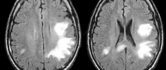
It is possible to identify signs of slight dilation of the lateral ventricles using such a modern diagnostic examination method as magnetic resonance imaging. On the resulting images of brain structures, you can see in detail the area of expansion, the area of damage, and the involvement of neighboring brain tissues in the process.
Increased intracranial pressure will also be diagnosed using the following procedures:
- echoencephaloscopy;
- electroencephalography;
- ophthalmoscopy;
- examination of cerebrospinal fluid - identification of previous neuroinfections;
- blood tests - general, biochemical, for autoimmune processes.
Only after careful comparison of all information from diagnostic procedures, a neurologist will be able to assess the severity of dilatation of the lateral ventricles, establish the root cause of the pathological condition and select optimal therapeutic measures.
Treatment tactics
In itself, the expansion of the size of the ventricles of the brain does not require intervention - if there are no clinical signs of intracranial pressure failure. Whereas in the case of a violation of liquorodynamics and symptoms of deterioration of well-being formed against this background, doctors will recommend conservative therapy:
- diuretics – removing swelling from brain tissue;
- neuroprotectors – correction of the conduction of nerve impulses;
- vasoactive agents – improving brain nutrition;
- nootropics – improvement of local blood circulation;
- sedative medications – normalization of the psychosomatic background;
- anti-inflammatory/antibacterial drugs – if the disorder is caused by an infectious process.
Neurosurgical intervention will be required if ventricular dilatation is caused by brain tumors or thromboembolism of cerebral vessels. If necessary, ventriculostomy is performed to create a new connection between the cavities of the brain.
Prognosis and prevention
The consequences of asymmetry of the lateral ventricles are varied. Their severity and severity directly depend on the size of the pathological expansion and the age of the patient. Thus, in mild forms of the disorder, children experience short-term developmental delays, both intellectual and physical. With timely medical care, hydrocephalus is completely eliminated.
Whereas with severe dilatation of the cavities, various neurological diseases are formed - for example, cerebral palsy, or persistent mental disorders. There is no specific prevention of ventricular asymmetry, since it is almost impossible to predict its occurrence. However, experts point out that when the expectant mother strives for a healthy image, she contributes to the birth of a baby with normal sizes of brain cavities. To do this, you need to give up bad individual habits even before pregnancy, eat right, get good sleep, and avoid psycho-emotional and stress overload.
Provoking diseases
The main disease causing this pathology is hydrocephalus. It can interfere with the absorption of cerebrospinal fluid. This leads to its accumulation in the lateral ventricles.
Excessive formation of cerebrospinal fluid is also observed with serious lesions of the central nervous system. Poor circulation is also associated with the formation of cysts, tumors and other neoplasms.
A common cause of hydrocephalus is a defect of the Sylvian aqueduct. If this defect was discovered during the prenatal period, termination of pregnancy is recommended. At the birth of a child, complex systematic treatment will be required.
Another cause is aneurysm of the vein of Galev and Arnold-Chiari syndrome. However, in children, the disease can be caused by rickets or due to the specific structure of the skull, so observation by a specialist is important if there is a predisposition to the disease.