- home
- Articles about cholelithiasis
- This is useful to know
- About the structure of the human body
- Catalog of useful medical articles
›
›
›
›
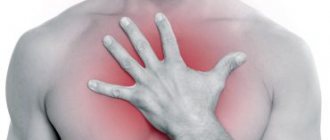
The heart is located in the center of the chest.
It is densely surrounded by other organs and anatomical structures. In this article we will look at what is below the heart. Sections of the article
Diaphragm
What organ is located under the heart?
Left lobe of the liver
Stomach
Pancreas
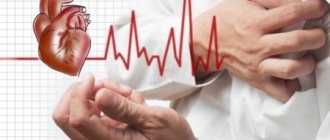
Causes of chest pain on the left
Cardiac ischemia
Currently, ischemic heart disease is a common cause of pain in this localization.
In a stable form, the pain syndrome begins during physical activity, fast walking. The functional class of stable angina is determined by the duration of the distance traveled, after which symptoms develop. Unpleasant sensations are caused by insufficient blood supply to the myocardium. The pain in the left side of the chest has a compressive, pressing nature, forcing the patient to stop walking or other physical activity. The symptom is accompanied by tightness in the chest and the inability to take a deep breath. Irradiation of pain into the shoulder blade and left arm is typical. With a complicated course of coronary artery disease and unstable angina, attacks of pain on the left side of the chest are observed more often, and are not always caused by physical activity. In severe situations, pain is observed at rest, accompanied by cyanosis and shortness of breath. Attacks of angina last from 5 to 30 minutes and are well controlled by taking nitroglycerin. Chest pain lasting more than half an hour against the background of a rapidly deteriorating condition is a serious symptom and requires immediate medical attention.
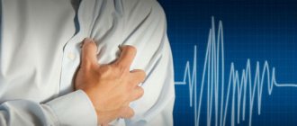
Myocardial infarction
Pain in the left side of the chest resembles a typical angina attack, but lasts 30 minutes or more. Pain during myocardial infarction is not relieved by nitroglycerin, and the patient gets worse every minute. A sharp pallor of the skin and cyanosis of the nasolabial triangle are noticeable, cold sticky sweat appears on the forehead. The pain is so severe that it becomes difficult to breathe, and fainting often occurs. If a person remains conscious, he may experience extreme feelings of fear and panic. If assistance is not provided in a timely manner, the pathology can be fatal.
Inflammatory heart diseases
Non-coronary causes cause acute stabbing or constant nagging pain to the left of the sternum, the intensity of which depends on the extent of the inflammatory process. With infective endocarditis, pain develops against a background of general weakness and sweating, and an increase in body temperature to febrile. The heart hurts constantly, the discomfort intensifies with deep breaths, but does not change during physical activity. In elderly people and patients with immunodeficiency, the symptoms are mild, pain is accompanied by shortness of breath and heaviness in the chest.
A similar clinical picture occurs with myocarditis. With infectious-allergic inflammation of the heart muscle, symptoms appear 2-3 weeks after tonsillitis or tonsillitis. I am concerned about moderate pain in the chest on the left, a feeling of palpitations and interruptions in the functioning of the heart. In acute myocarditis, pain occurs simultaneously with fever, weakness, and nocturnal hyperhidrosis. The chronic process is characterized by moderate discomfort in the heart area, shortness of breath and increased fatigue.
Cardiomyopathy
The severity of pain depends on the form of the disease. With hypertrophic cardiomyopathy near the sternum on the left, periodic stabbing or squeezing pains are observed, the appearance of which does not depend on external factors. With dilated cardiomyopathy, aching pain occurs in the heart, which intensifies during habitual physical activity. A person periodically feels shortness of breath and lack of air, and in the mornings they are bothered by weakness and dizziness. As the pain intensifies, signs of heart failure increase - swelling in the legs, ascites.
Arrhythmia
Heart rhythm disturbances are manifested by periodic stabbing or squeezing pain in the left chest, which usually begins without warning or exogenous influences. The pain during arrhythmia is sharp and severe, the patient freezes for a short time in one position, and grabs the heart area with his hand. At the same time, tachycardia and palpitations are determined. As a result of ineffective blood circulation, severe weakness and darkening of the eyes, pallor of the skin, and lightheadedness occur. Symptoms often disappear on their own within a few minutes.
Heart defects
Anatomical defects in the structure of the heart and efferent vessels cause anginal pain in the chest - squeezing or pressing, radiating to the clavicle area, poorly relieved by nitroglycerin. In addition to pain, signs of circulatory failure are detected: shortness of breath during exercise, dry cough, swelling of the neck veins. In children, chest pain on the left side can be caused by congenital developmental abnormalities that appear as the child grows. Causes that provoke cardiac pain syndrome:
- Mitral valve prolapse
. - Acquired defects
: mitral, aortic, tricuspid, combined forms. - Congenital “white” defects
: atrial and ventricular septal defects, coarctation of the aorta, patent ductus arteriosus. - Congenital “blue” defects
: tetralogy of Fallot, Ebstein’s anomaly.
Hypertonic disease
Chest pain develops in the later stages of hypertension, when degenerative changes occur in the heart muscle and coronary vessels. The pain syndrome is usually associated with spasm and obliteration of the arteries supplying the heart, and is manifested by compressive sensations that do not have a clear localization. During a hypertensive crisis, the pain intensifies and is accompanied by a rapid heartbeat and flashing “spots” before the eyes. Frequent dull or pressing pain in the heart is often caused by the secondary addition of atherosclerosis.
Dissecting aortic aneurysm
The severity of symptoms in dissecting aneurysm correlates with the degree of damage to the aorta. When only the inner lining of the vessel ruptures, patients experience sharp pain in the chest on the left and a faint state. In case of ongoing rupture and dissection of the aortic membranes, severe pain occurs in the chest cavity, which radiates to the back and shoulder girdle area. The pain syndrome is so intense that the person often loses consciousness due to shock. Continued bleeding causes a drop in blood pressure, collapse and deep fainting.
TELA
With massive thromboembolism of large trunks of the pulmonary artery, sharp stabbing pain in the chest suddenly appears without clear localization. Pain syndrome with pulmonary embolism is accompanied by a feeling of lack of air, shortness of breath, and cyanosis of the skin. If help is not provided, loss of consciousness and cardiac arrest occurs. Blockage of small vessels and focal pulmonary ischemia are causes of moderate chest pain, which intensifies and reaches its maximum after a few days. Pain during a pulmonary infarction is combined with hemoptysis and severe cough, which indicates the development of infarction-pneumonia.
Systemic connective tissue diseases
Many rheumatic processes occur with damage to the heart, which causes pain on the left side of the chest. Painful sensations due to collagenosis occur periodically and are of a pressing, stabbing or squeezing nature. Irradiation of pain syndrome is uncharacteristic. The symptom is not associated with stress or other external factors. The pain is accompanied by palpitations, shortness of breath, and fatigue. Most often, a similar clinical picture occurs with rheumatism. Less commonly, similar symptoms are caused by systemic lupus erythematosus, dermatomyositis, and vasculitis.
Respiratory diseases
Visceral pain is often associated with inflammatory damage to the left lung, pleura and bronchial tree. In this case, constant pain is typical, which is aggravated during bouts of coughing and deep breathing. Other signs of an infectious process are also typical: sputum production and febrile body temperature. The most common causes of pain in the left chest are:
- Bronchitis
. With purulent inflammation, mild pain in the chest is bothered, accompanied by discomfort behind the sternum and sore throat. Painful sensations occur against the background of a deep cough with scanty sputum. - Pneumonia
. In the focal form of the disease, moderate dull pain is felt in the left chest, which intensifies with coughing. Lobar pneumonia is characterized by severe diffuse chest pain, which is accompanied by high fever and difficulty breathing. - Pleurisy
. Pain syndrome localized in the inferolateral surface of the chest on the left is typical for inflammation of the pleura. The pain intensifies with coughing and body movements, patients are forced into a forced position on the painful side. - Tuberculosis
. Painful sensations of medium intensity, diffuse. When the pleura is involved in the pathological process, the pain intensifies and becomes clearly localized. The symptom is combined with shortness of breath, cough, scanty sputum, and profuse night sweats are characteristic.
Hiatal hernia
When the hernial protrusion is small, periodic discomfort occurs in the middle of the sternum and to the left of it. As the size of the hernia increases, sharp pains in the chest and epigastrium appear, which develop after eating, lifting weights, or against a background of stress. The duration of the symptom is from minutes to several hours. Along with the pain, dysphagia increases, even swallowing water becomes difficult. Constant intense pain on the left side is typical for a strangulated hernia, which requires emergency medical attention.
Pathology of the abdominal organs
The proximity of the nerve endings and segments of the spinal cord responsible for visceral innervation explains why there is pain in the chest area on the left when the digestive system is damaged. Such pain can be disturbing in acute pancreatitis and splenitis, since these organs are located in the abdominal cavity on the left, directly under the diaphragm. Symptoms sometimes occur with hematomas and ruptures of the spleen. The pain syndrome is also caused by organic causes - the inflammatory process spreads from the abdominal organs to the chest through the diaphragm.
Depression
Heart pain is one of the “masks” of a depressive state, which is provoked by disturbances of autonomic innervation and the development of cardioneurosis. Symptoms appear for no apparent reason and are detected in people of any age, more often in women. The pain is variable and usually appears in the early morning hours. Patients with depression tend to describe their discomfort at length and in detail. The pain is often preceded by a feeling of “heart sinking” and heaviness in the chest. The pain syndrome cannot be relieved with standard cardiotropic drugs.
Rare causes
- Pathology of nervous structures
: intercostal neuralgia, herpes zoster. - Diseases of the musculoskeletal system
: myalgia and myositis, inflammation of the costal cartilages on the left, chest injuries. - Left-sided pneumothorax
. - Damage to the stomach and esophagus
: GERD, hyperacid gastritis, peptic ulcer.
Myocardial infarction
If an attack of angina pectoris lasts a long time, then the heart muscle, not receiving nutrients and oxygen, begins to quickly deteriorate, its necrosis (death) occurs, this is myocardial infarction. That is why you should never tolerate pain due to angina pectoris.
Myocardial infarction is manifested by attacks of severe pain in the chest, which are not relieved by nitroglycerin. The duration of this pain is from 20-30 minutes to several hours. In such cases, the sooner the patient receives medical assistance, the greater the chance of recovery.
Diagnostics
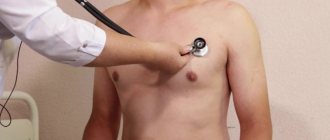
If a patient begins to have pain in the chest on the left, he needs to consult a therapist. First of all, regardless of the patient’s age, the specialist excludes all cardiac causes of pain, after which instrumental examinations of other organs of the chest cavity are performed. To clarify the etiological factor of the disease, advanced laboratory tests are indicated. In diagnostic terms, the most valuable are:
- ECG
. An electrocardiogram is recorded at the time of an attack of pain and signs of myocardial ischemia are revealed - a rise in the ST interval, a widening or change in the shape of the T wave. In the case of myocardial infarction, a typical “cat’s back” pattern is detected on the ECG. To confirm the anginal cause of pain, a troponin test is performed and the level of creatine phosphokinase is determined. - Echocardiography
. Ultrasound diagnostics of the heart allows you to identify signs of inflammatory processes and degenerative changes in the heart valves. Left ventricular contractility and ejection fraction are measured to rule out heart failure. Additionally, Doppler ultrasound and color mapping of the great vessels are prescribed. - Radiography
. If pneumonia is suspected, chest x-rays in the frontal and lateral planes are required. If a suspicious shadow appears on the image, it is informative to perform a CT scan of the chest cavity for detailed visualization of the affected area. X-rays can also help look for signs of a diaphragmatic hernia. - Ultrasonography
. An abdominal ultrasound is necessary to exclude inflammatory processes in the spleen and pancreas. The structure and homogeneity of the organ parenchyma and the presence of focal formations are taken into account. Ultrasound of the veins of the lower extremities is required to detect thrombotic masses, a common cause of pulmonary embolism. - Blood
tests . A general blood test is not informative enough; it usually reveals leukocytosis and an increase in ESR, which indicates an acute pathological process in the body. In a biochemical study, attention is paid to the levels of acute phase indicators. To diagnose atherosclerosis and the ischemic cause of left chest pain, the lipid profile is assessed. - Bacteriological research
. In case of pneumonia or bronchitis, sputum samples should be collected for bacterial culture on nutrient media, then the isolated microorganisms are tested for sensitivity to antibiotics. To confirm the diagnosis of infective endocarditis, it is necessary to culture pathogens in two or more blood samples taken 12 hours apart. - Invasive diagnostics
. To clarify the condition of the vessels, coronary angiography with a contrast agent is indicated, during which the patency of the arteries, the presence of atherosclerotic plaques and thrombotic masses are studied. Angiography is used to select the method of myocardial revascularization. In case of pleurisy, a pleural puncture is performed to take exudate for analysis.
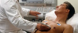
Echocardiography
Gastritis, ordinary and not very
The most common disease that causes pain in the epigastric region is gastritis, that is, inflammation of the gastric mucosa. Depending on the nature of the disease, gastritis can be acute or chronic.
The danger of chronic gastritis is that most cases of stomach cancer develop against the background of a long course of this seemingly harmless disease.
The type of gastritis is divided into superficial gastritis, in which only the mucous membrane becomes inflamed, and atrophic - in this case, against the background of inflammation, the stomach glands that produce hydrochloric acid gradually die.
| How can you examine the stomach? | |
| Unfortunately, not every person can safely undergo gastroscopy. The test is not a pleasant one. Therefore, now one of the most common (albeit paid) services is gastroscopy in a dream. If you have no contraindications to general anesthesia, then you can sleep through the entire examination with a clear conscience and not get negative impressions. Another innovative research method involves the patient swallowing an endoscopic capsule. A small device goes on a journey through the digestive tract and “makes a movie” along the way. Due to the fact that such a study is the least traumatic and most informative (the esophagus, stomach and entire small intestine are examined), it costs much more than all alternatives. | |
Unfortunately, it is possible to identify the type of gastritis and, accordingly, decide on treatment only after an examination, colloquially called “gastroscopy” - during this procedure, an endoscope is inserted into the upper parts of the gastrointestinal tract.
In fact, the study is called “esophagogastroduodenoscopy”, because during the procedure the doctor examines the patient’s esophagus, stomach, and duodenum. It is very important to carry out this study in as much detail and quality as possible, since gastritis very often “adjacent” to duodenitis (inflammation of the duodenum) and pathology of the esophagus.
Treatment
Help before diagnosis
A person with pain syndrome needs to be seated, freed from restrictive clothing, and provided with access to fresh air. If the patient suffers from cardiovascular pathology and is prescribed treatment, in case of exacerbation it is necessary to take nitroglycerin or other drugs as recommended by the doctor. In all situations where there is pain in the left chest, consultation with a specialist is required who can exclude or confirm serious cardiac and respiratory diseases.
Conservative therapy
For severe pain caused by myocardial infarction or pulmonary embolism, narcotic analgesics are indicated to help prevent the development of painful shock. Further therapy depends on the diagnosis. In case of cardiac pathology, the time of initiation of treatment is important; a delay of several hours can cause serious complications. For etiotropic treatment of diseases that manifest themselves as pain in the left areas of the chest, medications such as:
- Antianginal agents
. As first aid for a painful attack of angina, nitroglycerin is prescribed under the tongue or intravenously. Beta blockers, calcium antagonists, myotropic antispasmodics increase blood supply to the myocardium and reduce its need for oxygen. - Anticoagulants
. The drugs improve the rheological properties of blood and inhibit the activity of the hemostatic system, thereby reducing the risk of thrombosis and myocardial infarction. Heparin and its low molecular weight fractions are used, and acetylsalicylic acid is taken in low doses for a long time. - Thrombolytics
. The indication for their use is pain in the left chest due to a heart attack in the first 6 hours after the onset of symptoms. The drugs increase the activity of the fibrinolytic system and stimulate thrombus lysis. They contribute to the complete restoration of blood flow in the affected vessel. - Lipid-lowering drugs
. The drugs normalize the concentration of cholesterol and low-density lipoproteins, which cause the appearance of atherosclerotic deposits in the vessels. They are prescribed in long courses in combination with a special diet that includes limiting animal fats. - Antibiotics
. For pneumonia and exudative pleurisy, antibacterial agents from the group of cephalosporins and macrolides are effective. For severe lobar pneumonia, combinations of two medications are indicated. To treat infective endocarditis, penicillin is administered over a course of at least 4 weeks. - Antidepressants
. To eliminate the psychogenic cause of left chest discomfort, selective serotonin receptor agonists are recommended. Medicines normalize mood and eliminate apathy. If necessary, the drugs are combined with weak psychostimulants and tranquilizers.
Surgery
To restore arterial patency in patients with coronary artery disease and myocardial infarction, endovascular technologies are used - angioplasty and stenting under the control of selective angiography. In case of extensive changes in the intima of the vascular wall, coronary artery bypass grafting is performed, which improves blood supply to the heart. For diaphragmatic hernias, the following surgical interventions are used: suturing the hernial orifice and strengthening the ligamentous apparatus (crurorrhaphy), fixation of the stomach in the abdominal cavity (gastropexy), elimination of gastroesophageal reflux.