Electroencephalography (EEG) is a method that is used to study the state of the human brain and is based on recording its electrical activity. This examination allows you to detect the spread of pathological processes and signs of epilepsy.
The average duration of the procedure is about an hour, but it is highly informative and makes it possible to track functional changes in the brain, the dynamics of the disease, and evaluate the impact of the therapy.
Since the procedure does not cause any pain or discomfort, EEG can be called one of the most accurate and gentlest methods of examining the brain.
EEG principle
The human brain consists of millions of special cells called neurons. Each of them generates its own electrical impulse. Within individual areas of the brain, impulses must be consistent. They can also strengthen each other or make each other weaker. Their strength and amplitude are not stable and are constantly changing.
Content:
- How EEG works
- Historical reference
- Significance of EEG
- Benefits of an Electroencephalogram
- Indications for the procedure
- Contraindications
- How to prepare for an EEG
- Performing an EEG
- Purpose of EEG video monitoring
- Conclusion of electroencephalography
This is the bioelectrical activity of the brain. To register it, special electrodes are applied to the intact scalp, which will pick up vibrations, amplify them and record them in the form of special curves, so-called waves. The latter, depending on their shape, frequency and amplitude, are divided into five types: α- (alpha), β- (beta), δ- (delta), θ- (theta) and μ- (mu) waves. Each of the waves reflects the work of a specific part of the brain and is named by the first letter of its Latin name.
Their registration in real time is the essence of encephalography.
Interpretation of survey results
Even a qualified specialist is not always able to accurately name the cause of the patient’s poor health and immediately make a diagnosis. The doctor may be alerted to focal changes on the EEG. In this case, magnetic resonance imaging will be required to exclude a tumor or cyst.
When you contact a medical doctor, you will be able to undergo all examinations as quickly as possible, without delaying your visit to specialists.
If you need to do an EEG of the brain and you want to do it using modern expert-class equipment and in the shortest possible time, call the phone number listed on the website or leave a request in the feedback form.
Medical staff will answer your questions and conduct all necessary examinations within one business day. Let's take care of your health together!
Historical reference
One of the founders of the electroencephalography method is considered to be the physiologist and psychiatrist from Germany Hans Berger. In 1924, using a device for measuring small currents called a galvanometer, he was the first to perform a procedure resembling an EEG recording.
Later, a special device called an encephalograph was created to conduct electroencephalography. Today, there are stationary encephalographs, which make it possible to conduct research exclusively in a special room, and portable ones, which are designed for long-term monitoring and can be moved.
It is noteworthy that initially EEG was considered exclusively as a method for identifying mental disorders in a person. Only over time it became clear that the technique also makes it possible to detect deviations not related to psychiatry.
The legislative framework
The procedure for undergoing medical examinations is constantly changing.
So, the last of them happened in 2021. The new procedure was introduced by order of the Ministry of Health No. 344. This document extends its force to the territory of the entire Russian Federation.
The new rules came into force in March 2021. The previous procedure for undergoing medical examinations was canceled by this order.
Read the explanation of the categories of the new driver's license here.
It will apply to both driver candidates and those who are re-obtaining their licenses. The examination itself is carried out to identify medical contraindications for driving the vehicle.
The presence of a contraindication is established in accordance with Government Decree No. 1604.
Significance of EEG
EEG is a highly informative diagnostic method that allows:
- assess the nature of brain dysfunction and how severe they are;
- determine the localization of the pathological focus;
- clarify information obtained during other diagnostic procedures (for example, computed tomography);
- monitor how effective the therapy is;
- identify areas of the brain in which epileptic activity is present;
- assess brain function in the periods between attacks;
- identify the causes of panic attacks and fainting;
- study the sleep-wake cycle.
It should be taken into account that if a person has seizures, the study will be informative only if it is carried out after about a week.
Graph of brain biopotentials
An encephalogram allows you to assess the degree of damage to brain structures and indicate the localization of the pathological process. Its technique consists of placing electrodes on certain areas of the skull, taking their readings and recording the results on special paper. Biopotentials (rhythms) look like curves of a certain shape (Table 1). Its decoding is carried out by a specialist.
Table - Potential graph
There are also µ- and λ-rhythms, but they are extremely rarely used in encephalography. For the convenience of conducting research, modern devices are equipped not with separate electrodes, but with silicone caps, on which contacts are already attached in the right places.
Indications:
- sleep disorders (insomnia/excessive sleepiness);
- vascular lesions of the brain;
- DEP (diencephalic encephalopathy);
- injuries of the skull and soft tissues of the brain;
- headaches associated with liver disease (hepatic encephalopathy);
- suspicion of epilepsy and epileptiform seizures;
- neoplasms;
- neuroinfections, neurotoxicosis;
- suspected neurotoxin poisoning;
- coma;
- degenerative changes;
- dysfunctional disorders;
- monitoring the effect of anticonvulsants, dose adjustment, etc.
Many disorders can be confirmed by other modern studies. For epilepsy, the EEG remains irreplaceable, often the only diagnostic procedure. As well as a method of monitoring treatment.
Benefits of Electroencephalography
Today, electroencephalography is widely used in neuropathological practice. It makes it possible to clarify a huge number of problematic situations that are associated with the diagnosis and differentiation of neurological diseases. One of the undeniable advantages of encephalography is the fact that it not only helps to identify certain problems, but also helps to distinguish true disorders from hysterical manifestations or simulation.
In addition, the procedure is not as expensive as examination using a tomograph or other similar devices. EEG equipment is available in most hospitals.
The procedure does not have any negative impact on human health and condition. The patient remains fully functional. The study can be carried out even on patients in serious condition, children and adults of any age, since it does not cause deterioration of the condition, discomfort or pain.
Photo gallery
A headshot and other photos on the topic are shown in the gallery:
Cap with electrodes
EEG device
EEG for an adult
EEG for a child
Indications for the procedure
Today, electroencephalography is widely used in the practice of neurologists to solve a number of problems.
EEG is recommended:
- for prolonged insomnia and other sleep disorders, including apnea, somnambulism and talking during sleep;
- during a convulsive attack;
- for diseases of the thyroid gland;
- with signs of developmental disorders in children or mental disorders in adults;
- after recently experienced traumatic brain injury;
- in case of pathological changes in the vessels of the neck and head, brain tumors detected during ultrasound;
- with frequent migraines, complaints of regular dizziness, feeling of constant fatigue;
- for panic attacks, autism, Asperger's syndrome, stuttering, nervous tics, delayed speech and mental development in children;
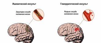
- for meningitis, encephalitis, after a stroke and micro-stroke;
- after neurosurgical operations.
Contraindications
It is noteworthy that there are no absolute contraindications for electroencephalography. If a person suffers from convulsive attacks, is diagnosed with coronary heart disease, hypertension, or mental disorders, an anesthesiologist must be present during the diagnosis.
The procedure should be postponed if there are open wounds, traumatic injuries, postoperative sutures or any signs of an inflammatory process in the area where it is necessary to install electrodes. Also, the study is not carried out for patients with ARVI.
Stages
Algorithm for conducting routine EEG:
- The patient sits on a chair or lies on the couch, relaxes, and closes his eyes.
- Electrodes are applied to the head. Places of contact with the skin are lubricated with gel or isotonic solution.
- After turning on, the device begins to read information and transmit it to the monitor in the form of a graph. This records background activity.
- Conducting functional tests necessary to assess the brain's response to stressful situations.
- Completion of the procedure. The electrodes are removed, the doctor makes a description and prints out the results.
How to prepare for an EEG
It should be noted that there are no special restrictions that must precede the procedure. However, there are a number of rules that are recommended to be followed to ensure that the examination is successful and informative.
First of all, inform your doctor if you are taking any medications. You may need to stop taking them for a while or change the dosage.
At least twelve hours before the procedure, or even better, a day before, eliminate from your diet foods that contain caffeine, carbonated drinks, chocolate and cocoa, as well as foods that contain them, foods with energy components, for example, taurine. You should also avoid products with sedative effects.
Wash your hair before the procedure. It is not recommended to use additional styling products (oils, gels, balm, varnishes, etc.) as this may affect the quality of contact of the electrodes with the skin.
If the main purpose of the procedure is to detect seizure activity, you need to sleep before the study.
In order for the result to be as reliable as possible, the patient should avoid stressful situations and refrain from driving for twelve hours before the procedure.
Eating is recommended two hours before the procedure.
If the patient is prescribed EEG sleep monitoring, the night before should be sleepless. Immediately before the procedure, the subject will receive a special sedative, which will enable him to fall asleep during the electroencephalography.
If a child is to undergo an examination, parents should first psychologically prepare him for the manipulations, explaining that there will be no pain or discomfort. You can practice putting on a pool cap under the pretext of playing pilots or astronauts, teach your child to breathe deeply, showing him how to do it by personal example. On the eve of the procedure, the child should wash his hair without using any additional styling products. Before leaving the house, the baby should be fed and calmed down. Just in case, parents are advised to take with them tasty food and drink, and a favorite toy that will help calm and distract the baby.
Please note that if the above rules are not followed, the EEG result may not be very accurate or very informative. In this case, the procedure will have to be repeated.
Preparation
Specific preparation for the event. It is recommended to wash your hair the day before the procedure. Before the procedure, remove hairpins and elastic bands if you let your hair down long.
If an EEG is performed on a small child, psychological preparation is needed. You will need to explain to your baby the need to wear an electrode cap or separate electrodes; you can play with him at home.
In some cases, sleep restriction may be necessary. You should not use sedatives before the procedure. An infant can take a bottle of milk. You should have wipes with you to remove any remaining gel.
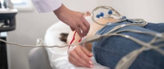
Performing an EEG
Electroencephalography is performed in a special room, which is completely isolated from light and sound. The patient sits in a chair or is asked to lie on the couch. A special cap with electrodes is first placed on his head. During the procedure, the patient is alone in the room, contact with doctors is maintained using a camera and microphone. If the diagnosis is performed on a child, one of the parents remains in the office.
Before starting the procedure, the patient is asked to close and open his eyes several times to adjust the equipment. During the diagnosis, the eyes must be closed. If during the procedure the patient needs to change position or visit the restroom, he can inform the doctors about this, after which the diagnosis will be suspended.
It is extremely important that the patient lies with his eyes closed and does not move during the procedure. In the event that a person opens his eyes slightly or moves, the doctor makes a corresponding note, since these actions must be taken into account when deciphering the electroencephalogram.
After the resting EEG is recorded, so-called “stress tests” are performed. Their goal is to test how the brain will react to situations that are stressful for it.
So, a hyperventilation test can be performed. The patient is asked to breathe frequently and deeply for three minutes. Photostimulation with a stroboscopic light source can also be used. It flashes frequently, and this allows you to evaluate how the brain reacts to bright light.
Stress tests can trigger convulsions or an epileptic seizure. The doctors who conduct the study have the appropriate skills to provide emergency care to the patient if necessary.
After the study is completed, the physician should remind the patient to resume taking medications that were stopped the day before the EEG.
The total duration of the procedure is from forty minutes to two hours.
Types of research methods
Several EEG research methods are used:
- routine;
- with deprivation;
- long;
- night.
Depending on the duration and purpose, computer encephalography is divided into types:
- Electroencephalogram of the brain - used in the initial stages of the examination. Both background activity and stress tests (hyperventilation, sharp sounds, flashes of light) are recorded.
- EEG monitoring is a long-term recording of brain activity. It is used when it is necessary to cover all possible physiological states of the central nervous system (sleep, wakefulness, mental work, emotions).
- Rheoencephalography is a study of cerebral vessels. Diagnostics is based on recording the changing value of electrical resistance of tissues when a weak high-frequency current is passed through them. Provides information about the tone and elasticity of the vascular wall, the amount of pulse blood supply.
Routine method
The routine method consists of short-term (approximately 15 minutes) recording of brain biopotentials. This is necessary to examine and evaluate the dominant rhythms, the presence of pathological potentials and paroxysmal activity.
Functional tests are also carried out, during which the reaction to:
- opening - closing eyes;
- clenching a fist;
- hyperventilation - forced breathing;
- photostimulation - blinking LEDs with eyes closed;
- sharp sounds.
The video shows an EEG with functional tests. Filmed by the channel “Clinic Doctor SAN”.
Encephalography with deprivation
Deprivation encephalography is performed with complete or partial sleep deprivation. Determines epileptic activity in situations that did not arise during provocative tests.
The patient either does not sleep all night or wakes up 2–3 hours earlier than usual. A routine EEG will be performed no earlier than 24 hours after the initial awakening.
Long-term EEG recording
Long-term recording of parameters during sleep is often carried out after deprivation EEG, since sleep is a powerful activator for the detection of epiactivity.
Only by performing a sleep EEG can a differential diagnosis of epilepsy with cognitive impairment be made. Therefore, this type of examination is prescribed if the doctor suspects that changes in the brain occur while the patient is sleeping.
Night EEG
Night EEG recording occurs in a hospital setting as follows:
- begins a few hours before bedtime;
- covers the period of falling asleep and the entire night's sleep;
- ends after natural awakening.
If necessary, additionally:
- video monitoring;
- electrooculography (EOG);
- recording of a cardiogram (ECG);
- electromyograms (EMG);
- spirography.
Purpose of EEG video monitoring
One of the types of electroencephalography is EEG video monitoring. This is a long-term recording of electroencephalography, usually lasting several hours, which can be performed during sleep. The duration of the procedure in each specific case is determined by the attending physician and the personnel of the laboratory that conducts the examination.
EEG video monitoring is prescribed if a short standard procedure does not reveal pathologies, but they are present.
Also, this type of examination allows you to evaluate the EEG during wakefulness and sleep.
Many patients are interested in the question of whether they should necessarily sleep during the study. The answer to this question cannot be unambiguous, because it depends on the specific situation. So, for example, if the reason for the examination is a tic that bothers the patient while awake, there is no need to sleep during the examination.
At the same time, EEG video monitoring during sleep sometimes helps to identify conditions that neither the patient nor his loved ones may even be aware of.
The peculiarity of this procedure is that it can be carried out not only during the day, but also at night. In the event that sleep EEG is required, night monitoring is more rational. Not everyone can fall asleep without problems during the daytime.
At the same time, we should not forget that carrying out a multi-hour procedure in a completely isolated room can be extremely tiring for the patient, especially if we are talking about a child. Most pathologies can be detected during a relatively short recording of a conventional EEG.
Best materials of the month
- Coronaviruses: SARS-CoV-2 (COVID-19)
- Antibiotics for the prevention and treatment of COVID-19: how effective are they?
- The most common "office" diseases
- Does vodka kill coronavirus?
- How to stay alive on our roads?
Also, overnight EEG video monitoring is much more expensive.
Price
The cost of EEG video monitoring depends on the qualifications of doctors, the quality of equipment, the quality of service and the room in which the patient is located during the study, and the clinic’s experience in the field of neurophysiology. The clinic's specialization in neurology provides a great advantage for the patient who wants to receive an accurate and definitive diagnosis.
Medical has been operating since 1988 - it has vast experience and a scientific base, as well as the best specialists. Video-EEG monitoring is carried out on elite neurophysiological equipment “Grass Telefeaktor” in special rooms with all the necessary equipment in compliance with world standards of EEG video monitoring
The diagnostic price may include consultation with specialists with analysis of the electroencephalogram in complex cases - participation in deciphering the results of the medical consultation examination. The cost also depends on the time of the study - multi-day EEG video monitoring is more expensive.
The diagnostic price may include consultation with specialists with analysis of the electroencephalogram in complex cases - participation in deciphering the results of the medical consultation examination. The cost also depends on the time of the study - multi-day video-EEG monitoring is more expensive.