Two types of hypertensive crisis
Doctors distinguish two types of hypertensive crisis. A first-order hypertensive crisis can appear already at the first stage of hypertension, and it is expressed in an increase in blood pressure, especially systolic, with an increase in pulse pressure. In this case, symptoms such as:
- general nervous agitation, increased irritability, sweating, red face,
- crawling sensation, general trembling,
- severe headaches, dizziness, flushing of the head,
- sudden deterioration of vision, appearance of fog before the eyes,
- noise in ears,
- increased heart rate, loud heart sounds.
A hypertensive crisis of the second order can develop against the background of hypertension in the last stages, and is expressed in an intensification of all the above symptoms. The headache comes on suddenly and is localized in the back of the head. Bradycardia, weakness, nausea and vomiting are observed. Pain in the heart area of a compressive nature, accompanied by a lack of air.
Breathing rhythm disturbance
Stroke
Hepatitis
Diabetes
Encephalitis
Thyrotoxicosis
16238 08 April
IMPORTANT!
The information in this section cannot be used for self-diagnosis and self-treatment.
In case of pain or other exacerbation of the disease, diagnostic tests should be prescribed only by the attending physician. To make a diagnosis and properly prescribe treatment, you should contact your doctor. Irregular breathing: causes, diseases in which it develops, methods of diagnosis and treatment.
Definition
Respiration is a set of physiological processes whose ultimate goal is the delivery and consumption of oxygen, as well as the removal of carbon dioxide.
The respiratory organs include the airways and lungs. In addition, the muscular and nervous systems take part in the breathing process. Thanks to the diaphragm, intercostal and some other muscles, respiratory movements are performed, a change in the volume of the chest occurs, which is a necessary condition for the expansion and collapse of lung tissue during inhalation and exhalation. The respiratory center is located in the central nervous system, namely in the medulla oblongata. The key feature of the functioning of the respiratory center is the automatic generation of impulses in it - that is, the person does not think about the fact that he needs to take a breath. At the same time, the activity of the respiratory center can be suppressed by higher-lying parts of the brain; for example, a person can voluntarily hold his breath while diving.
It is the respiratory center of the medulla oblongata that plays a key role in maintaining the rhythm of breathing.
Types of breathing rhythm disturbances
The following types of breathing are distinguished:
- eupnea
– normal breathing rhythm (in a healthy adult it is about 14–18 respiratory movements per minute); - hyperpnea
– deep and rapid breathing, observed during muscular work, emotional stress, thyrotoxicosis, anemia, acidosis, decreased oxygen content in the inhaled air; - tachypnea
- an increase in the frequency of respiratory movements due to pronounced stimulation of the respiratory center during hypercapnia, hypoxemia; observed with increased body temperature, congestion in the lungs, etc.; - bradypnea
- a decrease in the frequency of respiratory movements, which occurs when the respiratory center is damaged and depressed against the background of hypoxia, edema, ischemia and exposure to narcotic substances; - apnea
– temporary cessation of breathing (considered the extreme severity of bradypnea); - Cheyne-Stokes breathing
- characterized by alternating groups of respiratory movements with increasing amplitude and periods of apnea; - breathing according to the Biota type
- characterized by alternating periods of apnea with groups of respiratory movements of equal amplitude; - Kussmaul breathing
is noisy and deep breathing, which is characterized by separate convulsive contractions of the main and auxiliary respiratory muscles. This type of breathing indicates significant hypoxia of the brain.
Possible causes of respiratory rhythm disturbances
The immediate cause of respiratory rhythm disturbances is a change in the functioning of the respiratory center. The respiratory center has numerous connections with other parts of the nervous system and with receptors (nerve endings) that perceive changes in the internal environment of the body: changes in the concentration of oxygen and carbon dioxide in the blood, blood acidity, etc. For example, with a decrease in the concentration of oxygen in the blood, an increase in the concentration of carbon dioxide and acidification of the blood, receptors that activate the respiratory center are excited. In turn, an increase in the frequency and depth of breathing compensates for changes in the gas and acid-base composition of the blood.
The respiratory center can be directly affected by certain brain-toxic substances circulating in the blood.
Disturbances in the functioning of the respiratory center occur as a result of structural changes in the corresponding area of the brain during strokes, cerebral edema, etc.
Diseases that cause breathing problems
Among the diseases that lead to the accumulation of toxic substances in the blood, which can subsequently irritate or damage the respiratory center, it is worth mentioning the following:
- diabetes mellitus in the stage of decompensation
, when carbohydrate metabolism products accumulate in the blood; occurs at the onset of previously undiagnosed diabetes mellitus or with insufficient control of the disease during insulin therapy and glucose-lowering therapy; - liver failure
, developing acutely against the background of hepatitis (viral, alcoholic, autoimmune etiology, etc.), toxic liver damage (for example, paracetamol poisoning), as well as chronic intoxication with liver failure against the background of cirrhosis. Toxic damage to the brain in liver diseases is primarily due to the fact that the pathologically altered liver is not able to perform a barrier function (retain and neutralize harmful substances), and therefore toxins accumulate in the blood; - renal failure
, which can manifest acutely or have a long-term chronic course, develops against the background of inflammatory diseases (glomerulonephritis, tubulointerstitial nephritis, etc.), toxic damage to the kidney (for example, tubular necrosis) and other diseases; - Severe infectious diseases
can cause severe intoxication or lead to direct brain damage, for example, in viral or bacterial encephalitis.
Respiratory rhythm disturbances can occur during poisoning with opium-type drugs that cause bradycardia and apnea.
Structural changes in the brain are observed during strokes (acute cerebrovascular accident due to hemorrhage or blockage of cerebral vessels by a blood clot), neoplasms, and cerebral edema.
Separately, it is worth mentioning Pickwick's syndrome, which develops in patients with severe obesity, one of the manifestations of which is episodes of respiratory arrest during sleep.
Which doctors should I contact if my breathing rhythm is disrupted?
Irregular breathing is a serious symptom, so often the first specialist to assist a patient is an emergency physician or resuscitator.
If a violation of the breathing rhythm does not pose a threat to life, then diagnostic measures are carried out by a doctor of a narrower specialty: a hepatologist (a specialist in liver diseases), a nephrologist (a specialist in kidney diseases), a toxicologist, an infectious disease specialist, etc.
Diagnostics and examinations for respiratory rhythm disorders
Diagnosis begins with a survey of the patient, during which the doctor establishes possible causes and risk factors for the development of arrhythmic breathing.
Laboratory tests include determination of blood glucose levels, arterial blood gases and the acid-base status of the blood CBS.
First aid rules for hypertensive crisis
First of all, the patient needs to be put to bed, but so that the head and upper body are higher than the legs. Before the ambulance arrives, the patient must be given a drug that lowers blood pressure, for example, captopril at a dosage of 50 mg. You can also give an intramuscular injection of papaverine 2-4 ml of a 2% solution. If there is pronounced shortness of breath, lack of air, or cardiac pathologies appear, then the patient should be given an aminophylline tablet, as well as 40 drops of Corvalol or Valocordin.
You can urgently call a doctor to your home in Yeysk at the Sensitive LCC by the numbers listed on our website.
The effectiveness of shock wave therapy
Services of a private clinic in Yeysk
Rest in Yeysk, and quality medical care
Examination of temporary disability in LKK "Sensitive"
Medical certificate for entry into civil service
New rules for a medical certificate for weapons
Why enter into a voluntary health insurance agreement?
"Infanrix" - a three-component vaccine
Pulse study and what is a high pulse
Necessary preface from the site creators
Patients often want to know
what a high pulse is
? There are 2 concepts, distinguish between them.
1) In propaedeutics of internal diseases under high pulse
understand the pathological feature of the pulse when
the artery wall rises high
at the moment the pulse wave arrives.
This happens, for example, with heart disease - aortic valve insufficiency. Read more on this page in the sections “ PULSE VALUE
” and “
PULSE SHAPE
”.
2) When patients search the Internet for the term “ high pulse”
", they usually mean
FREQUENT heart rate
.
In other words - tachycardia
, in which the heart rate in adults
exceeds 60 per minute
.
Read more about the causes of tachycardia on this page in the “ PULSE RATE
” section.
Pulse
is a vasodilation that periodically occurs simultaneously with left ventricular systole, visible to the eye or felt with the fingers.
Before starting to study it, it is necessary to assess the condition of the vascular wall
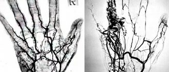
. To do this, use the 2nd and 4th fingers of the palpating hand to compress the radial artery, and with the 3rd (middle) finger, using sliding movements along and across it, study the properties of its wall (Fig. 50). Normally it should be soft but elastic. In some diseases (atherosclerosis), the arteries change and their walls become denser, and the course becomes tortuous.
Rice. 50. Study of the condition of the vascular wall of the radial artery.
The main method for determining pulse
is palpation.
It can be performed on the radial, carotid, temporal and other arteries (Fig. 51). The radial artery is most often palpated because it is located directly under the skin and can be easily felt between the styloid process of the radius and the tendon of the internal radial muscle. During palpation, the hands of the subject are grasped in the area of the wrist joint and, having felt the arteries, they are pressed with 2-3 fingers. The study on both arms simultaneously is due to the fact that the pulse value on them may not be the same due to different degrees of dilation of the arterial vessels. A different (unequal) pulse is observed when there is a narrowing of the lumen or an anomaly in the location of one of the radial, brachial or subclavian arteries, or when the subclavian artery is compressed by an aortic aneurysm, tumor, or enlarged lymph nodes. With mitral stenosis, the pulse may also be uneven, since the sharply enlarged left atrium compresses the subclavian artery, resulting in a decrease in blood flow and pulse filling on the left ( Savelyev-Popov symptom
).
Rice. 51. Pulse study
: a, b, c - on the radial, carotid and temporal arteries, respectively; d - on the dorsal artery of the foot.
After comparing the pulse value on both hands, you should move on to studying its properties on one hand (if the pulse is different on both hands, on the one on which its value is greater).
PULSE RHYTHM
determined by the work of the left ventricle of the heart.
It can be correct (regular, rhythmic)
and
incorrect (irregular, arrhythmic)
. The first indicates rhythmic contractions of the heart and is characteristic of its normal functioning. The second is observed with atrial fibrillation and occurs as a result of random vibrations of the arterial wall.
Sometimes, against the background of a normal rhythm, additional weak pulse waves are felt, followed by an extended pause (compensatory pause). This is the so-called extrasystole
(extraordinary contraction of the heart). In some cases, it occurs so quickly after the main contraction of the heart that its cavities do not have time to fill with blood and it contracts empty - blood does not enter the aorta, and therefore no pulse wave occurs. When palpating the pulse, this is perceived as its loss.
Extrasystoles can occur after each normal contraction of the heart (bigeminy), after two (trigeminy), after three (quadrigeminy) contractions, etc. This correct alternation of normal and additional contractions is called allorhythmia
.
In addition, periodic loss of pulse without extrasystolic (extrasystolic) contraction is possible. It is observed with incomplete atrioventricular block. These are the so-called Samoilov-Wenckebach periods
.
The rhythm of the pulse during inhalation and exhalation may be different (increases during inhalation, slows down during exhalation). This respiratory arrhythmia
can also be observed in healthy people.
With adhesive and effusion pericarditis (sticking of pericardial layers or accumulation of exudate between them), during inspiration, pulse waves almost completely disappear. This pulse is called paradoxical
.
PULSE RATE
normally corresponds to the heart rate and is on average 60-80 beats per minute. The pulse is usually counted for a minute (in case of arrhythmia it is necessary) or half a minute. In the latter case, the result obtained is doubled.
With tachycardia
(the number of heartbeats is more than 90 per minute)
a rapid pulse
. This happens with increased temperature, thyrotoxicosis, myocarditis, and heart failure.
In case of bradycardia
(heart rate less than 60 per minute)
a rare pulse
. An extremely rare pulse (40 beats per minute or less) occurs with complete blockade of the atrioventricular node.
In some cases, for example, with atrial fibrillation and some extrasystoles, the amount of blood ejected into the aorta by the left ventricle is so small that individual pulse waves do not reach the periphery. The difference between the number of heart contractions and pulse waves is called pulse deficit
. With it, the number of heart contractions is always greater than the number of pulse waves. To identify a pulse deficiency, it is necessary to count the number of heart contractions during auscultation and pulse waves when palpating the pulse within a minute. However, since the number of heart contractions in arrhythmias (for example, atrial fibrillation) may not be the same at different times, to more accurately determine the magnitude of the pulse deficit, the number of heart contractions and pulse beats should be counted in the same minute. This is done by two examiners.
PULSE VOLTAGE
can be different, which depends on the value of systolic blood pressure and is determined by the force with which it is necessary to press on the artery in order for its pulse fluctuations to disappear.
Solid pulse
characteristic of hypertension and sclerotic changes in the vascular wall.
A soft pulse
indicates a decreased tone of the vascular wall, which may be due to hypotension (decreased blood pressure), bleeding, etc.
PULSE FILLING
depends on the amount of blood ejected into the aorta by the left ventricle of the heart.
It can be good (full)
or
bad (empty)
. Poor filling is caused by the same reasons as a soft pulse.
PULSE VALUE
is determined by its tension and filling and depends on the degree of expansion of the artery during systole, as well as on its collapse during diastole.
With an increase in stroke volume of blood, a large fluctuation in pressure in the artery, and a decrease in the tone of the arterial wall, the magnitude of the pulse waves increases. Such a pulse is called large
.
It is characterized by high vibration amplitude. Therefore, it is also called high pulse
. A high pulse, for example, is observed with aortic valve insufficiency and thyrotoxicosis.
In cases of a decrease in stroke volume of blood, small fluctuations in pressure in the artery, or an increase in the tone of the vascular wall, the magnitude of the pulse waves decreases and the pulse becomes small
.
It is characterized by a low amplitude of pulse fluctuations, which is why it is also called low pulse
. Such a pulse is observed, for example, with stenosis of the aortic mouth, narrowing of the left atrioventricular orifice.
A barely palpable small soft pulse is called a threadlike pulse.
. It is observed with significant blood loss, acute cardiac and vascular failure.
In a healthy person, the pulse is rhythmic, the magnitude of the pulse waves is the same, i.e. the pulse is uniform
.
If the heart rhythm is abnormal, such as atrial fibrillation, the pulse waves may be uneven
, i.e. disordered, and of different sizes (due to unequal filling).
In case of severe myocardial damage, alternation of large and small pulse waves is possible (due to the weakness of the contractility of the heart). Then they talk about an intermittent (alternating) pulse
.
PULSE SHAPE
depends on the rate of change in pressure in the arterial system during systole and diastole.
If the pulse wave rises quickly and falls quickly, then the amplitude of the vascular wall vibration is always large. Such a pulse is called fast, galloping, fast, high
.
It is characteristic of aortic valve insufficiency. The opposite is the fast slow pulse
, when the pulse wave slowly rises and slowly falls. Such a pulse can also be of small filling. The amplitude of vibration of the vascular wall is small. This pulse is typical for narrowing of the aortic mouth.
If, following the pulse expansion of the radial artery, a second slight expansion is felt (a second weak pulse wave), then they speak of a dicrotic pulse
. It is observed when the tone of the arteries decreases, which happens with fever and infectious diseases.
Explanation for medical college students: