Norm of leukocytes by trimesters of pregnancy
The rate of leukocytes in the blood during pregnancy is higher than in women in general.
Doctors do not rule out slight fluctuations of several units. This is a normal variant and does not require treatment. The maximum number of leukocytes in the blood is observed in the first weeks after fertilization. The immune system reacts sharply to the appearance of a foreign cell with a different genetic makeup, so white blood cells and antibodies are produced. But thanks to changes in hormonal levels, the embryo is not rejected and attaches to the wall of the uterus, where its further development occurs.
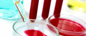
Doctors rarely look at the white blood cell count by day of the week of fetal development. More often the indicator is observed in trimesters. These data are presented in the table.
| Index | 1st trimester | 2nd trimester | 3rd trimester |
| Leukocytes, ×10 9 /l | 4 — 10 | 4 — 10,5 | 4 — 10,5 |
After the completion of the first trimester, the level of the indicator increases slightly. But until the end of gestation, it remains the same and does not change.
High white blood cell count in pregnant women
In most cases, an increased number of leukocytes during pregnancy is observed for physiological reasons. A woman works a lot, leads an active lifestyle, and eats poor quality food. She may experience various types of infections. Therefore, doctors recommend adhering to the following rules:
- to sleep more;
- rest, limit physical activity;
- sports should be carried out with the permission of a doctor; heavy loads are not recommended;
- walks in the open air;
- proper nutrition containing nutrients, microelements, vitamins, minerals;
- lack of stress, tension, limitation from worries.
If a woman has infectious diseases, they are treated immediately. There is a high risk of spread of a pathogenic microorganism to the fetus through the placenta. Self-medication is prohibited. Doctors prescribe drugs for leukocytosis, which are allowed during pregnancy.
Reasons for low levels of leukocytes in the blood
Less commonly, there are cases when the level of leukocytes in the blood decreases. The following reasons are given for this:
- intoxication due to the spread of a pathogenic microorganism, consumption of low-quality food;
- exposure to radiation;
- eating food containing minimal amounts of nutrients;
- taking medications (glucocorticosteroids, antibiotics);
- long-term allergies causing depletion of immune cells;
- disruption of the hematopoietic system.
It is prohibited to use immunomodulators on your own. They lead to a threat to the development of the fetus and the formation of internal organs.
Clinical and laboratory features during pregnancy
Pregnancy
is the period of time during which the fetus develops inside a woman's uterus, ending with the birth of the child.
During pregnancy, numerous physiological changes occur to meet the needs of the growing fetus and the mother's body, which adapts to them. Most of these changes begin soon after conception and continue until late pregnancy. Physiological adaptation is reflected in changes in the values of laboratory parameters. Some of the changes are well known, for example, a decrease in hematocrit and hemoglobin, creatinine; others, on the contrary, are known to a lesser extent and therefore their observation in the analysis result form can lead to incorrect interpretation.
The cardiovascular system
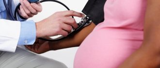
Changes in the functioning of the cardiovascular system occur among the very first. Their profound restructuring begins already in the early stages of gestation. The main events are physiological vasodilation and fluid retention in the body. Peripheral vasodilation leads to a decrease in vascular resistance and an increase in cardiac output, an increase in heart rate, and an increase in venous pressure.
Blood pressure decreases in the first and second trimesters, but rises to levels in non-pregnant women in the third trimester.
Water balance
Low blood pressure during pregnancy leads to activation of the renin-angiotensin aldosterone system, resulting in increased release of antidiuretic hormone. There is a tendency to retain water and sodium, and the likelihood of edema formation increases.
Hematological changes
Pregnancy is accompanied by stimulation of hematopoietic processes. There is a general increase in plasma, red blood cell (RBC) counts, and total circulating blood volume. Plasma volume increases during normal pregnancy. An increase of 15% occurs in the first trimester; in the second trimester, this trend continues, reaching a maximum at 32 weeks. By this time, the plasma volume has increased exactly by half.
The number of red blood cells increases, but occurs more slowly compared to plasma, making the blood more dilute and leading to the “physiological anemia” of pregnancy. There is a decrease in hemoglobin and hematocrit and mean erythrocyte hemoglobin content (MCH). The maximum decrease in hemoglobin levels is observed at 32–34 weeks of pregnancy.
During pregnancy, the volume of red blood cells changes. During the first 8 weeks of pregnancy, MCV decreases, by week 16 it returns to normal values, as in non-pregnant women, and then MCV increases.
White blood cell (WBC) levels increase by an average of 20%. The following changes are noted in the leukocyte formula: the absolute number of neutrophils (band and segmented) increases, the number of lymphocytes decreases.
The platelet count (PLT) changes ambiguously. Pregnancy is associated with increased platelet turnover.
An increase in blood volume is accompanied by an increase in ESR. In the first trimester, the ESR value is 15 mm/hour, in the second - 25 mm/hour, in the third - 40 mm/hour.
Hemostasis indicators
Throughout the entire period of pregnancy, the body prepares for the upcoming blood loss, so certain changes occur in the hemostatic system.
Changes in the blood coagulation system during pregnancy lead to a physiological state of hypercoagulability or increased susceptibility to thrombosis. In the third trimester, coagulation activity is twice as high as normal. The procoagulant activity of the hemostasis system increases, on the other hand, the activity of the fibrinolysis system decreases.
The concentrations of factors VII, VIII, IX, X, XII and von Willebrand factor increase.
Factor XI values decrease to 60–70% of those in non-pregnant women.
Fibrinogen levels increase significantly - up to 50%.
Levels of protein S and antithrombin III gradually decrease during pregnancy, while protein C activity remains unchanged.
Plasma fibrinolytic activity decreases throughout pregnancy but returns to normal within one hour after delivery.
Thrombin is generated as the gestational age increases. The D-dimer value increases throughout pregnancy. Only 3–5 days after delivery, the D-dimer value returns to its original values.
A shortening of APTT is observed in the second and third trimester and is associated with an increase in the activity of procoagulants in the blood. In the third trimester, a shortening of prothrombin time is observed.
Carbohydrate metabolism
Pregnancy is a diabetogenic condition because it is associated with the development of insulin resistance. An increase in the level of estrogen and progesterone in the initial stages leads to hypertrophy of pancreatic cells that secrete insulin. As a result, insulin secretion and tissue sensitivity to it increase in the early stages.
In the second trimester, insulin resistance begins to appear, reaching a peak in the third trimester. This results from the secretion of contrainsular hormones: human placental lactogen, growth hormone, progesterone, cortisol and prolactin. These hormones cause a decrease in the sensitivity of peripheral tissues to insulin, especially in adipose tissue and skeletal muscle, by interfering with insulin receptor signaling.
Insulin levels during pregnancy increase during fasting, after eating.
Fasting glucose levels often decrease due to:
- increasing storage of tissue glycogen reserves;
- increased use of peripheral glucose;
- decreased liver glucose production;
- glucose absorption by the fetus.
Insulin resistance and relative hypoglycemia lead to an increase in the process of lipolysis - the formation of fats, which primarily allows the use of fat as fuel, preserving glucose and amino acids for the fetus. The placenta allows the transfer of glucose, amino acids and ketones to the fetus, but is impermeable to large lipids. If a woman's endocrine pancreatic function is impaired and she cannot overcome the insulin resistance associated with pregnancy, she develops gestational diabetes.
During pregnancy, physiological transient glycosuria is often observed, which is associated with an increase in the glomerular filtration rate and an increase in the permeability of the renal tubular epithelium.
Protein metabolism
Throughout pregnancy, a woman needs more protein, since anabolic processes prevail over catabolic processes.
There is a physiological decrease in blood albumin; dilution of the blood also contributes to a decrease in the proportion of albumin.
The proteinogram in the first and second trimester has the following features: a decrease in albumin levels, a slight increase in the a-2 and b-1 globulin fraction. In the third trimester, there is a sharp increase in the a-1 globulin fraction.
And due to increased protein metabolism, a positive nitrogen balance is observed. Due to an increase in GFR by 75%, creatinine and urea levels decrease. A decrease in creatinine occurs mainly in the first and second trimesters, when intensive growth of the uterus is observed. The level of urea falls due to increased protein utilization, especially pronounced in the third trimester.
Lipid metabolism
The general effects of altered lipid metabolism during pregnancy are the accumulation of fat reserves in the mother's body in the first half and increased mobilization of fats in the second half of pregnancy.
Hypercholesterolemia is caused by increased production of sex steroid hormones, changes in metabolism in the liver and adipose tissue. Elevated triglyceride values provide the mother with energy needs. An increase in LDL cholesterol is associated with an increase in progesterone, in addition, LDL cholesterol is a source of placental progesterone. Increased concentrations of estrogen during pregnancy cause an increase in total cholesterol, LDL cholesterol and triglycerides.
Regional fat deposition in the mammary glands and subcutaneous fat is associated with increased conversion of carbohydrates into fats under the influence of insulin.
Mineral metabolism
Pregnancy causes an increase in the need for iron by 2-3 times for the synthesis of hemoglobin, for the production of certain enzymes. The need for folic acid increases 10–20 times, the need for vitamin B12 doubles.
The third trimester marks the peak demand for calcium.
During pregnancy, the concentration of total calcium in the blood serum decreases due to a decrease in the proportion of blood albumin, but the level of ionized calcium remains unchanged.
Treatment tactics
Treatment of leukocytosis during pregnancy is prescribed carefully. It is important to take into account the level of high indicators, the degree of the inflammatory process, and the nature of pregnancy. If the indicators are elevated, according to studies of vaginal microflora, local therapy or observation is limited. Typically, after childbirth, the vaginal microflora returns to normal. In other cases, the following drugs are indicated:
- antibiotics;
- uroseptic drugs for inflammation of the kidneys and urinary system;
- immunomodulators to enhance local and systemic immunity.
In the treatment of this condition in women, preference is given to herbal-based drugs to minimize side effects and risks to the growing fetus. The total duration of treatment is 7–14 days, after which a control analysis of vaginal secretions and other biological fluids is indicated. In severe cases, therapy is carried out in a hospital setting.
Causes of leukocytosis
Leukocytosis can develop in pregnant women for various reasons. The most common causes of such a disorder are: acute respiratory viral infections, acute respiratory infections and other inflammatory diseases that a woman could contract during pregnancy; severe pathologies, such as viral pneumonia or chickenpox, dangerous not for the woman herself, but for the child in her womb; an allergic reaction to certain medications prescribed by a doctor; receiving injuries, burns and other traumatic injuries (scratches with suppuration, abscesses, fractures and others).
Also, the reasons that a woman has developed leukocytosis may be associated with severe processes in her body, for example, with the disintegration of malignant tumors or internal bleeding. In some cases, pregnant women develop leukocytosis against the background of increased psycho-emotional stress. Therefore, doctors recommend that pregnant women avoid stress, as well as physical and mental fatigue. Classification of leukocytosis Sometimes leukocytosis is physiological and not pathological, that is, it occurs as a reaction to the action of certain environmental factors.
Symptoms of leukocytosis in pregnant women
Leukocytosis during pregnancy most often occurs in a latent (asymptomatic) form; it is discovered accidentally during a clinical blood test. But, despite this, the following clinical signs can be identified that indicate an increase in leukocytes in the blood of pregnant women:
- Dizziness;
- Moderate or high hyperthermia (up to 40 degrees);
- Hyperhidrosis (excessive sweating);
- Bruising, hematomas on the skin;
- Fainting conditions;
- Weight loss;
- Decreased visual acuity;
- Tingling of fingertips;
- Labored breathing;
- Causeless fatigue, apathy;
- Painful sensations in the abdominal area.
Reviews
Daria 05/10/2017
Girls, has anyone had it diagnosed during pregnancy? What consequences? Did they find the cause and were the leukocytes significantly elevated?
Ilyushina Mama 05/19/2017
They didn’t give me one, but they increased it 3 times. Then I read that this can happen during pregnancy
Elena Liukkonen 06/22/2017
From the beginning of pregnancy until the 22nd week, my leukemia was within 14. And then it sharply increased to 19 and the ESR also increased. The gynecologist ordered a test for c-reactive protein and it was normal. Then they said that there was an increase in leukemia and ESR during pregnancy, there was no inflammation.
Hat, 06/25/2017
Hello everyone! I’m writing straight from the hospital ward! On the 17th, I was admitted for preservation (28 weeks) with the threat of premature birth! They gave me magnesia for 5 days and injections for 7 days, and the rest of the time I’m just lying there without procedures! They don’t want to discharge me because . They say that I have an increased number of leukocytes (leukocytosis). There is no treatment for it and in response to all questions they yell that no one is keeping me here and I can leave, but they won’t pay me any money and won’t give me sick leave! I want to ask you about this leukocytosis, because I didn’t really find anything on the Internet! Thanks in advance.
Rules for taking the analysis
In order for the analysis results to be reliable, the following rules must be observed:
- Leukocytes in the blood - normal by gender and age, increased and decreased values
- The day before donating blood, exclude fatty and spicy foods from your diet;
- Blood sampling is carried out on an empty stomach, before 12 noon;
- Before the study, avoid physical and emotional stress;
- Before blood collection, you should have a good night's sleep;
- 10-15 minutes before taking the test, you need to sit in a calm, relaxed atmosphere.
Preventive actions
Increases in white blood cell counts in body fluid tests can be prevented by following simple clinical guidelines:
- sexual contact using a condom;
- frequent and adequate genital hygiene;
- eliminating bad habits;
- protective regime, prevention of hypothermia, colds;
- timely treatment of viral infections;
- adequate asepsis of pustular lesions on the skin and mucous membranes.
In the early stages of gestation, it is important to carry out sanitation of the oral cavity in order to exclude periodontal disease, periodontitis, deep caries, and stomatitis. Pregnancy reduces a woman’s immunity, so even minor inflammatory foci can lead to the spread of infection throughout the body.