General information
Hemorrhagic syndrome or a tendency to hemorrhage (bleeding) is bleeding of the skin and mucous membranes caused by pathological changes in one or more components of hemostasis .
They usually consist of damage to the vascular walls, disturbances in the structure, functional abilities and number of platelets, and deviations in coagulation blood stasis. In addition to superficial hemorrhages, this pathological condition is characterized by nasal, gastrointestinal and uterine bleeding, as well as bleeding gums. In addition, hemorrhages can occur in internal organs, including brain tissue, retina and joint cavities.
When determining the causes of hemorrhagic syndrome, it is important to consider that acquired and hereditary types of pathology may occur a little more often. Acquired disorders are usually caused by secondary thrombocytopenia and thrombocytopathy , disseminated intravascular , prothrombin factor deficiency, and hemorrhagic vasculitis . Hereditary pathological changes are usually represented by hemophilia A and B , von Willebrand disease and vascular form of telangiectasia .
Pathophysiology
RG Hart et al. [22] hypothesized that the use of NAC merely unmasks ICH that would otherwise remain asymptomatic, especially in patients with hypertension or cerebrovascular disease; Several observational studies support this assumption. First, MRI in gradient echo mode shows that microbleeds, which are one of the risk factors for ICH during the use of antithrombotic drugs [23, 24], can be detected even in people without acute neurological symptoms - usually elderly and suffering from hypertension [25 ].
Secondly, cerebral amyloid angiopathy, which is most often found in people over 65 years of age, along with age, is one of the risk factors for both spontaneous and warfarin-associated ICH [26, 27].
Third, data from the SPIRIT (Stroke Prevention in Reversible Ischemia Trial) and EAFT (European Atrial Fibrillation Trial) studies show that patients with pre-existing cerebrovascular disease have a significantly higher risk of warfarin-associated ICH [28, 29], and the presence of focal or diffuse hypodense changes in the deep layers of the white matter (so-called leukoaraiosis) is an independent predictor of warfarin-associated ICH [30]. In addition, the localization of spontaneous and warfarin-associated ICH is the same [4, 12]. Thus, the reasons underlying both types of hemorrhage may be the same, and the use of NAC acts only as an aggravating factor.
According to JJ Flibotte et al. [19], no relationship was found between the initial size of the hematoma and warfarin use. Predictors of a larger initial hematoma volume are hyperglycemia (p < 0.0001) and lobar localization of hemorrhage (p < 0.0001).
It is known that in almost half of patients with spontaneous ICH there is a slow increase in the size of the hematoma during the first 12–14 hours, which is accompanied by worsening neurological deficit [3, 7, 18, 19, 31–33]. It is believed that hematoma growth may be the result of ongoing bleeding, recurrent bleeding, or secondary bleeding around the primary lesion.
Pathogenesis
There are several pathogenetic mechanisms for the development of hemorrhagic syndrome:
- Along the path of coagulopathy - in individuals with hereditary or secondary (acquired) deficiency of plasma blood clotting factors - procoagulants , as a result of their insufficient activation, synthase disorders, and also, on the contrary, excessive activation of anticoagulants .
- Loss of platelet link - a pathological condition occurs as a result of insufficient number of platelets (with immune conflicts, lack of vitamin B12 , aplastic anemia , tumor processes in the bone marrow and mechanical destruction as a result of splenomegaly ) or in case of a violation of their adhesive-aggregation properties in various thrombocytopathies.
- Hyperfibrinolytic conditions are associated with excessive fibrinolysis.
- Caused by pathologies of the vascular wall or its increased permeability.
Typical location of hemorrhages
Most often, and this is about 55% of cases, hemorrhages occur in the putamental zone. Putamental bleeding occurs due to rupture of degenerated lenticulostriate arteries, which causes blood to enter the brain shell. The culprit of pathogenesis with such localization is usually long-standing hypertension. In some cases, bleeding from the putamentum breaks into the ventricular system, which is fraught with tamponade of the gastrointestinal tract and acute occlusive-hydrocephalic crisis.
The next most common location is the subcortical region (subcortical). Subcortical GIs are observed in 17%-18% of cases. As a rule, the leading sources of such hemorrhage are ruptured AVMs and aneurysms against the background of increased pressure. The subcortical zones involved in the hemorrhagic process are the frontal, parietal, occipital or temporal lobe.
The third most common place, where brain hemorrhage is determined in 14%-15% of cases, is the optic thalamus, or thalamus. Thalamic hemorrhages occur due to the release of blood from the blood vessel of the vertebrobasilar region. Pathogenesis can be associated with any etiological factor, however, as always, the involvement of hypertensive syndrome is significantly more often noted.
In fourth place (7%) in terms of frequency of development are pontine GIs. They are concentrated in the back of the brain stem, that is, in the pons. Through the bridge, the cortex communicates with the cerebellum, spinal cord and other major elements of the central nervous system. This department includes the centers for controlling breathing and heartbeat. Therefore, the bridge is the most dangerous localization of hemorrhage, practically incomparable to life.
Classification
There are 5 types of hemorrhagic syndrome:
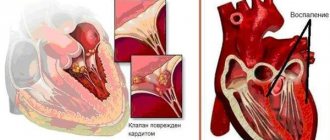
- Hematoma - caused by a deficiency of blood coagulation factors VIII, IX and XI, usually found in hemophilia A or B, manifesting itself as painful intense hemorrhages into the thickness of soft tissues (petechial rashes) and joints, which leads to gradual functional disorders of the musculoskeletal system, characterized by the appearance of delayed bleeding - a couple of hours after injuries, even hemorrhages in the brain tissue are possible - most often due to thrombocytopathies.
- Petechial-spotted, capillary, microcirculatory - the so-called bruise variety, which occurs with deficiency of blood coagulation factors II, V and X, is usually caused by thrombocytopenia , thrombocytopathy , such disorders of the coagulation system as hypofibrinogenemia , dysfibrinogenemia and hereditary deficiency of blood clotting factors.
- The mixed, microcirculatory-hematoma form is usually initiated by severe deficiency of factors of the prothrombin complex and factor XII-XIII, von Willebrand disease, disseminated intravascular coagulation syndrome, overdose of thrombolytics and anticoagulants, the appearance of immune inhibitors of factor VIII and IX in the bloodstream, externally manifested by a combination of petechial-spotted skin hemorrhages and individual large hematomas in the retroperitoneal space and intestinal wall; in extremely rare cases, hemorrhages occur in the joint cavity, and the resulting bruises are widespread and painful.
- The vasculitic purpuric form is caused by infectious or immune vasculitis and can easily transform into DIC syndrome; it is characterized by hemorrhagic rashes and the development of erythema with an inflammatory process, the addition of nephritis and the development of intestinal bleeding are possible.
- The angiomatous type of hemorrhagic syndrome can occur in the area of telangiectasia, angioma or arteriovenous shunt in the form of persistent local hemorrhages in areas of vascular pathologies.
Diagnosis of ICH
The appearance in a patient receiving NAC of a sharp headache predominantly in the occipital region, nausea, vomiting that does not bring relief, non-systemic dizziness, as well as stunned consciousness is the basis for immediately excluding the diagnosis of ICH. Symptoms of hemorrhage usually develop suddenly [3, 7, 17–19], and meningeal syndrome and low-grade fever may develop first. Already in the first hours, signs of ICH can be determined using computer or magnetic resonance imaging (MRI). A highly sensitive technique is MRI using gradient echo [1, 20, 21].
Causes
Hemorrhagic syndrome is usually caused by various clotting factor deficiencies and thrombocytopathy, including:
- hemophilia A or B;
- injuries, hypothermia and overheating;
- ARVI and acute bacterial infections;
- preventive vaccination;
- acute radiation injuries;
- von Willebrand disease;
- systemic lupus erythematosus;
- thrombocytopenic purpura or Werlhof's disease ;
- DIC syndrome;
- HIV infection;
- severe liver damage;
- infectious and immune vasculitis;
- hypo- or dysfibrinogenemia.
The psychogenic form of hemorrhagic syndrome is usually represented by neurotic bleeding or can be caused by Munchausen syndrome .
It must be remembered that due to the general availability of medications, hemostasis disorders - the development of hemorrhagic syndrome can be provoked by taking medications - non-steroidal anti-inflammatory drugs, glucocorticoids, anticoagulants and antiplatelet agents, which interfere with blood clotting and the ability of platelets to aggregate.
Monitoring hemostasis parameters
To monitor the level of anticoagulation, the INR level is traditionally determined. However, this indicator is sensitive to changes in the levels of factors VII, X and prothrombin, but not to factor IX [48, 58–61]. Therefore, even with normalization of the INR, a high risk of bleeding may remain. Thus, M. Makris et al. [58] found that 800 mL FFP reduced mean INR from 6.73 to 2.38, while mean factor IX levels remained essentially unchanged (baseline 26.45 IU/dL, post-treatment 27.36 IU/dl). The use of INR to monitor the degree of hypocoagulation in patients receiving rVIIa therapy is also problematic. Pharmacological doses of rVIIa will always reduce the INR regardless of the levels of other coagulation factors [59, 61]. In this situation, the thromboelastography method may be more appropriate. In this case, the profile of thrombus formation in whole blood is recorded, which gives a more complete picture of the state of the coagulation system [53, 62, 63].
Symptoms
In the complex of symptoms of hemorrhagic syndrome there are various manifestations, the most pronounced are externally noticeable changes, as well as:
- petechiae - a pinpoint rash that occurs when there are changes in vascular-platelet hemostasis, consists of multiple elements no larger than 3 mm in size, does not rise above the skin and is not palpable, most often violet, purple or red;
- bruising and hematoma formations - can be subcutaneous, muscular, etc. most often caused by coagulation disorders, characterized by pain and a gradual increase in size;
- dissecting hematoma is usually caused by changes in coagulation hemostasis and is a false channel filled with blood of varying length, located in the media of the aorta;
- the presence of many superficial skin hemorrhages ( ecchymoses ) of small size - no more than 3 mm, which can also be observed on the mucous membranes;
- “delayed bleeding” - occurring several hours late;
- hemarthrosis - hemorrhages in the joint cavities, which cause an increase in the volume of the joint, local hyperemia and increased temperature of the skin, as well as a pronounced pain syndrome ;
- bleeding even with minor injuries - cuts or scratches.
Clinical manifestations of HI
Shortly before the attack, pre-stroke clinical symptoms may precede the attack (not always), by which one can suspect impending danger:
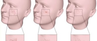
- tingling, numbness of one facial half;
- numbness in fingers or toes;
- sudden weakness, dizziness, noise in the head;
- severe pain in the eyes, spots, double vision, vision in red;
- sudden staggering when walking;
- causeless tachycardia;
- attacks of hyperhidrosis;
- increased blood pressure;
- unreasonable occurrence of nausea;
- inhibition in communication and perception of other people's speech;
- flushing of the face, hyperthermia.
A cerebral stroke with hemorrhage is still characterized by an immediate acute onset without warning signs, which occurs during or almost immediately after vigorous activity, a stressful situation, or excitement. A hemorrhagic stroke is indicated by classic symptoms that develop suddenly, are pronounced and rapidly progress:
- sharp and severe headache;
- uncontrollable vomiting;
- prolonged depression of consciousness, coma;
- blood pressure above 220 mmHg.
Common signs of shock are also noisy breathing, epileptic seizures, lack of pupillary response to light, and spastic miosis. Depending on the location of the lesion, there may be a rotation of the head and rotation of the eyeballs in the direction of the affected hemisphere or contralaterally. Having discovered signs of GI in a victim, a person nearby must immediately call an ambulance!
Acutely developed hemorrhage leads to the fact that blood flows freely into certain structures of the brain, saturating them and forming a cavity with a hematoma. The bleeding continues for several minutes or hours until a blood clot forms. Over a short period of time, the hematoma quickly increases, exerting a mechanical effect on the affected areas. It stretches, presses and displaces the nervous tissue, causing its swelling and death, which leads to an intensive increase in neurological deficit (respiratory depression, loss of sensitivity of one half of the body, speech disorders, loss of vision, paresis of the swallowing muscles, etc.).
The size of the blood accumulation can be small (up to 30 ml), medium (from 30 to 60 ml) and large (more than 60 ml). The volumes of spilled liquid can reach critical levels, up to 100 ml. Clinical observations show that with intracranial hemorrhages exceeding 60 ml, the pathology ends in death in 85% of patients within 30 days.
Tests and diagnostics
The basis for suspecting a violation of the vascular-platelet and coagulation processes of hemostasis are skin hemorrhagic manifestations, their nature and appearance. Differential diagnosis is carried out taking into account the number of peripheral blood platelets and the obtained data from simple coagulation tests, including the Rumpel-Leede-Konchalovsky test, blood coagulation rate, bleeding time, etc.
A fairly important aspect of the examination is determining the root cause of the pathological condition, that is, the disease that caused the hemorrhagic syndrome.
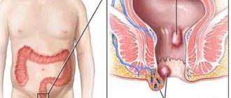
Mallory-Weiss syndrome and differences from gastrointestinal bleeding
The most important task of the diagnostician is the ability to timely recognize Mallory-Weiss syndrome , which is manifested by vomiting blood, causing mild gastrointestinal bleeding and melena . Factors contributing to the development of this condition include repeated vomiting as in bulimia , alcoholism , also a hiatal hernia, chronic inflammatory diseases of the upper part of the digestive tube, severe cough, portal hypertension , etc.
With gastroesophageal rupture-hemorrhagic syndrome, both rupture of only the lining integumentary structures and more deeply lying submucosal and muscular structures can occur with complete rupture of the esophagus and the risk of developing peritonitis , mediastonitis and pneumothorax . In this case, bleeding in the mildest form of the pathology can resolve spontaneously after a few days; therapy usually includes the use of cold and antacids ; in more severe cases, endoscopic treatment or gastrotomy may be necessary.
Hemorrhagic syndrome in children
Hemorrhagic syndrome is manifested by bleeding, spontaneous or caused by hereditary and external factors. To avoid the consequences and complications of a hemorrhagic condition in children, it is important to identify the disease in time and seek qualified help from a hematologist. Alarm bells for parents include symptoms such as:
- small pinpoint red bloody skin rashes such as petechiae or bruising;
- formation of hematomas even with minor injuries;
- causeless bleeding from the nasal passages and gums;
- a history of internal esophageal, uterine, gastrointestinal or other bleeding;
- pain and stiffness of joint movements, possibly resulting from hemorrhage into the joint cavity;
- pain, more often in areas of local lesions;
- manifestations of anemic syndrome - pallor, increased fatigue, irritability, etc.
Early hemorrhagic disease of newborns
Even healthy children at birth have reduced levels of vitamin K-dependent factors II, VII, IX and X, which generally form the prothrombin complex. of vitamin K itself , which is formed in the intestines due to the bacterial flora, which has not yet been formed in newborns in the first days of life, is reduced. The maximum deficiency of vitamin K-dependent factors is usually observed on the second to fourth day. Only functionally sufficiently mature liver cells in the direct presence of vitamin K participate in their synthesis. This deficiency is more pronounced in premature infants and they are assigned hemorrhagic status.
Predisposing factors are:
- phenobarbital , aspirin , carbenicillin and sulfa drugs shortly before giving birth
- the presence of problems in a pregnant woman with the process of absorption of vitamin K caused by intestinal disorders - enteropathy, diseases of the biliary tract, dysbacteriosis ;
- cases of placental pathology and acute toxicosis in pregnant women.
Hemorrhagic syndrome in newborns is manifested by melena, vomiting with blood, hemorrhages in the skin and mucous membranes, bleeding of the lungs and navel, hematuria , cephalohematoma , decreased body weight, intracranial hemorrhages, in the adrenal glands and liver, and anemia. Severe bleeding can be life-threatening for the newborn.
Late hemorrhagic disease of the newborn
The late form of hemorrhagic disease of the newborn may also be called vitamin K-deficiency coagulopathy . It develops at 2-8 weeks of life, in more rare cases it can last up to six months. Usually occurs against the background of another disease or nutritional deficiency of vitamin K and is accompanied in almost half of the cases by extensive intracranial hemorrhages, as well as extensive ecchymoses and bleeding from injection sites. All therapeutic measures in such cases are aimed at preventing hypovolemic (hemorrhagic) shock .
How to treat?
In cases of a mild form of the disease, treatment of hemorrhagic vasculitis can be done without compulsory hospitalization of the patient, but in any case it will require bed rest. During the acute period, a hospital stay under the constant supervision of doctors is necessary. An acute disease is usually cured within about 2 months; if the disease is protracted, the duration of treatment can increase to six months.
Therapy includes medication as indicated. In severe cases of the disease, glucocorticosteroids may be prescribed, as well as plasmapheresis or transfusion therapy for children. In the presence of allergic manifestations, antihistamines are prescribed. The diet for hemorrhagic vasculitis is aimed at excluding potentially allergenic foods that can worsen the patient’s condition - citrus fruits, strawberries, chocolate, coffee, eggs, etc. For severe renal and abdominal syndrome, special diets are prescribed.
Diet for hemorrhagic syndrome
Diet Table No. 1
- Efficacy: therapeutic effect after 3 weeks
- Terms: 2 months or more
- Cost of products: 1500 - 1600 rubles. in Week
Most often, when hematological disorders are detected, dietary Table No. 1 , which is characterized by a fairly high calorie content and fractional nutrition. The menu usually consists of such dishes and ingredients as:
- vegetable soups with breadcrumbs;
- steamed poultry breast;
- boiled fish;
- eggs;
- semi-viscous porridge with milk;
- berries and fruits – ground in jelly and compotes;
- There are practically no restrictions on the types of oil, the main thing is that it is natural, unsalted and in moderation.
Excessively salty, peppery, fried, fresh dough, fatty meat, canned food, snacks, carbonated drinks and coffee should be excluded from the diet. “Specific prohibitions” include the need to avoid white cabbage, mushrooms, sorrel, spinach, onions and cucumbers.