Article for the "bio/mol/text" competition: Cells of the immune system travel through the lymph and bloodstream in search of an antigen that can be recognized and begin a protective immune response. But a significant portion of T-lymphocytes are not found in the blood or lymph nodes, but in organs that are not related to the immune system. This article explains what tissue-resident T cells do, how they get there, and the medical benefits of studying them.
Note!
This work took first place in the “Best Article on Immunology” category of the “bio/mol/text” competition 2015.
The sponsor of the nomination “Best article on the mechanisms of aging and longevity” is the Science for Life Extension Foundation. The audience award was sponsored by Helicon.
Competition sponsors: Biotechnology Research Laboratory 3D Bioprinting Solutions and Scientific Graphics, Animation and Modeling Studio Visual Science.
An adequate protective reaction when infected with a pathogenic virus is to destroy the infected cells, preventing the infection from spreading throughout the body and killing more cells. A cell infected with a virus can notice the virus in itself and begin autophagy or apoptosis - or receive instructions for programmed cell death from a T-killer cell.
The cytotoxic T-lymphocyte, or killer T-lymphocyte, is the pinnacle of the evolution of adaptive immunity, because to recognize a fragment of the virus (antigen) on an infected cell, it uses a T-cell receptor, which randomly and independently assembles on each T-cell in the thymus. The T-cell receptor assembly mechanism, which has no analogues outside the adaptive immune system of vertebrates, uses the advantages obtained by vertebrates during genome duplications in the process of evolution and proceeds with the participation of special recombinase proteins, once borrowed from DNA transposons (see more in Chudakov’s article “ Analysis of individual repertoires of T-cell receptors").
Classical human immunology is based on the study of immune cells in the blood simply due to the fact that a blood test can be taken from any patient and examined in normal and pathological conditions. It is on blood cells that the classification of T-lymphocytes was built: division into T-killers and T-helpers, which check the antigen specificity of T-killers, give them a “license to kill” and are able to control the entire course of the immune response through soluble signaling molecules - cytokines. As well as the later isolation from the T-helper branch of a group of regulatory T cells that suppress excess adaptive immunity.
But as yogurt commercials remind us, a significant portion of the immune system's cells are concentrated around the lining of the digestive tract and in other tissues. While in 5–6 liters of blood of an adult there are about 6–15 billion lymphocytes, the number of T cells found in the epidermis and skin is estimated at 20 billion [1], and in the liver of an adult male there is another 4 billion [2 ]. Is studying blood cells enough to fully describe the functions of T cells if there are more T cells in peripheral organs than in the bloodstream? And are classical subpopulations sufficient to describe all the types of T cells found in the human body?
Life cycle of a T lymphocyte
Each T cell, after assembling a T-cell receptor, is tested for the functionality of the randomly assembled receptor (positive selection) and the lack of specificity for the body's own antigens (negative selection), that is, for the absence of an obvious autoimmune threat. The stages of selection occur in the thymus gland, thymus; in this case, more than 90% of precursor cells die, failing to correctly assemble the receptor or undergo selective selection. Surviving T cells proliferate and exit the thymus into the bloodstream - these are naïve T cells that have not encountered the antigen. The naive T cell circulates through the blood and periodically enters the lymph nodes, where in the T-cell zone it contacts specialized antigen-presenting cells.
After meeting the antigen in the lymph node, the T cell acquires the ability to divide again - it becomes the precursor of memory T cells (TSCM, stem cell memory T cells). Among the clone of its descendants, central memory cells (TCM), short-lived effector cells that carry out the immune response (SLEC or TEMRA cells), and effector memory precursor cells TEM, which in turn produce TEMRA when dividing, appear [3]. All these cells leave the lymph node and travel through the blood. Effector cells can then leave the bloodstream to carry out an immune response in the peripheral tissue of the organ where the pathogen resides. What then - another journey through the blood and lymph nodes?
Figure 1. Effector T cell emigration into tissue during viral infection. Inflammatory signals from infected epithelial cells with the participation of resident cells are transmitted to the vascular endothelium; endothelial cells attract effector T cells with chemokines CXCL9, CXCL10. Rolling: When moving along a postcapillary venule in tissue, the effector cell slows down, forming temporary contacts between E-selectins and P-selectins on endothelial cells. Arrest: The effector cell adheres tightly to the endothelium when LFA-1 and other alpha integrins interact with ICAM-1/VCAM-1/MAdCAM-1 (on the endothelium). Transmigration: The effector T cell binds endothelial JAM-1 with molecules PECAM, CD99, LFA-1 and penetrates through endothelial cells into the submucosa. Figure from [3].
The process of leukocyte transmigration.
The cells of the stroma, that is, the base of the lymph node, secrete signaling substances in order to call the T cell to the lymph node - chemokines. Lymph node chemokines are recognized by homing receptors CCR7 and CD62L. But effector cells lack both of these receptors. Because of this, it has long been a mystery how effector cells can get from peripheral tissue back to the secondary lymphoid organs - the spleen and lymph nodes.
At the same time, evidence began to accumulate of differences in membrane marker repertoires and transcriptional profiles between memory T cells in the blood (TEM) and memory T cells in other organs, which did not fit into the concept of constant migration of T cells between tissues and blood . It was decided to isolate a new subpopulation: resident memory cells that inhabit a specific organ and do not recycle - TRM cells [4].
Figure 2. Scheme of the transition of descendants of activated T lymphocytes between populations. Figure from [14].
How do T cells work?
When a virus or other invader enters a person's system, the body mounts an immunological response. Some cells have receptors that can identify an attacking substance and can activate various cells in the body to find and destroy harmful invaders.
People with strong immune systems usually have high numbers of white blood cells and T cells. Patients with autoimmune diseases or those suffering from cancer often undergo blood cell counts, which can tell doctors how well these and other beneficial cells survive and fight their disease.
Origin of tissue resident T cells
Where do tissue resident cells first appear? These are the descendants of effector cells that have lost their ability to recycle. Some tissues peripheral to the immune system, for example, the mucosa of the small intestine, the abdominal cavity, allow effector T lymphocytes to penetrate freely; others - very limited, a large flow of effector T cells into these tissues is observed only during an inflammatory reaction. The second type of tissue includes those separated by a barrier from the immune system, for example, the brain and spinal cord, as well as many others: peripheral ganglia, mucous membranes of the genital organs, lungs, epidermis, eyes. The difference between the two tissue types is the expression of additional homing molecules for effector T cells, for example, the epithelial infiltration adhesion molecule MadCAM-1 [3].
Figure 3. “To home or not to home?” - difficult choice of effector cell. To home is the process of homing, or migration of T cells, for example, to the place most familiar to naive cells - the lymph node. The alternative is to not travel through the body and become a resident tissue cell.
What else is prescribed with this study?
Activated lymphocytes (T-lymphocytes, T-helpers, T-cytotoxic cells, immunoregulatory index, T-activated, NK- and B-activated cells)
17.54. Venous blood 3 days
6,500 RUR Add to cart
Humoral immunity (immunoglobulins IgA, IgM, IgG, IgE, circulating immunocomplexes, complement components C3, C4)
17.51. Venous blood 8 days
RUB 3,890 Add to cart
Immune status (screening) (Phagocytic activity of leukocytes, cellular immunity, total immunoglobulin IgE, immunoglobulins IgA, IgM, IgG)
27.960. Venous blood 3 days
RUR 7,640 Add to cart
Immune status expanded
17.61. Venous blood 14 days
RUR 22,310 Add to cart
Cellular immunity (T-lymphocytes, T-helpers, T-cytotoxic cells, Immunoregulatory index, B-lymphocytes, NK-T cells, NK cells, Leukocyte formula)
17.50. Venous blood 3 days
RUB 5,200 Add to cart
Resident T cells in aging human tissues
A map of the relationships between the presence of individual subpopulations of T cells in different human organs, oddly enough, was compiled only in 2014. Donna Farber's team at Columbia University Medical Center in New York compared the phenotypes of T cells isolated from the blood and tissue of organ donors of all age groups from 3 to 73 years, from a total of 56 donors [5]. Analysis of T cell subpopulations using flow cytometry confirmed many data obtained using methods with lower resolution and lower statistics, and some features of the description of the immune system transferred from mouse immunology to humans, for example, a decrease in the content of naïve T lymphocytes with aging in all organs .
The decrease in the number of naïve T cells with age is associated with rapid aging of the thymus gland, in which future T cells undergo the stages of T-cell receptor assembly, testing the functionality of the receptor, and selection for a lack of autoimmune potential. It is important not only to reduce the absolute number of naïve T cells, but also to reduce the diversity of the T-cell receptor repertoire, and therefore the ability to form an adaptive immune response to a previously unfamiliar infection [6]. For naive killer T cells, a progressive decline in abundance in the blood and lymph nodes was confirmed, although for naive helper T cells the negative correlation of abundance with age in this study was significant only in secondary lymphoid organs, but not in the blood.
Isolation of memory T lymphocytes, memory effector cells and short-lived effector cells from the mucous membranes of the lungs, small and large intestines, inguinal and mesenteric lymph nodes of organ donors made it possible for the first time to assess the dynamics of these populations in human tissues during aging. The proportion of central memory cells expectedly increases over the course of life, in accordance with the increase in the number of infections that the body has encountered and entered the memory library of the immune system. The percentage of terminally differentiated effector T-killer cells (TEMRA) also increases, but only in the lymph nodes and spleen; in nonlymphoid tissues, TEMRA abundance decreases. TEM effector memory cells rapidly fill the niche for T cells in the child's tissues, quickly displacing naïve T cells by about 12 years of age. Short-lived terminally differentiated killer T cells are most often found in the blood, spleen and mucous membranes of the lungs at any age, but among T helper cells this subpopulation is represented by a vanishingly small number of cells. Similarly, there are few central memory cells among T-killers; they are predominantly located in the mucous membranes of two barrier tissues: the lungs and intestines.
In broad strokes, the distribution map of human T-lymphocytes can be outlined as follows: naïve T-cells travel through the blood and periodically enter secondary lymphoid organs; TEMRA killers are found in the blood, spleen and lungs. Apparently, central memory cells are characterized by a more individual distribution across tissues than other subpopulations: in any case, patterns of dynamics during aging of different tissues could not be identified. Effector memory cells, including the TRM subpopulation, dominate among T cells of mucosal barrier tissues. In general, with aging of T-cell immunity in non-lymphoid tissues, there is a greater age-related change in T-cell types [5]. The stability of tissue cells is easier to explain if we understand which of the TEM effector cells remain in the tissue, become resident TRMs, and what events their life consists of after they stop traveling throughout the body.
Figure 4. Circulation pathways of T lymphocytes of different subpopulations. Tnaive - naive T cells, together with the TCM subpopulation, move through the blood and enter the T-cell zone of various lymph nodes; they are found in tissue capillaries, but do not exit into the tissue (red trajectory). Effector T cells (blue) move through the lymph and bloodstream; when they enter a lymph node, they do not enter the T-cell zones (center of the lymph node) - the trajectory is purple. Resident T-cells of tissues (shown in green in the skin and in different colors in mucous membranes) move only within the tissue - green trajectory. Figure from [9], with modifications.
Helper T cells:
The task of helpers is also quite obvious at first glance. These are helper cells (“help” means “to help”). Who or what do they help? They induce and stimulate an immune response: under their influence, cytotoxic lymphocytes enhance their work. Helpers also transmit information about the presence of a foreign protein in the body to B-lymphocytes, which secrete protective antibodies against them. Finally, helpers have a stimulating effect on the work of phagocytes, mainly monocytes.
T helper cells
How to distinguish resident tissue cells from admixtures of blood cells?
Resident T cells can be correctly, but inconveniently, determined each time by the ability of an individual cell to migrate to lymph nodes, so it is necessary to compile a list of characteristic features that can be used to determine membership in this subpopulation. Resident T lymphocytes in the body's natural barrier tissues (for example, in the lungs and small intestinal mucosa) are somewhat similar to classical effector blood cells: they express the activated cell marker CD69, and the expression is stable throughout life during adulthood and aging and is characteristic of all non-lymphoid tissues . But in addition, CD69 colocalizes with the marker CD103, which denotes a group of adhesion molecules - integrins, which promote the attachment of resident T cells to the epithelium and to fibroblasts in the submucosa of the selected organ. For effector T cells in secondary lymphoid organs, the expression of CD103 integrins is completely uncharacteristic: TEM cells constantly maintain a motile phenotype.
The map compiled by Donna Farber's team has a major flaw: it is unclear how cleanly T-lymphocytes can be isolated from an organ, and what proportion of the analyzed cells are actually blood T-lymphocytes from capillaries inside the organ.
The issue of contamination with blood cells is especially acute for the lungs; it is no coincidence that the subpopulation composition of T cells in the lungs is unexpectedly similar to T cells in the blood and lymph nodes. The issue of blood cell contamination was elegantly solved for mouse T cells: experimental mice were infected with lymphocytic choriomeningitis virus after transplantation of a transgenic P14 T cell clone specific for this virus. As a result, during infection, the majority of circulating cells were represented by the virus-specific P14 clone, and its presence in tissues could be monitored by immunofluorescence with a P14-specific antibody. Before the mice were killed, they were injected with an antibody to the killer T-cell marker anti-CD8, which quickly spread through the bloodstream and bound to all killer T-cells in the blood (but not in the tissues). By microscopy of organ sections, it was easy to distinguish resident killer TRMs from cells only recently released from the blood into the organ, labeled with an anti-CD8 antibody [7]. The number of resident cells calculated by this method was 70 times higher than the numbers determined by flow cytometry; a difference of less than twofold was observed only for resident cells of the lymph nodes and spleen: it turns out that standard methods for isolating lymphocytes from organs are poorly suited for the analysis of killer resident cells and significantly underestimate the population size.
What are the types of T cells?
There are several different types of T cells in the body, each with a specific function. T helper cells are able to quickly divide and help the body's immune response to an attack by creating a useful protein called a cytokine. Another type, often called a memory cell, helps the body fight repeated infections by preventing a person from catching the same disease more than once.
One of the most useful types of these cells also has a huge disadvantage. Cytotoxic cells destroy tumors or mutated cells, like soldiers repelling invaders. Unfortunately, when transplanted, this type of cell may incorrectly identify the transplanted organ as a threat and attack it. Cytotoxic cells are considered one of the main factors in transplant rejection.
The work of resident T cells: do not confuse tourism with emigration
In a normal situation, mouse resident tissue cells hardly move within non-lymphoid tissue and are quite firmly attached by adhesion molecules to the stroma of the organ. When resident macrophages of the same tissue initiate an inflammatory response by secreting cytokines, TRMs become more motile and patrol the nearby epithelium in search of infected cells.
If the inflammatory reaction intensifies, then the cells understand this as a signal for reinforcement: TCM and TEM cells newly arriving from the blood are connected to the work of patrol TRM. These blood cells are much more mobile and move better in the epithelium: does this mean that it is in the blood that T-killer T cells are ready to act among TEM, and CD8+ TRMs perform helper and regulatory functions in the tissue?
On the one hand, T-helpers are more tissue-specific in the spectrum of T-cell receptors, that is, there is very little overlap between the repertoires of T-cell receptors of cells taken from different tissues, while cells of the same T-killer clone are found in different tissues among TEM[ 5]. The range of functions and repertoire of antigen specificity of TRM remains to be studied, but TRM killers definitely have the ability to destroy infected tissue cells. Moreover, the affinity of virus-specific T-cell receptors (TCRs) of resident killer cells is higher than that of virus-specific central memory cells in a mouse model of polyomavirus infection in brain tissue [8].
However, the size of the T-cell population depends not only on the specificity of T-cell receptors for infections that previously occurred in a given organ, but also on the homeostatic proliferation of T-cells - the proliferation of more successful cells to fill the organ's capacity according to the number of T-lymphocytes. By using the markers CD28 and CD127 on the cell surface, it is possible to distinguish cells recently and long ago activated through the T-cell receptor from those that received only a homeostatic signal to proliferation from the growth factor IL-7. With tissue aging, homeostatic cell proliferation begins to prevail over the proliferation of TCR-activated cells.
NKT cells, a large type of liver resident cell that is also found in other tissues, often function independently of T-cell receptors. They can be activated by NK cell receptors through recognition not of individual antigens, but of general molecular patterns of danger and tissue stress. When activated, CD8+ NKT cells release cytotoxic granules and lyse suspicious tissue cells, for example, single tumor cells and virus-infected cells that express and display MHC-like stress molecules on the outer membrane. With aging, the tendency of TRM to be activated without the T cell receptor through NK cell receptors or cytokine signals can lead to erroneous lysis of tissue cells, insufficient control of chronically infected or degenerating areas of the epithelium.
Pathological manifestations associated with the work of resident T cells include organ-specific autoimmune syndromes and chronic tissue inflammation syndromes. Examples of chronic inflammation supported by resident T lymphocytes are contact dermatitis and psoriasis, and the mechanism is the release of inflammatory factors IL-17 by resident killer T cells and IL-22 by resident T helper cells of the dermis. CD8+ effector killer T cells located in the brain are similar in their set of membrane marker molecules to the TRM of the skin, intestines and lungs and are able to push the development of intermittent multiple sclerosis with periodic releases of inflammatory cytokines; It is unclear, however, whether there is a normal TRM population in the brain or whether these are T lymphocytes remaining in the tissue after a neurotropic viral infection [9].
The functions of resident memory cells normally, in the absence of infection or chronic inflammation, may include cross-talk (mutual regulation primarily through the secretion of cytokines and costimulatory molecules) with non-classical, poorly understood lymphoid cells such as mucosal-associated gamma/delta T cells, carrying an alternative version of the T-cell receptor assembly; or innate lymphoid cells (ILC), which share common features of the epigenetic landscape with T and B lymphocytes, but do not have T/B or NK cell receptors [10, 11].
TRM cells come into contact with antigen-presenting cells of tissues - these are dendritic cells of the skin and resident tissue macrophages. Resident myeloid cells in different tissues are differentiated and slightly similar to each other. For example, marginal zone macrophages of the spleen, liver macrophages, and microglia (brain macrophages) will differ greatly in both morphology and range of functions. In addition to detecting antigens in tissue, resident macrophages are involved in regulating the processes of aging and tissue self-renewal, in particular, they secrete growth factors and cytokines that stimulate the division of tissue stem cells. In adipose tissue, for example, macrophages stimulate the differentiation of new fat cells, but when they enter an activated M1 state, they trigger inflammation and, instead of differentiating, cause existing fat cells to enlarge and swell. Concomitant changes in adipose tissue metabolism lead to the accumulation of fat mass and in recent years have been associated with the mechanisms of development of obesity and type II diabetes. In the skin, cytokines released by macrophages and resident gamma/delta T cells stimulate stem cell division during the regeneration of the epidermis and hair follicle stem cells [12, 13]. It can be assumed that TRM helper cells, when patrolling the epithelium and forming contacts with tissue macrophages, can modulate the spectrum and volume of growth factors secreted by the latter for stem cells, inflammatory cytokines and epithelial remodeling factors, and thereby participate in tissue renewal.
Figure 5. Proposed functions of tissue resident T lymphocytes. Some functions can be performed in interaction with resident macrophages (see explanations in the text).
T cells are considered extremely important in the fight against autoimmune diseases
T cells are considered extremely important in the fight against autoimmune diseases, especially in the cases of human immunodeficiency virus (HIV) and acquired immunodeficiency syndrome (AIDS). Many of the treatments currently used for these viruses involve stimulating the production of these cells and their response to combat the harmful effects of these conditions. Naturally occurring "killer" T cells are unable to effectively fight HIV, so scientists have developed a variety of techniques to improve the cells and make the cells' receptors more sensitive to this deadly virus.
Because the study of cellular function is still relatively new, many scientists still do not understand what T cells are and how they work. As researchers continue to study these beneficial cells, many believe it may be possible to reduce cellular transplant rejection and improve the treatment of autoimmune diseases.
What can the study of Trm give to medicine?
Understanding how resident T cells work is absolutely essential for fighting infections that do not enter the bloodstream directly, but enter the body through barrier tissues—that is, for the vast majority of infections. Rational design of vaccines for protection against this group of infections can be aimed specifically at enhancing the first stage of protection with the help of resident cells: a situation in which optimally activated antigen-specific cells eliminate the pathogen in the barrier tissue is much more beneficial than triggering acute inflammation to call T lymphocytes from the blood, since there is less tissue damage.
The T-cell receptor repertoire of cells associated with mucosal barrier tissues is considered to be partially degenerate and public, that is, identical for many individuals in a population. However, biases in the isolation of T cells from organs, data bias resulting from the selection of only certain Caucasian donors into cohorts, and the overall small amount of accumulated sequencing data do not provide confidence in the public availability of T-cell receptor repertoires of TRM cells. While this would be convenient, vaccine design could be reduced to finding and modifying the most affinity and immunogenic peptides from a pathogen to interact with one of the publicly available TCR variants in the pathogen's barrier tissue.
Of course, understanding what T-cell receptors TRM cells carry on their surface is not enough to effectively manipulate immune responses in tissue. It is necessary to study in detail the factors influencing the colonization of tissues by certain T-cell clones and to understand the mechanisms of activation of local tissue immunity and induction of TRM tolerance. How are the niches of T-lymphocytes populated in the mucous membranes of a child before encountering a large number of pathogens and, accordingly, before the formation of a significant pool of effector memory T-cells - precursors of resident cells and central memory cells? Why and how, instead of the classical activation of lymphocytes, ignoring and tolerance reaction to microbes of non-pathogenic mucosal flora is formed? These questions are on the agenda in the study of resident cells of the immune system.
Determining the patterns of T-lymphocyte homing into specific tissues may provide an advantage in cellular immunotherapy of tumor diseases. Theoretically, killer T cells of the required tumor antigen specificity, activated in vitro, should kill the patient's tumor cells. In practice, such immunotherapy is complicated by the fact that tumor cells are able to suppress immune responses and render T-killer cells approaching the tumor into an inactive state of anergy. Often, anergic T-lymphocytes, primarily the TRM of a given tissue, accumulate in and around a growing tumor. Of the many active tumor-specific T cells injected into a patient, few will reach the target, and even these may be virtually useless in the immunosuppressive tumor microenvironment.
Deciphering the mechanisms that drive specific T cell clones to specific tissues could allow laboratory-engineered T cells to be targeted more effectively to tumors and usher in the era of accessible, personalized immunotherapy.
Killer T cells:
Killer T cells are the most famous subpopulation of lymphocytes. They have the ability to destroy defective cells of the body by coming into direct contact with them. They are also called cytotoxic lymphocytes: “cyto” in translation means “cell”, the meaning of the word “toxic” does not need to be explained.
Killer T cells, which strictly carry out immune surveillance, react aggressively to foreign proteins. They are the ones who cause graft rejection during organ transplantation. For this reason, when a person is transplanted with any organ, he is given special medications for some time that suppress the immune system: they reduce the increased content of lymphocytes and disrupt their interaction. Otherwise, any such operation would end in the rejection of a new organ or tissue, and perhaps even in the death of the patient undergoing such an intervention.
The mechanism of operation of these cells is interesting. Unlike phagocytes, which actively attack, devour and digest foreign particles, T-killers behave at first glance quite restrained. With their appendages they touch the object, and then break contact and “go about their business.” The cell that the lymphocyte touches dies after some time... Why?
The fact is that during their “kiss of death” T-killers leave particles of their membrane on the surface of the cell they destroy. At points of contact, the particles “corrode” the surface of the object of attack. As a result, a through hole is actually formed in a cell doomed to death. It loses potassium ions, sodium ions and water enter it - since the cell barrier is broken, its internal environment begins to communicate directly with the external one... As a result, the cell swells with water that has penetrated inside it, cytoplasmic proteins come out of it, organelles are destroyed... It dies, and then phagocytes approach it and devour its remains. This is the terrible punishment the body prepares for all cells that were recognized by the immune system as “wrong” or foreign.
Literature
- Clark RA (2010). Skin resident T cells: the ups and downs of on site immunity. J. Invest. Dermatol. 130 (2), 362–370;
- Husband and wife, one of Satan?;
- Iijima N. and Iwasaki A. (2015). Tissue instruction for migration and retention of TRM cells. Trends Immunol. 36, 556–564;
- Schenkel J. M. and Masopust D. (2014). Tissue-resident memory T cells. Immunity. 41, 885–897;
- Thome JJ, Yudanin N., Ohmura Y., Kubota M., Grinshpun B., Sathaliyawala T. et al. (2014). Spatial map of human T cell compartmentalization and maintenance over decades of life. Cell. 159, 814–828;
- Where did vision come from?
- Steinert EM, Schenkel JM, Fraser KA, Beura LK, Manlove LS, Igyártó BZ et al. (2015). Quantifying memory CD8 T cells reveals regionalization of immunosurveillance. Cell. 161, 737–749;
- Frost EL, Kersh AE, Evavold BD, Lukacher AE (2015). Cutting edge: resident memory CD8 T cells express high-affinity TCRs. J. Immunol. 195, 3520–3524;
- Park CO and Kupper T S (2015). The emerging role of resident memory T cells in protective immunity and inflammatory disease. Nat. Med. 21, 688–697;
- Schluns KS and Klonowski K. Diverse functions of mucosal resident memory T cells. E-book, 2015;
- Godfrey DI, Uldrich AP, McCluskey J, Rossjohn J, Moody DB (2015). The burgeoning family of unconventional T cells. Nat. Immunol. 16, 1114–1123;
- Castellana D., Paus R., Perez-Moreno M. (2014). Macrophages contribute to the cyclic activation of adult hair follicle stem cells. PLoS Biol. 12 , e1002002;
- Rodero M. P. and Khosrotehrani K. (2010). Skin wound healing modulation by macrophages. Int. J. Clin. Exp. Pathol. 3 (7), 643–653;
- Farber D., Yudanin N., Restifo N. P. (2014). Human memory T cells: generation, compartmentalization and homeostasis. Nat. Rev. Immunol. 14, 24–35..
What processes are T cells involved in?
A T cell is a type of lymphocyte or white blood cell that is involved in the function of the immune system. T cells can bind to various cells in the body, killing infected cells and attacking antigens that can cause disease. A decrease in T cell levels may indicate that someone has a disease that causes immune suppression, or that someone is taking medications that suppress immune system function, as is done in preparation for an organ transplant.
The mystery of T-cell immunity to coronavirus has been revealed
T-cell immunity is now being studied all over the world. This is perhaps the main hope of humanity for liberation from Covid “captivity”.
In order to get to know him better, here is a short introduction from general immunology.
What are "T cells"?
Perhaps even small children today know about antibodies to coronavirus. If they are not there, it means that you have not yet recovered from the disease, and you have no protection against the insidious virus.
And that's... the wrong answer. The fact is that antibodies are a temporary substance; a year after the illness, their number decreases significantly, and then they may disappear altogether. But there is another branch of specific immunity: cellular immunity, which is more thorough, deep, and remains for a long time after an illness (or vaccination). There is scientifically proven evidence that for some diseases, for example, polio, yellow fever and a number of other infections, this basic T-cell immunity can last for decades, or even a lifetime. As for the new coronavirus SARS-CoV-2, how our T-cell immunity behaves with it is still unknown, but scientists have great hopes for this particular segment of the immune system.
Based on their general rules of immunology, if a person has (or had) antibodies to coronavirus, they could not have arisen without T-lymphocytes. The fact is that at the very beginning of the disease, about a week before the first symptoms appear, the virus that has entered the body is “met” by T cells. Together, they “evaluate” it and, if they “understand” that there are many foreign particles that have entered the body, and they are not similar to the body’s own proteins, they “decide” whether the unknown invaders should be destroyed. If the decision is made, the T cells entrust the dirty work to the B cells (transmit the signal). B cells begin to produce antibodies to the new virus. First, they synthesize them internally, and then expose B-cell receptors to the surface, which, like antennas, take the direction of viral particles, and if they find them, they break away from the mother cell and engage in battle with the enemy on their own. These receptors are the same antibodies, or immunoglobulins G (IgG) known to us. Ideally, there should be a lot of them (high titer of antibodies) and they should be as specific as possible (able to effectively bind a specific virus). Such antibodies adhere to the virus from all sides, forming a kind of conglomerate that attracts the attention of all participants in the immune system, in particular, neutrophils and macrophages come to the rescue.
An interesting fact that I had never heard of was noted by a senior researcher at the Center for High-Precision Editing and Genetic Technologies for Biomedicine of the Pirogov Russian National Research Medical University, Professor Yuri Lebedev. It turns out that antibodies not only kill the virus, but also eat it, turning it into nutrients for the body. Yes, yes, you can also eat coronavirus, although not through the digestive tract, but through the blood.
Having coped with the virus, the antibodies themselves gradually disappear (dilute). The “headquarters leadership” in the form of T cells is also being reduced, but not completely.
“Some T-cells are preserved, turning into memory T-cells, which continue to be present in the bloodstream, in the lymph and at the borders of the mucous membranes, they are always in motion,” explains Yuri Lebedev. “And if suddenly the body encounters the same virus again, lymphocytes very quickly recognize their old enemy and immediately launch a response system. Now Covid-specific T cells no longer need time to “think about” the strategy, which initially took 5-6 days. The decision to attack is “made” within 24 hours! In response to the alarm, B cells also rise, and antibodies begin to enter the bloodstream on the day of infection.
“There are no people without IgG antibodies to coronavirus”
Experts determine antibody titer in two main ways: by mass and by the so-called likelihood coefficient. The amount is measured by the number of micrograms of immunoglobulin antibodies in one milliliter of blood. For an encouraging result, there must be at least 200 of them. As for the CP, it shows the level of excess of the same immunoglobulins in the blood above the conventional background. An excess of coronavirus IgG over the conventional background by 10 or more times is considered protective.
– But what is “background”? Where does it come from? — I ask Lebedev a question.
– No matter how you define it, there are no people who will not have IgG antibodies at all.
– Even to the new coronavirus?
– Even coronaviruses, they should be mentioned in the plural. There were many coronaviruses on Earth before SARS-CoV-2, including some very similar to the current one. When antibodies specifically to the new coronavirus are determined, they try to find and measure the amount of all antibodies that are capable of somehow recognizing it. As a result, test systems detect many different antibodies to coronaviruses, and not necessarily those with a specific S-protein (which appeared specifically in the “crown” in order to increase pathogenicity for humans).
How old coronaviruses save Muscovites
Now there is very encouraging information from the Vice-Rector for Scientific Work of the Russian National Research Medical University named after. Pirogov Denis Rebrikov, based precisely on the fact of the presence in our body of echoes of “old” coronaviruses. Large-scale testing of Muscovites for T-cell immunity conducted within the university walls showed that about half of us have so-called pre-existing immunity to SARS-CoV-2 (even without having had it)!
“There is not just one coronavirus; in our region alone, at least four constantly present species are known,” says Denis Vladimirovich. – We are constantly sick with coronaviruses in Moscow. And our study of Muscovites for the presence of T-cell immunity to SARS-CoV-2 revealed that approximately half of the residents of the metropolis have it. Moreover, we are not talking about SARS-CoV-2-specific T cells. It turned out that the new coronavirus is recognized by lymphocytes “trained” to the previous types of coronaviruses. Some proteins of the “old” coronaviruses, which we most often carry as acute respiratory viral infections, intestinal flu, etc., are so similar to the proteins of the new one that the T cells multiplied on them are able to protect us from SARS-CoV-2.
– Can we assume that the T-cells of old coronaviruses protect us one hundred percent from the new virus?
– The strength of pre-existing T-cell immunity is different for everyone, and it is incorrect to talk about 100% protection. But it can be argued that a significant number of Muscovites have not yet fallen ill with COVID-19, including due to the presence of a good T-cell response to “old” (ordinary) coronaviruses.
– And if a person has had the “old” coronavirus, do they still need to be vaccinated against the new one?
– Just in case, of course, it’s better to get vaccinated. Moreover, the test for T-cell immunity has not yet become routine and it cannot be determined quickly. It is always better to have a full set of antiviral “weapons”, and vaccination is a very powerful element of protection.
– Can this information be extended to other regions and countries?
– Unfortunately, Moscow data cannot be directly extrapolated to the whole of Russia. Moscow, like probably other large cities, is unique in its diversity of viruses and population density; it is a real seething viral cauldron, where people constantly re-infect each other at a high speed due to the high population density. In small towns and rural areas, the epidemiological profile is completely different, and there the proportion of people with pre-existing protection may be significantly lower. But as a result of such a “viral cauldron”, it was the residents of the metropolis who received such a unique bonus in the form of T-cell immunity to the new coronavirus.
– How many people have been tested and how did you identify T cells for the SARS-CoV-2 coronavirus and older coronaviruses?
– In Moscow, the program for assessing T-cell immunity is headed by Professor of the Russian Academy of Sciences, Doctor of Biological Sciences, Director of the Research Institute of Translational Medicine of the Russian National Research Medical University named after. Pirogova Dmitry Chudakov. Over the past six months, his laboratory has been able to test several thousand patients using high-throughput sequencing methods.
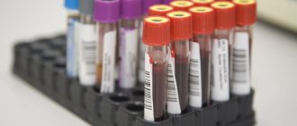
His colleague from the National Medical Research Center for Hematology of the Russian Ministry of Health, Grigory Efimov, in parallel, created a tool for direct assessment of T-cell immunity: the so-called MHC tetramer for identifying Covid-specific T cells. Under a microscope, it looks like a ball of artificial immunoglobulins-MHC with a piece of viral protein. Inside such a ball there is either a fluorescent tag or a tiny nanomagnet. Now imagine many such nano-sized “balls” collected into a single suspension. If you add such a suspension to a sample, 1-2 milliliters of venous blood, then hundreds and thousands of T cells that have already encountered the virus will stick around the balls and can be “pulled out” from the blood by a fluorescent or magnetic tag.
T cell repertoire
The already mentioned Russian professor Dmitry Chudakov is the author of determining the profile of T-cell immunity. He is the only one in the world who effectively determines the specificity of T cells based on their DNA and RNA sequences. This is no longer so much a method for determining the presence or absence of T-cell immunity to a specific virus, but a qualitative reconstruction of the entire repertoire of T-cells in the body.
“Our goal is to learn to determine from T-cells what pathogens a person has encountered throughout his life,” explains Chudakov’s colleague Yuri Lebedev. – That is, to determine the entire T-cell repertoire of the body. Imagine how great it will be if, using T cells, we can determine not only the entire immune status of a person, but also his immune memory, identifying people with known protection against serious infections (or without it)! We hope that we will be able to answer all these questions in the near future.
– I heard that you recently launched work on long-term tracking of T-cell immunity in two Muscovites who recovered from coronavirus in 2021. Do you intend to monitor its survivability for all subsequent years?
- Certainly. This is the essence of long-term control. But then we will know how many T-cells for the current disease will remain in them in a year, in 5 years, etc.
– How much time has passed since their recovery? Do they still have antibodies to coronavirus?
– Both were mildly ill at the beginning of March 2021. Six months after complete recovery, the number of anti-Covid antibodies fell by 2 times, and it continues to slowly decline. But coronavirus-specific memory T cells (such well-trained sentinels for secondary infection) have taken their place in the repertoire. Now, almost a year after the first meeting with the virus, every thousandth of the many millions of T-cells in both of these Muscovites is guarding their health...
Scientists are confident that if you combine two technologically different approaches to studying T cells, you can give each person a very good characterization of their resistance to the new coronavirus SARS-CoV-2.
Now there is a discussion among experts about how long the vaccination will work: a year or two. But at the T-cell level, it can become lifelong. If this is proven, then the need for repeated vaccination will disappear completely, unless, of course, the virus mutates too quickly.
What happens when T cells mature?
Another important event occurs in the thymus when T cells mature.
The thymus undergoes a process known as “negative selection,” in which T cells that may cause autoimmune reactions are allowed to die off. It is designed to prevent the immune system from attacking the body at random. Sometimes this process gets disrupted, causing autoimmune diseases, which can cause serious medical problems. A T cell can do anything, depending on its type. Some bind to cells and kill them if the cells become infected, while others store a memory of specific antigens so that the body can respond quickly if those antigens are identified. Helper T cells detect situations that require a response from the immune system and trigger various signals to be sent to the rest of the body. Regulatory T cells are flushed out after infection, trying to remove cells that have developed an autoimmune response.
Differentiation in the thymus
Stages of differentiation of T lymphocytes
All T cells originate from red bone marrow hematopoietic stem cells, which migrate to the thymus and differentiate into immature thymocytes
. The thymus creates the microenvironment necessary for the development of a fully functional T cell repertoire that is MHC-restricted and self-tolerant.
Thymocyte differentiation is divided into different stages depending on the expression of various surface markers (antigens). At the earliest stage, thymocytes do not express the co-receptors CD4 and CD8 and are therefore classified as double negative (DN) (CD4-CD8-). At the next stage, thymocytes express both coreceptors and are called double positive (DP) (CD4+CD8+). Finally, at the final stage, there is a selection of cells that express only one of the coreceptors (Single Positive (SP)): either (CD4+) or (CD8+).
The early stage can be divided into several substages. Thus, at the DN1 (Double Negative 1) substage, thymocytes have the following combination of markers: CD44+CD25-CD117+. Cells with this combination of markers are also called early lymphoid progenitors (ELP). As ELPs progress in their differentiation, they actively divide and finally lose the ability to transform into other cell types (for example, B lymphocytes or myeloid cells). Moving to the DN2 (Double Negative 2) substage, thymocytes express CD44+CD25+CD117+ and become early T-cell progenitors (ETPs). During the DN3 substage (Double Negative 3), ETP cells have a CD44-CD25+ combination and enter the process of β-selection.
β-selection
T-cell receptor genes consist of repeating segments belonging to three classes: V (variable), D (diversity), and J (joining). In the process of somatic recombination, gene segments, one from each class, are joined together (V(D)J recombination). Random combination of V(D)J segment sequences results in unique variable domain sequences for each receptor chain. The random nature of the formation of variable domain sequences allows the generation of T cells capable of recognizing a large number of different antigens, and, as a result, provide more effective protection against rapidly evolving pathogens. However, this same mechanism often leads to the formation of nonfunctional T-cell receptor subunits. The genes encoding the β-subunit of the receptor are the first to undergo recombination in DN3 cells. To exclude the possibility of the formation of a non-functional peptide, the β-subunit forms a complex with the invariable α-subunit of the pre-T-cell receptor, forming the so-called. pre-T cell receptor (pre-TCR). Cells that are unable to form a functional pre-TCR die by apoptosis. Thymocytes that have successfully passed β-selection move to the DN4 substage (CD44-CD25-) and undergo a process of positive selection
.
Positive selection
Cells expressing pre-TCR on their surface are still not immunocompetent, since they are not able to bind to molecules of the major histocompatibility complex. Recognition of MHC molecules by the T-cell receptor requires the presence of CD4 and CD8 coreceptors on the surface of thymocytes. The formation of a complex between the pre-TCR and the CD3 co-receptor leads to the inhibition of β-subunit gene rearrangements and at the same time causes activation of the expression of the CD4 and CD8 genes. Thus, thymocytes become double positive (DP) (CD4+CD8+). DP thymocytes actively migrate to the thymic cortex, where they interact with cortical epithelial cells expressing proteins of both classes of MHC (MHC-I and MHC-II). Cells that are unable to interact with the MHC proteins of the cortical epithelium undergo apoptosis, while cells that successfully complete this interaction begin to actively divide.
Negative selection
Thymocytes that have undergone positive selection begin to migrate to the corticomedullary border of the thymus. Once in the medulla, thymocytes interact with the body’s own antigens, presented in complex with MHC proteins on medullary thymic epithelial cells (mTECs). Thymocytes that actively interact with their own antigens undergo apoptosis. Negative selection prevents the emergence of self-activating T cells that can cause autoimmune diseases, being an important element of the body's immunological tolerance.