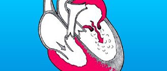
Puncture is the collection of biomaterial by puncturing an organ with a hollow needle. In cardiac surgery, this method is used to puncture the pericardium, a dense membrane around the heart filled with fluid.
The procedure is prescribed for therapeutic purposes and for diagnostics. This is a complex technique that is used only when absolutely necessary.
It gives doctors complete information about the condition of the heart and pericardial region, sometimes even saving the patient’s life. It is better to learn about the specifics of preparation and possible complications in advance.
Indications for use
The procedure is carried out for diagnostic or therapeutic purposes. In the first case, it is required to identify the pathogen during inflammatory reactions occurring in the heart muscle and adjacent areas. Executed as planned.
Puncturing is used in situations such as:
- the appearance of blood in the chest cavity if the patient has received a deep wound;
- pericarditis - excessive fluid formation due to hemorrhage in the heart, autoimmune and infectious diseases;
- purulent pericarditis - puncture is also performed for diagnostic purposes to isolate the pathogen;
- accumulation of air in the pericardial region due to injuries of the pleura or trachea;
- cardiac tamponade, which occurs due to compression of the pericardial area by pathological fluid. The heart loses its ability to contract, so immediate puncture is required.
Any condition that is caused by the accumulation of fluid or air can cause cardiac tamponade.
If traditional treatment methods do not allow the pathological condition to be stopped, a puncture of the pericardium is mandatory.
Possible complications
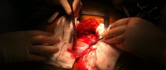
During such manipulations there may be complications. The heart or lungs may be damaged by the puncture instrument, this is considered the most serious consequence of the procedure.
This happens when the patient or the doctor himself makes sudden movements during manipulations; insufficient examination also leads to such consequences. In this case, the patient receives emergency care immediately; especially difficult situations require urgent surgical intervention.
Also, when inserting a needle, infection is possible if the instruments or chest have not been treated with an antiseptic.
If the exudate is removed too quickly, the body experiences stress, and the heart does not have time to adapt to the changed pressure. This is fraught with disturbed heart rhythm. It is important to monitor your well-being after pericardiocentesis; you should contact a cardiologist if:
- chills and fever appeared;
- there are chest pains that do not go away;
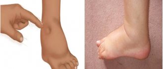
- causeless cough, shortness of breath, difficulty breathing occurs;
- blood is released from the puncture site;
- severely dizzy, nauseous;
- swelling and redness appeared around the puncture area.
Excess weight, bad habits, and the patient's state of shock increase the risk of complications. If the patient does not warn the doctor about the medications he is taking, blood clotting may be reduced, leading to serious consequences.
Contraindications
It is prohibited to use this surgical intervention in the following cases:
- low level of platelets in the blood;
- coagulopathy;
- post-traumatic hemopericardium;
- limited effusion;
- pathological fluid as a result of metastasis from an existing tumor;
- aortic dissection.
There are no serious contraindications to pericardial puncture. Sometimes doctors perform the procedure with one of the listed restrictions if there is a risk of developing dangerous complications.
Preparation stage
The stages of preparation for puncture of the cardiac membrane are different for planned and emergency interventions. It is mandatory to:
- Blood test to determine platelet levels.
- Chest x-ray to check fluid levels.
- ECG to determine the functioning of the heart.
- An echocardiogram to reveal the size and shape of the heart muscle.
- Determination of puncture method.
- Test for allergic reactions to painkillers.
A few days in advance, the patient informs the doctor about all the medications he is taking. A week before the date of puncture of the cardiac membrane, the intake of NSAIDs and drugs that thin the blood is limited.
Surgery is performed in the morning on an empty stomach. The last meal and liquid intake during planned intervention is no later than 22:00. Additionally, patients with diabetes need to check with the surgeon in what sequence and at what time to use medications.
Recovery after puncture
The patient remains in the hospital for observation for some time. After discharge, the doctor gives recommendations for a speedy recovery. For the first time after pericardiocentesis, it is strictly forbidden to lift weights, overexert yourself, or have sex. Smoking and alcohol are excluded if possible altogether, at least for 2 weeks.
When you are discharged, you should consult with your doctor about what medications you can take. A painkiller will be prescribed; you need to drink it in a clearly indicated dosage. At the first sign of complications, you should consult a doctor or call an ambulance.
For quick rehabilitation, a healthy diet is recommended; as a rule, diet No. 15 is prescribed. From time to time you need to go for leisurely walks and breathe fresh air. In winter, be sure to dress warmly; in summer, avoid overheating and sunbathing on the beach. Stressful situations are especially undesirable in case of heart disease and should be avoided as much as possible. For those who are especially sensitive, a cardiologist may recommend sedatives. You can return to an active lifestyle after your doctor approves it.
Process
The following is used as a pericardial puncture kit:
- an antiseptic to prevent infectious agents from entering the wound;
- syringes with thin needles for injecting local anesthetic;
- a monitor that records the activity of the heart muscle;
- clamp;
- an antiseptic solution that is injected into the affected area;
- a medicine for draining a postoperative wound.
For this technique, a 0.5% Novocaine solution or a 1% Lidocaine solution is used as an anesthetic at the choice of the anesthesiologist and according to health indications.
When performing a puncture technique, the following actions are carried out:
- The doctor treats the puncture site with an iodine solution.
- An anesthetic is injected into the puncture site.
- The doctor connects the ECG monitor using a clamp. Required for safety during the procedure.
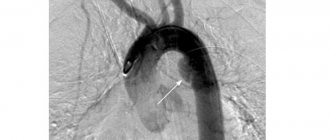
- The Larrey method - the injection is carried out in the angle formed by the xiphoid process of the sternum and the anterior part of the seventh rib. According to Marfan - under the xiphoid process along the nipple line. The needle is attached to the syringe.
- The needle is inserted behind the sternum, parallel to it, sharply upward to a depth of 3-4 cm.
- Periodically, the surgeon pulls the plunger toward himself to locate the needle.
- The doctor carefully monitors the readings reflected on the monitor. As soon as the ST segment rose on the cardiogram, the needle touched the myocardium. A change in the QRS complex indicates contact with the epicardium.
- The fluid is pumped out at the puncture point.
- If the reason for the procedure is purulent exudate, the cavity is sanitized with antiseptics. The volume of injected solution should not exceed the amount of pumped out liquid.
- Next, a broad-spectrum antibiotic is injected into the cavity.
- It is possible to install a Teflon catheter, which is designed to drain the area affected by inflammation.
- A bandage is applied and secured with adhesive tape.
In a small child, an intervention is performed using anesthesia administered through a mask, followed by a transition to endotracheal. Body position – lying down with the patient’s head elevated to 45 degrees.
Principles for diagnosing acute pericarditis
P
Ericarditis is a manifestation or complication of many diseases, including infectious diseases, pneumonia, coronary heart disease (CHD) and non-coronary myocardial diseases, systemic connective tissue diseases, tumors and allergic processes. In some cases, pericarditis may be the main manifestation of the disease itself.
Features of the diagnosis of pericarditis
The development of instrumental diagnostic methods has significantly increased the possibilities of diagnosing pericarditis. Some techniques for physically identifying signs of the disease, which at one time helped to recognize pericarditis, have lost their meaning. Echocardiography (ECHOCG) began to play a particularly important role in verifying changes in the pericardium. Nevertheless, the possibilities of instrumental examination should in no way supplant the classical methods of diagnosing pericarditis - they only usefully complement them, sometimes ahead of the clinical detection of the disease and shifting diagnostic judgments to the desired level. Thus, the unexpected detection of a layer of fluid in the pericardium during echocardiography raises the question of the nature of the effusion, the very presence of which was so difficult to establish just 20 years ago. At the same time, incorrect interpretation of instrumental data not only makes it difficult to establish an etiological diagnosis, but also generates a significant number of errors that negatively affect the course of the disease and the choice of treatment tactics.
Another reason that allows us to pose the problem of diagnosing pericarditis in relation to the present stage of its tasks and opportunities is a change in the structure of heart diseases and the pericardium itself in recent decades, a decrease in the proportion of infectious (especially purulent) pericarditis and a progressive increase in the number of allergic, autoimmune, and oncological lesions of the heart. shirts.
The degree of interest of surgeons has seriously changed, on the one hand, in determining the indications for surgical treatment of patients with diseases of the heart and blood vessels and, on the other hand, in the timely detection of pericarditis after cardiac surgery. Expanding technical capabilities of surgery and increasing the number of cardiac surgical interventions require increasingly reliable and accurate characterization of pericardial diseases.
Primary diagnosis of dry pericarditis
Complaints from patients with dry pericarditis are usually associated with a feeling of dull, monotonous pain to the left of the sternum. Pain
with pericarditis, it has a more gradual onset, is monotonous, lasts for several hours, is not relieved by nitroglycerin, and is temporarily weakened by the use of non-narcotic analgesics. There may be complaints of palpitations, shortness of breath, dry cough, general malaise, chilling, which bring the picture of the disease closer to the symptoms of dry pleurisy. The dependence of pain on breathing, movements, and changes in body position is characteristic. The patient cannot take a deep breath, breathes shallowly and frequently.
Pericardial friction rub is of great diagnostic value.
, which in patients under medical supervision allows even painless forms of pericarditis to be diagnosed. At the height of pain, the friction murmur is gentle, limited in extent, and difficult to distinguish from a short systolic murmur. With an increase in fibrinous deposits on the pericardial layers, the noise becomes rough and is heard over the entire zone of absolute dullness of the heart. It can be two- or three-phase, as it occurs even during atrial systole and in the fast diastole phase. All components of such noise are similar in character and strength; it is compared to the rhythm of a steam locomotive. The friction murmur is always limited to the zone of absolute dullness of the heart or localized in some part of it. A distinctive feature of pericardial murmur is its poor conductivity; it “dies where it was born.”
When, during acute pericarditis, the subepicardial layers of the myocardium are involved in the inflammatory process, this is reflected in ECG changes
. An early sign of acute pericarditis is a concordant rise in the ST segment, covering all standard leads within 1-2 days (the greatest rise is noted in lead II). The ST segment smoothly turns into a high positive T wave. After 1-2 days, the ST interval drops below the isoelectric line, becomes convex upward, then within a few days returns to the isoelectric line, despite the ongoing inflammatory process in the pericardium. The T wave, which is positive and even slightly enlarged in the early stages of pericarditis, then flattens and after 10-15 days becomes negative or biphasic in those leads in which the ST segment dynamics occurred.
Depending on the etiology of dry pericarditis, in some cases a rapid positive dynamics of the process is noted, a friction noise is heard for only a few hours (epistenocardiac), in others the course of pericarditis becomes protracted or recurrent, in others there is a transformation into effusion pericarditis.
Exudative pericarditis
Exudative pericarditis means total involvement of the heart lining in the inflammatory process
. Liquid effusion can accumulate after the stage of dry pericarditis or, bypassing it, with rapidly beginning total pericarditis (allergic) and with primary chronic “cold” (tuberculosis, tumor).
With the slow accumulation of fluid, the volume of the pericardial sac gradually increases, the pericardial pockets are filled, the outer layer of the pericardium is stretched, and intrapericardial pressure sometimes does not increase even with large effusions (up to 2-3 l).
With large effusions, percussion determines the expansion of cardio-pericardial dullness in all directions. The boundaries of dullness change depending on the position of the patient’s body: when he stands up, the zone of dullness in the second and third intercostal spaces decreases by 2-4 cm on each side (shifts medially), and dullness in the lower intercostal spaces expands by the same amount. Therefore, having noted the boundaries of cardiac dullness when the patient is lying on his back, the study is repeated in a standing position. Absolute dullness in the lower parts comes close to the boundaries of relative dullness, and there is a sharp transition to tympanitis over the compressed lung.
Heart sounds, even when a large effusion accumulates in the pericardial sac, often remain clear and well audible, but only medially from the apex beat.
The radiologist may suspect the presence of fluid in the pericardium based on the increase in the size of the “heart” shadow. However, since an increase in the shadow of the heart can also occur as a result of its dilatation, simply establishing an increase in the “heart” shadow is not enough to resolve the issue of fluid accumulation in the pericardium. The difficulty lies in the fact that radiographically, behind the shadow of the fluid-filled pericardial sac, the shadow of the heart itself is not visible.
An early radiological sign of the accumulation of exudate in the cardiac membrane is not so much an increase in size as a change in the silhouette of the “heart” shadow
.
The triangular shape of the shadow occurs with long-term chronic pericardial effusions due to loss of elasticity of the outer layer of the pericardium. The spherical shape of the shadow speaks in favor of a fresher and increasing effusion. A characteristic sign of exudative pericarditis is a weakening of the pulsation of the shadow contour
. The aortic pulsation remains clear. In case of a recurrent course of the process with the formation of adhesions, radiographic examination may reveal jaggedness of the cardiac contours.
The possibilities for early diagnosis of acute pericarditis have increased with the widespread use of echocardiography. The layer of fluid anterior and posterior to the cardiac contour is confidently visualized as an echo-negative space. Often there is also compaction of the pericardial layers and heterogeneous shadows of fibrinous deposits, and with large effusions, characteristic vibrations of the heart inside the stretched pericardial sac, depending on the respiratory phases.
Echocardiographic overdiagnosis of pericardial effusion is observed in the case of left-sided pleural effusion, in persons with a giant left atrium with severe mitral stenosis, when a duplication of the left atrium is formed behind the left ventricle, with pronounced fatty deposits near the heart, and with the location of the lumen of large vessels.
Cardiac tamponade
With the rapid accumulation of effusion in the pericardial cavity, cardiac tamponade develops, tachycardia occurs, and pulse filling decreases.
There is no congestion in the lungs due to an obstruction to the blood supply to the right heart. The presence of congestive rales in the lungs contradicts the diagnosis of cardiac tamponade. The left heart becomes empty as you inhale, and the pulse volume decreases. This phenomenon is called paradoxical pulsus
. The paradoxical nature of the pulse is of decisive diagnostic importance.
Echocardiogram confirms cardiac tamponade
a decrease in the size of its cavities, overflow of the hepatic veins, and sometimes prolapse of the mitral valve leaflets (disappear after unloading puncture). Echocardiographic signs of cardiac tamponade also include bowing of the wall of the right ventricle and its diastolic collapse: the wall of the right ventricle is pressed against the interventricular septum in diastole. On inhalation, an increase in the size of the right ventricle and a decrease in the size of the left ventricle can be detected; on exhalation, the opposite phenomena occur - an increase in the size of the left ventricle and a decrease in the size of the right ventricle occur - an echocardiogram equivalent of paradoxical pulsus.
Doppler ultrasonography allows us to judge the increase in pressure in the right atrium and right ventricle and the filling pressure of the right ventricle (sometimes equal to the left ventricular filling pressure).
However, echocardiographic signs of tamponade are not as informative as clinical symptoms, especially with a negative conclusion. The higher the intrapericardial pressure, the higher the venous pressure, the peripheral and cervical veins swell. The liver enlarges and becomes painful on palpation, especially its left lobe. Since in certain positions the basin of the superior vena cava is partially unloaded, the patient with increasing cardiac tamponade takes a characteristic position in bed. Usually he sits, his torso is tilted forward, his forehead rests on a pillow (Breitman pose), or he freezes in a deep bow position. There are painful attacks of weakness with a small, barely perceptible pulse, and the patient experiences a feeling of fear of death. The skin is covered with cold sticky sweat, cyanosis increases, the extremities are cold, and consciousness is disturbed at times. There are vital indications for pericardial puncture. The faster tamponade develops, the more dangerous the delay; sometimes the count is not days, but hours or minutes.
Pericardial effusion and pericardial effusion
Echocardiography makes it possible to establish initial forms of pericarditis that were previously inaccessible for diagnosis. These small, usually spontaneously resolving effusions should in no way be identified with exudative pericarditis (as is sometimes described in the echocardiogram report): often it is a non-inflammatory effusion (hydropericardium) or an initial form of the catarrhal process. It has become obvious that dry pericarditis is not the initial form of pericarditis. Its development indicates the transition of the inflammatory process from catarrhal to “lobar” with the entry of fibrinogen into the exudate and the loss of fibrin while maintaining effective suction of liquid fractions through the lymphatic vessels.
Echocardiography reveals an increase in the amount of intrapericardial fluid up to 100 and even 500 ml. With a targeted examination of patients with acute myocardial infarction, effusion can be detected in 1/3 of cases during the first week of the disease - much more often than signs of dry epistenocardial pericarditis occur.
To the appearance of hydropericardium
may give general or local reasons. General diseases include diseases that disrupt the oncotic properties of the blood and the permeability of vascular membranes, heart failure, hydremic, cachetic, and arrowroot conditions. They, as a rule, lead to the accumulation of transudate also in other serous cavities and to anasarca. In severe forms of myxedema, effusion almost always forms in the pericardium. Usually it is small. The involvement of the pericardium in the process has been described in ankylosing spondylitis, systemic lupus erythematosus, Reiter's syndrome, and rheumatoid arthritis.
There is usually no pain in the area of the heart and no friction noise with hydropericardium, but sometimes a touch noise similar to a short light friction is heard.
Pericardial puncture
The final diagnostic and highly effective treatment measure in the clinic of effusion pericarditis remains puncture. It allows you to conduct a cytological examination, perform bacteriological, immunological and biochemical tests.
Based on the nature of the contents obtained , hydropericardium, cholesterol pericarditis, chylopericardium are established, and supuration of effusion is detected
(beginning of purulent inflammation).
Indications for pericardial puncture
: cardiac tamponade (vital indications, puncture is performed urgently); purulent nature of the process and prolonged resorption of exudate (therapeutic and diagnostic); effusion pericarditis, the nature of which needs clarification or verification (diagnostic).
Several ways of needle insertion (trocar or catheter with stylet) have been proposed. At present, only two have retained their significance: 1) in the angle between the cartilage of the VII rib and the xiphoid process to the left of it (according to Larrey) or downward from the xiphoid process (according to Marfan); 2) 2-3 cm medially from the left border of absolute dullness in the fifth or sixth intercostal space (according to Kurshman), if the apical impulse is clearly defined medial and above this point.
It is advisable to insert a string through the needle, and then a catheter along it. This allows you not only to completely drain the pericardial cavity and introduce oxygen into it, but also to leave the catheter in the cavity for 72 hours for subsequent manipulations (only for puncture using lower approaches!).
Punctures through the intercostal spaces are strictly contraindicated, no matter how they are performed, even under ultrasound control.
Etiological diagnosis of acute pericarditis
Although the detection of even initial forms of pericarditis has been significantly simplified due to the introduction of instrumental methods for examining patients, their etiological diagnosis remains complex, and in many cases, only speculative.
Nonspecific coccal pericarditis
confidently diagnose purulent effusion, based on the predominance of neutrophils in the effusion, according to bacterial culture. In other cases, the diagnosis is made presumably on the basis of the development of pericarditis in connection with acute pneumonia or as a complication of sepsis, infective endocarditis, or mediastinitis.
Specific bacterial pericarditis
recognized by the general symptom complex of the disease, they are always difficult for etiological diagnosis in cases of isolated damage to the pericardium. Meanwhile, in some cases, of all the serous membranes, one pericardium is affected.
Tuberculous pericarditis
occurs more often in persons with a hyperergic tuberculosis process of another localization or in those who have had tuberculosis in the past. Pain in the heart area is rare. Low-grade fever, night sweats, and dry cough are observed. The course of the disease is long and torpid, intrapericardial effusion can be large without the development of tamponade. Sometimes the effusion persists stably for years, almost without being accompanied by fever and inflammatory changes in the blood (“cold” flow). The myocardium is not involved in the process, and ECG changes do not occur. They attach importance to high tuberculin tests. In later stages, radiologically it is possible to detect areas of calcification.
Pericardial tuberculosis is severe and often unfavorable
– one of the forms of organ tuberculosis: high temperature, leukocytosis, night sweats, rapid accumulation of exudate in the pericardial cavity, loss of body weight. The process, even with active treatment, often leads to constriction after 1.5-2 months and then requires urgent pericardiectomy.
Viral pericarditis
- a complication of a viral infection, although relatively recently the viral origin of a significant number of acute benign pericarditis, currently identified primarily as allergic and autoimmune, was assumed. The viral etiology of pericarditis is assumed when the disease begins with pharyngitis, rhinitis, herpes, focal or interstitial pneumonia, or herpangina, myalgia, pleurisy, serous meningitis (ECHO virus, Coxsackie virus). Coxsackie-III is the most cardiotropic. Myopericarditis caused by this strain is dangerous due to severe myocarditis; pericarditis with this infection always occurs in combination with myocarditis. Infectious mononucleosis, involving the pericardium, is recognized by enlargement of the lymph nodes, liver and spleen, polymorphic roseola rash, leukopenia, and mononuclear blood reaction are characteristic. The course is sometimes recurrent.
Suggest a rheumatic etiology of pericarditis
possible on the basis of concomitant myocarditis, polyarthritis and other clinical manifestations of rheumatism, prolongation of the PQ interval on the ECG, increased hyaluronidase activity of serum, increased content of g-globulins and immunoglobulins, high titer of antistreptolysin.
At a time of high incidence of rheumatism, signs of pericarditis usually appeared at 1-2 weeks of joint attack, and with relapses of polyarthritis - at 3-4 weeks. In the cardiac form of rheumatism, pericarditis develops from the first days of clinical manifestations of the disease. Involvement of the pericardium in rheumatic carditis indicates a high degree of activity of the process and gives grounds to diagnose its stage III (pancarditis). Dry pericarditis occurs in rheumatism three times more often than exudative pericarditis. Large pericardial effusions are an exception; cardiac tamponade almost never develops in adults. The increase in the boundaries of dullness is due not only to the accumulation of effusion, but also to dilation of the heart itself. Effusive pericarditis in rheumatism is an unfavorable prognostic sign, especially if the effusion becomes hemorrhagic.
Rheumatism is one of the common causes of intrapericardial adhesions. The rheumatic etiology of adhesive pericarditis is judged not only by anamnesis, but also by the presence of rheumatic heart disease in the patient.
Allergic pericarditis
characterized by an acute onset with sharp pain in the heart area and a tendency to relapse, occurring some time after exposure to a resolving factor (administration of serum or allergenic medication). They usually occur in the form of myopericarditis with the formation of serofibrinous effusion, skin rashes and other manifestations of a drug-induced disease or allergic condition.
Autoaggressive (alterogenic) pericarditis
associated with various damage to the cardiac membrane: post-infarction, post-commissurotomy, post-pericardotomy.
The most common post-infarction syndrome ( Dressler syndrome
) with clear, sometimes violent manifestations occurs in the 3rd week of acute myocardial infarction, when the highest titer of circulating antibodies to myocardial antigens is detected. Post-infarction syndrome can first form in a wide time range - from 10 days to 2 years after a heart attack, depending on the nature of the course of coronary artery disease and a number of associated factors, and with a re-infarction it often occurs from the first days of acute coronary syndrome.
Postcommissurotomy, postpericardotomy, and posttraumatic syndromes have similar pathogenetic mechanisms and a similar clinical course. All of these variants of autoimmune pericarditis can occur with simultaneous pleurisy and focal pneumonia, with high fever for several days. In case of relapses of alterogenic syndrome, patients may lack its central clinical sign – pericardial friction rub, if obliteration of the cardiac membrane has occurred. In these cases, the activation of the process is indicated by pain, ECG changes and other signs of relapse of post-infarction (or post-pericardotomy) syndrome, including processes of extra-cardiac localization - focal pneumonia, pleurisy, arthritis. Eosinophilia is detected in the blood; the effusion also contains many eosinophils and is sterile. Treatment with corticosteroid drugs is effective (diagnosis ex juvantibus).
Lupus pericarditis
develops more often in young women, occurs in the form of a dry, exudative (usually hemorrhagic) or adhesive process. As a rule, pleurisy and pneumonitis occur simultaneously. Sometimes percarditis occurs earlier than other manifestations of a systemic disease, begins acutely, and is characterized by a persistent relapsing course.
Uremic pericarditis
It can be dry, serofibrinous or hemorrhagic, has few symptoms, and is not accompanied by pain in the heart area. It is identified by the pericardial friction noise, which was considered the “death knell of brights.” Routine hemodialysis removes the gloomy prognostic significance of uremic pericarditis, but it has become a criterion for the urgency of hemodialysis.
Pericarditis may be caused by local radiation trauma.
, in particular, with gamma or radiotherapy on the mediastinal area in doses of 25-40 Gy for tumor processes. The difficult task of differential diagnosis of tumor (relapse) and post-radiation pericarditis arises. The latter is often delayed for 1-5 years after irradiation and takes the form of dry recurrent exudative or constrictive pericarditis.
Pericarditis due to tumors
usually hemorrhagic, but in 50% of cases, at the first puncture, an effusion not stained with blood is detected; it becomes hemorrhagic later. A large amount of effusion accumulates in the cavity. In the exudate of cancerous pericarditis, up to 90% of leukocytes are often lymphocytes, and conglomerates of tumor cells are found. Cytological examination is highly informative.