Medical editor: Strokina O.A. - therapist, functional diagnostics doctor. September, 2021.
Synonyms: effusion in the pericardial cavity, pericardial effusion
ICD-10 code: I30, I31.3.
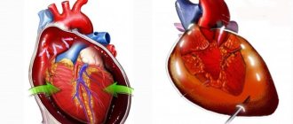
Effusion is an accumulation of fluid in the pericardial cavity. It can be acute or chronic. Acute cases occur with increasing chest pain and dry cough. The chronic form is often asymptomatic. There is no special treatment; therapy is aimed at eliminating the underlying disease.
The liquid accumulates in the form of:
- transudate - non-inflammatory effusion in chronic heart failure,
- with pulmonary hypertension,
In rare cases, air or other gas is found in the pericardial cavity, which occurs as a result of the activity of certain types of bacteria or due to an open chest injury.
The nature of the fluid (transudate or exudate) in the pericardial cavity is determined using laboratory tests by calculating its density, the amount of proteins and other elements. Based on the difference in these elements, the laboratory doctor makes a conclusion about the inflammatory or non-inflammatory nature of the effusion.
Kinds
Pericardial effusion is classified according to:
- degree of increase in its volume - acute or chronic (more than 3 months);
- distribution of contents in the cavity - encysted (localized) or surrounding;
- quantity determined using echocardiography (ultrasound of the heart) - small (less than 10 mm), medium (10-20 mm), pronounced (more than 20 mm);
- influence on hemodynamics - no effect, cardiac tamponade, constriction.
Encapsulated or localized pericarditis occurs when there is a large number of adhesions (connective tissue growths) in the cavity, formed due to inflammatory processes between the two layers of the pericardium. They just limit the fluid.
Massive effusions often occur with the tumor nature of the underlying disease.
What are the types of pericarditis?
According to the pathophysiological mechanism, pericarditis is divided into:
- exudative pericarditis (when inflammatory fluid accumulates in the cavity - exudate);
- adhesive pericarditis (when “dry” inflammation predominates and adhesions form);
- constrictive pericarditis (when compression of the heart occurs and calcification of the pericardial walls).
This division is very arbitrary, because at different stages of the pathological process the type of inflammation can transform. And the formation of areas of calcification is the outcome of any pericarditis in the absence of adequate therapy.
Causes and risk factors for effusion
Effusion in the pericardial cavity can form due to a large list of diseases:
- infectious - bacterial (pathogens of pneumonia, meningitis, syphilis, gonorrhea, tuberculosis),
- viral (Coxsackie, Epstein-Barr, mumps, rubella, HIV);
- non-infectious - autoimmune (systemic lupus erythematosus, systemic scleroderma, rheumatoid arthritis),
- tumor (metastatic tumors in lung and breast cancer),
- traumatic,
- metabolic (renal failure - accumulation of protein metabolism products in the body, myxedema - extreme degree of thyroid hormone deficiency).
Separately, it is worth highlighting pericarditis, which is characterized by large volumes of effusion:
- tumor;
- tuberculosis;
- cholesterol;
- uremic (occurs with renal failure);
- myxedema (myxedema is an extreme form of hypothyroidism, thyroid insufficiency).
Large volumes of effusions have a very high risk of developing cardiac tamponade with subsequent possible death.
Risk factors
In addition to the listed reasons for the development of effusion, risk factors can also include certain categories of patients:
- people with immunodeficiency;
- patients who are refractory to anti-inflammatory therapy (NSAIDs: indomethacin, ibuprofen, diclofenac, etc.) within 1 week after starting pericarditis therapy;
- HIV-infected.
Causes
Hydropericardium is not considered as an independent disease; it is a complication of other pathological processes in the body. Therefore, there are quite a few possible reasons for the development of this condition. These include:
- Heart failure - effusion is formed due to congestion.
- Intoxication with chemical reagents (mercury, heavy metal salts), tumor decay products.
- Cachexia. In malnourished patients, hydropericardium may develop due to a decrease in the size of the heart, which ceases to sufficiently fill the pericardial cavity.
- Inflammatory diseases of the urinary system and renal failure. The volume of fluid in the pericardial sac increases due to disturbances in acid-base and water-salt metabolism, and may also be a manifestation of swelling of the internal organs at the last stage of kidney dysfunction.
- Severe anemia - disruption of cellular respiration processes leads to the formation of transudate.
Hydropericardium is also one of the symptoms of such congenital heart disease as left ventricular hypoplasia. In addition, this pathology can accompany chest injuries.
Symptoms of effusion pericarditis
Clinical pericardial effusion occurs acutely or chronically.
Acute form
Acute effusion pericarditis is characterized by the following symptoms:
- chest discomfort or pain;
- heartbeat;
- dyspnea;
- dry cough;
- Pericardial friction rub is not typical.
Chest pain is usually progressive and lasts for several hours. They have a clear dependence on breathing (pain intensifies with inspiration, especially with deep inspiration), changes in body position, movements, which is very similar to the clinical symptoms of pleurisy (inflammation of the pleura - the outer lining of the lungs). It is also possible for pain to radiate to the left supraclavicular region, shoulder and neck.
In acute effusion pericarditis, fluid accumulates quickly, pressure in the pericardial cavity increases at a high rate, which sharply increases the risk of cardiac tamponade occurring within minutes or hours.
Chronic form
Chronic effusion pericarditis is often asymptomatic because the pericardium has time to adapt to the accumulation of fluid - it gradually stretches. However, even for this scenario, there is an end point when the outer lining of the heart is no longer able to expand. In this case, intrapericardial pressure increases and symptoms of the disease begin to appear, which sooner or later will develop into cardiac tamponade.
Life-threatening symptoms
Before tamponade develops, it is important to note signs of a hemodynamically significant effusion that precedes a life-threatening condition. Its symptoms:
- Muffled heart sounds.
- Disappearance of pericardial friction noise, if it was heard initially.
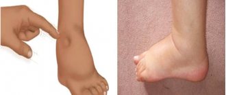
- Swelling of the neck veins.
- A decrease in systolic (upper) blood pressure during inspiration by more than 10 mmHg during quiet breathing.
If the listed symptoms are detected, the doctor must act very quickly: perform an echocardiogram on an emergency basis and schedule a consultation with a thoracic or cardiovascular surgeon to determine further treatment tactics.
Signs of effusion in localized pericarditis
For encysted or localized pericarditis, other symptoms are also characteristic, which very rarely occur:
- hiccups;
- nausea;
- swallowing disorder;
- hoarseness of voice.
The appearance of these symptoms depends on the location of pericarditis and its pressure on nearby anatomical structures.
Radiation diagnosis of pericarditis of tuberculous etiology
Ultrasound machine HS40
Top seller in high class.
21.5″ high-definition monitor, advanced cardio package (Strain+, Stress Echo), expert capabilities for 3D ultrasound in obstetrics and gynecology practice (STIC, Crystal Vue, 5D Follicle), high-density sensors.
Introduction
Damage to the inflammatory process of the pericardium occurs in 3-6% of autopsies, i.e. it occurs relatively often, but during life it is poorly recognized even in hospital conditions. This is due to the wide variety of forms and clinical manifestations of the disease, often masked by symptoms of concomitant diseases. This diversity is presented in detail in complex classifications that reflect the etiology and clinical and morphological features of pericarditis [1].
In recent years, there has been an increase in the incidence of tuberculosis and its more severe course, which is accompanied by an increase in complications. In this case, tuberculous pericarditis is suffered by up to 1/10 patients with pulmonary tuberculosis [5]. Timely diagnosis of tuberculous pericarditis is of great importance, especially with constriction of the heart, since surgical intervention will help to radically change the prognosis.
The frequency of tuberculous pericarditis, according to pathological studies, ranges from 1.1 to 15.8% [3]. Of course, the number of actual cases of illness is greater than the number of deaths.
Tuberculosis can cause any known form of pericarditis. Clinical symptomatology depends on the course of pericarditis (acute, chronic), on the amount and nature of fluid in the pericardial lining, on complications of pericarditis and mainly on the nature of the underlying tuberculous process [5,6]. As is known, pericarditis can be the first clinical manifestation of tuberculosis and occur against the background of tuberculous damage to other organs. In the first case, pericarditis is characterized by a certain set of symptoms, in the second, it is masked by the underlying tuberculosis disease.
Radiation diagnostic methods (traditional X-ray diagnostics, ultrasound methods, computed tomography and radionuclide diagnostics) today are the main ones in identifying pathology of the cardiopulmonary system in general and the pericardium in particular. Despite the fact that recently, among the methods of radiological studies in the diagnosis of pericarditis, the emphasis has shifted towards ultrasound location, traditional X-ray diagnosis of this pathology in patients with tuberculosis has not lost its importance and still remains the primary stage (verification fluorography) [4]. In the presence of clinical manifestations and suspicion of cardiac pathology (since all patients are admitted to the hospital with already available X-ray data), a preliminary assessment of the cardiovascular system can be given using a regular plain radiograph.
The main clarifying method for diagnosing cardiac pathology in our clinic is ultrasound, which is especially effective in identifying inflammatory processes in the pericardium, allowing one to assess the movement of the pericardial layers, their echogenicity, the presence of inclusions in the pericardial fluid, and hemodynamic disorders [1, 6, 7].
Materials and methods
From January 1997 to August 2001 at the Research Institute of Phthisiopulmonology MMA named after. THEM. Sechenov examined 3224 patients, of which 1896 (58.8%) had respiratory tuberculosis. Pericardial pathology during a comprehensive examination was identified in 240 (7.4% of the total number of patients), of which 108 were diagnosed with tuberculous pericarditis (45%). Since 1997, in patients with pulmonary tuberculosis, there has been a tendency to increase the number of pericarditis: 1997. - 4.9%, 1998 - 6.4, 1999 - 7.0, 2000 - 8.2%.
Pericarditis of tuberculous etiology was observed in primary tuberculosis in 20 (18.5%) and in secondary forms in 88 (81.5%) cases. In primary tuberculosis, the course of pericarditis was acute: in 85% (15 cases) dry pericarditis and in 15% (5 cases) exudative pericarditis. In secondary forms of tuberculosis, the clinical course of pericarditis was chronic: in 90.9% (80 cases) exudative and 9.1% (8 cases) constrictive pericarditis.
In primary tuberculosis, pericarditis, emanating from the lymph glandular component of the primary complex, occurred in the case of progression of the process during the period of generalization. The pericardium was affected predominantly by the lymphogenous route, and in the genesis of pericarditis, not only the infectious, but also the allergic factor (damage to the serous membranes like polyserositis) was important.
In secondary forms of tuberculosis, pericarditis developed as toxic-allergic (serous-fibrinous or serous) in most cases during the period of exudative-caseous exacerbation of the process. In patients with pleural empyema, damage to the pericardium occurred through contact.
The main methods of radiological diagnosis of pericarditis were ultrasound and x-ray.
Research results
Analysis of the results showed that each case of tuberculous pericarditis presents great diagnostic difficulties, especially if the tuberculous lesion is limited only to the pericardium and the effusion remains small.
Example 1 illustrates the presence of acute dry pericarditis in a patient with tuberculosis of the peripheral and intrathoracic lymph nodes (Fig. 1).
A 22-year-old man was treated as an outpatient in the consultation department for 2 months. In the last 2 days, complaints have appeared of a dull, pressing pain in the heart area, intensifying with inspiration and movement; shortness of breath. The examination revealed a systole-diastolic, not associated with heart sounds, pericardial friction murmur, which was heard in the second and fourth intercostal spaces on the left. The boundaries of relative cardiac dullness have not changed.
An X-ray examination of the chest organs did not reveal focal or infiltrative changes in the lungs, the roots were structural, the shadow of the heart was not changed (Fig. 1 a).
The electrocardiogram (ECG) data also turned out to be uninformative (Fig. 1b): only moderately pronounced diffuse changes in the myocardium of the left ventricle are determined (the T wave is flattened and smoothed in all leads).
An echocardiographic (ECHOCG) study revealed hyperechoic inclusions on the internal surfaces of the pericardial layers, the absence of echo-negative space behind the anterior wall of the right ventricle, behind the posterior wall of the left ventricle and the apex; parallel anteroposterior movement of the pericardial layers; impaired diastolic function of the left ventricular myocardium (Fig. 1c, d).
Rice. 1.
A patient with dry pericarditis.
A)
X-ray of the chest organs.
b)
Electrocardiogram.
V)
Echocardiogram.
G)
Echocardiogram.
During a cardiointervalographic study using the method of R.M. Baevsky determines the hyperdiastolic variant and the tension of the compensatory forces of the body (Table 1).
Table 1
. Indicators of the cardiointervalographic study method according to R.M. Baevsky.
| Mo1 | 0,80″ | Mo2 | 0,56″ |
| Amo1 | 23% | Amo2 | 42% |
| ΔX1 | 0,44″ | ΔX2 | 0,61″ |
| IN1 | 65 USD | IN2 | 468 USD |
Results of the rhythmogram analysis: blood pressure (BP) lying 120/80, standing 110/90 mm Hg. Art. Autonomic reactivity (VR) - 7.2 c.u.
The coefficient of compensatory capabilities of the left ventricular myocardium (L), calculated according to the formula we proposed using indicators of heart rate (HR), ejection fraction (EF) and tension index (SI) to identify hidden heart failure, was equal to 10.8 conventional units (cu).
An enzyme immunoassay of blood gave a weakly positive reaction, and according to the results of the NCT test (modified by M.P. Gracheva, 1986), data on active specific inflammation were obtained.
Thus, according to echocardiography and cardiointervalography studies, the patient was diagnosed with acute dry pericarditis with the presence of latent heart failure.
Example 2 illustrates the presence of acute exudative pericarditis with signs of cardiac tamponade in a patient with bronchial tuberculosis (Fig. 2).
Rice. 2.
A patient with exudative pericarditis.
a, b)
X-rays of the chest cavity.
V)
Electrocardiogram.
G)
Echocardiogram.
d)
Echocardiogram.
e)
Echocardiogram.
A 49-year-old man was admitted to the department with complaints of increasing shortness of breath, palpitations, and pressing pain in the heart area. The face was puffy, the skin was pale, and the neck veins were swollen. From the anamnesis it was known that the patient was treated in an anti-tuberculosis dispensary for 3 weeks and was sent to the clinic to clarify the diagnosis and treatment.
A survey X-ray of the patient's chest cavity organs reflects the classic signs of exudative pericarditis (Fig. 2a): all sizes of the cardiac shadow are increased to very significant, its contours are rounded in the shape of a “decanter”, the shadow of the vessels is shortened, atelectasis of the lower lobe of the left lung is observed (Evert’s symptom).
With exudative pericarditis, the ECG showed a total decrease in the voltage of the QRS complexes in all leads, atrial overload, arrhythmia (ventricular extrasystole), but these signs are not specific (Fig. 2 c).
The cardiointervalographic study data reflect the hyperdiastolic variant and the breakdown of the body’s compensatory forces (Table 2).
table 2
. Indicators of the cardiointervalographic study method according to R.M. Baevsky.
| Mo1 | 0,56″ | Mo2 | 0,52″ |
| Amo1 | 57% | Amo2 | 46% |
| ΔX1 | 0,10″ | ΔX2 | 0,12″ |
| IN1 | 1017.85 USD | IN2 | 737.2 USD |
Results of the rhythmogram analysis: blood pressure lying 180/116, standing 160/130 mm Hg. Art. BP - 0.72 USD
The coefficient of the body's compensatory capabilities is L=71 cu, which corresponds to severe heart failure.
An echocardiography study revealed 1.5 liters of fluid in the pericardial cavity (Fig. 2 d), compaction and thickening of the pericardial leaves up to 8 mm, separation of the leaves behind the anterior wall of the right ventricle up to 26 mm and the posterior wall of the left ventricle up to 12 mm were noted (Fig. 2 d, f), the presence of inclusions of fibrin and platelets in the pericardial fluid; Collapse of the anterior wall of the right ventricle and the anterior wall of the right atrium in diastole, a decrease in systolic and impaired diastolic function of the myocardium of the right and left ventricles, and regurgitant flows in the inferior vena cava and hepatic veins were present.
Culture of pleural fluid revealed Mycobacterium tuberculosis (++); An enzyme immunoassay of blood and the results of the NCT test provided data on specific inflammation.
Thus, the diagnosis of acute exudative pericarditis with signs of tamponade and severe heart failure was made according to X-ray examination, echocardiography and cardiointervalography study. Sufficiently convincing signs of pericarditis of tuberculous etiology were tuberculin skin tests, as well as enzyme immunoassays of blood and pleural fluid.
Example 3 illustrates the good result of specific therapy in a patient with chronic exudative pericarditis and pleurisy of tuberculous etiology (Fig. 3).
A 56-year-old woman was admitted to the surgical department with a diagnosis of chronic exudative pleurisy of tuberculous etiology, which she had suffered from for 5 years. The patient was admitted to the clinic to practice treatment tactics during an exacerbation with complaints of pain in the right side and behind the sternum, aggravated by coughing and changing body position; shortness of breath on exertion, low-grade fever for 2 weeks.
An X-ray examination of the chest organs revealed an effusion in the interlobar fissure, paramediastinal and in the infero-posterior parts of the right pleural cavity; the costophrenic sinus is not differentiated, the pulmonary pattern is emphasized, the structure of the right root is preserved. The left lung is unchanged. The size of the cardiovascular shadow is not increased. The cardiac arches along the left contour of the heart are not clearly differentiated (see Fig. 3 a, b).
ECG data revealed a decrease in the amplitude of the waves in all leads, an increase in the load on the right atrium and moderately pronounced diffuse changes in the myocardium of the left ventricle (see Fig. 3c). During the cardiointervalographic study, asympathicotonic autonomic reactivity and the hyperdiastolic variant of the cardiointervalographic study were determined (Table 3).
Results of rhythmogram analysis: blood pressure lying down 160/95, standing 150/100 mm Hg. Art. BP - 0.68 USD
The coefficient of the body's compensatory capabilities is L=41.3 cu, which corresponds to moderately severe heart failure.
Rice. 3.
A patient with chronic tuberculous pleurisy and pericarditis before and after treatment.
a, b)
X-rays of the chest cavity before treatment.
V)
Electrocardiogram before treatment.
G)
Echocardiogram before treatment.
d, f)
X-rays of the chest cavity after treatment.
and)
Electrocardiogram after treatment.
h)
Echocardiogram after treatment.
Table 3
. Indicators of the cardiointervalographic study method according to R.M. Baevsky.
| Mo1 | 0,66″ | Mo2 | 0,58″ |
| Amo1 | 57% | Amo2 | 48% |
| ΔX1 | 0,10″ | ΔX2 | 0,14″ |
| IN1 | 863.63 USD | IN2 | 591.13 USD |
An echocardiogram examination of the patient revealed: thickening and compaction of the pericardial leaves, areas of adhesion behind the posterior wall of the left ventricle, separation of the pericardial leaves behind the anterior wall of the right ventricle of 9 mm, unevenness of the internal contour of the pericardial leaves, a moderate increase in the amount of fluid in the pericardial cavity up to 110 ml with inclusions fibrin, early diastolic movement of the interventricular septum, impaired diastolic function of the left ventricular myocardium (type I), decreased systolic function of the myocardium of the right (EF=50%) and left (EF=56%) ventricles (see Fig. 3d).
After 3 months of anti-tuberculosis treatment, the patient showed significant positive dynamics. During an X-ray examination of the chest organs, the amount of fluid in the mediastinal pleura decreased significantly, and a tendency towards the organization of pleural effusion was noted. Noteworthy is the increase in the intensity of the pleural shadow adjacent to the cardiac shadow, which indirectly indicates the involvement of the pericardium along with the pleural layers in the process (see Fig. 3 e, f).
A cardiointervalographic study reveals a hypersympathicotonic variant of a cardiointervalographic study with sufficient autonomic reactivity (Table 4). The coefficient of compensatory capabilities of the body is L = 3.1 c.u., which corresponds to the norm.
Table 4
. Indicators of the cardiointervalographic study method according to R.M. Baevsky.
| Mo1 | 1,0″ | Mo2 | 0,86″ |
| Amo1 | 33% | Amo2 | 43% |
| ΔX1 | 0,24″ | ΔX2 | 0,12″ |
| IN1 | 137.5 USD | IN2 | 471.5 USD |
Results of the rhythmogram analysis: blood pressure lying down 140/80, standing 150/85 mm Hg. Art. VR - 3.4 USD
According to echocardiography, compaction of the parietal layer and still persisting thickening of the visceral layer of the pericardium, absence of adhesion areas behind the posterior wall of the left ventricle, a decrease in the amount of fluid in the pericardial cavity to 89 ml, normalization of the diastolic function of the left ventricle myocardium, an increase in the systolic function of the right myocardium (EF=62) were noted %) and left (EF=75%) ventricles (see Fig. 3 h). The absence of clear positive dynamics on the ECG (Fig. 3g) is characteristic of the course of chronic pericarditis.
Thus, according to echocardiography and cardiointervalography studies, the patient was diagnosed with chronic exudative pericarditis with moderately severe heart failure before treatment and in the stage of partial resorption without signs of heart failure during anti-tuberculosis therapy.
Another observation of ours demonstrates the difficulties of clinical and radiological diagnosis of constrictive pericarditis of tuberculous etiology.
Example 4. A 55-year-old patient was admitted to the therapeutic department for examination and treatment with a diagnosis of cirrhotic pulmonary tuberculosis, pulmonary heart failure (Fig. 4). His condition began to deteriorate 6 months before admission to the hospital. From the anamnesis it is known that the man has been ill with tuberculosis for more than 20 years, and for the last 5 years there have been no exacerbations.
Upon admission, the condition was serious. Complaints of severe shortness of breath at rest, dry cough, swelling. On examination, the skin is pale, ascites, swelling of the legs and feet. Auscultation - harsh breathing, no wheezing, no breathing in the upper left regions. Heart sounds are dull, the boundaries of the heart are not expanded.
On survey radiographs (Fig. 4 a, b), the upper lobe of the left lung is reduced in volume, intensely heterogeneously darkened due to gross fibrous changes and massive pleural layers, against which intensive, clearly defined foci of varying sizes are identified. An area of coarse linear fibrosis is also detected in the upper lobe of the right lung. The left root is pulled upward and is not structured. The mediastinal organs in the upper section are shifted to the left. The shadow of the heart is located vertically, the 2nd arch bulges on the right. Conclusion: pleuropneumocirrhosis of the lungs.
The patient underwent computed tomography (Fig. 4 d, e), which revealed no fresh infiltrative and focal changes in the lungs. Conclusion: CT scan shows cirrhotic pulmonary tuberculosis. Atherosclerosis of the aorta. Fluid in the pericardial cavity, pleural cavities, compaction of the pericardial layers, and pericardial adhesions are not detected.
The ECG shows pronounced sinus tachycardia (heart rate = 120 beats/min, deviation of the electrical axis of the heart to the right with a clockwise rotation around the longitudinal axis, systolic overload of the right parts of the heart, pronounced diffuse changes in the myocardium of the left ventricle (Fig. 4c).
Rice. 4.
A patient with constrictive pericarditis.
a, b)
Survey radiographs.
V)
Electrocardiogram.
G)
Computer tomogram.
d)
Computer tomogram.
e)
Echocardiogram.
In a cardiointervalographic study, there is an asympathicotonic autonomic reactivity and a hyperdiastolic variant of a cardiointervalographic study (Table 5). The coefficient of compensatory capabilities of the body L = 92 cu, which corresponded to severe heart failure.
Table 5
. Indicators of the cardiointervalographic study method according to R.M. Baevsky.
| Mo1 | 0,56″ | Mo2 | 0,48″ |
| Amo1 | 43% | Amo2 | 41% |
| ΔX1 | 0,08″ | ΔX2 | 0,12″ |
| IN1 | 479.9 USD | IN2 | 355.9 USD |
Results of rhythmogram analysis: blood pressure lying down 160/100, standing 150/115 mm Hg. Art. BP - 0.74 USD
An echocardiography study (Fig. 4f) revealed thickening up to 8 mm and compaction of the pericardial layers, absence of echo-negative space behind the posterior wall of the left ventricle and behind the anterior wall of the right ventricle, parallel anteroposterior movement of the pericardial layers, diffuse hypokinesis of the interventricular septum, dyskinesis of the interventricular septum, diffuse hypokinesis posterior wall of the left ventricle, a significant decrease in the systolic function of the myocardium of the left and right ventricles: LV ejection fraction (EF) = 39%, LV shortening fraction (SF) = 19, RV EF = 35, RV EF = 17%. It was not possible to assess left ventricular diastolic function due to severe tachycardia. Dopplerography revealed a decrease in early mitral flow at the beginning of inspiration, a slowdown in the time of isovolumic relaxation, and a decrease in early tricuspid flow with the beginning of expiration. The absence of changes in the diameter of the inferior vena cava during inspiration, regurgitant flows in the inferior vena cava and hepatic veins indicated the development of pulmonary hypertension (systolic pressure in the pulmonary artery was 84 mm Hg).
Thus, according to echocardiography and cardiointervalography studies, a diagnosis of constrictive pericarditis with signs of severe heart failure was made, and the patient was sent to the surgical department.
From example 4 it is clear that the duration of the period from the appearance of the first signs of the disease to the start of antibacterial therapy is of great prognostic significance.
Analyzing the above clinical observations, I would like to note that of the entire range of radiological research methods, ultrasound is the leading one in the diagnosis of pericarditis. It should be noted that a thorough analysis of clinical data, supplemented by such radiation, functional and laboratory research methods as radiography, ECG, cardiointervalography study, tuberculin skin tests, enzyme immunoassays of blood and pleural fluid, allowed us to avoid serious diagnostic errors and once again confirmed the well-known thesis domestic clinician prof. I. A. Kassirsky: “Yes to technology, no to technicalism.”
Conclusion
The development of medicine is inextricably linked with the improvement of instrumental methods of functional, ultrasound and x-ray diagnostics and the deepening of knowledge in these disciplines.
However, correct interpretation of instrumental data and obtaining a general idea of the pathology is impossible without a preliminary clinical and laboratory examination of the patient. Only in the hands of a clinician is it possible to correctly assess the data of the anamnesis, complaints and objective status in each specific case, and compare with them the information obtained using instrumental methods. The latter provide the most complete information about the disease.
Literature
- Diagnosis and treatment of internal diseases / Under. ed. HER. Gogin. M.: Medicine, 1991. T. I. S. 383, 388-390.
- Clinical guidelines for ultrasound diagnostics. T. 5. M.: Vidar, 1998.
- Malaya L.T. Diagnosis and treatment of heart and vascular diseases caused by tuberculosis. Kyiv, 1969. P. 43.
- Ratobylsky G.V., Ovchinnikov V.I. X-ray diagnosis of pericarditis in patients with end-stage chronic renal failure using computed tomography and color interpretation // Therapeutic archive. T. 61. 1989. N 6. P. 102-105.
- Sumarokov A.V., Moiseev V.S. Clinical cardiology: A guide for doctors. M., 1995. P.127.
- Khomenko A.G. Tuberculosis: A Guide to Internal Medicine. M.: Medicine, 1996.
- Feigenbaum H. Echocardiography. M.: Vidar, 1999.
- Keller E. Beitr. Klin. Tuberk., 1963. N 82. R. 213-219.
Ultrasound machine HS40
Top seller in high class.
21.5″ high-definition monitor, advanced cardio package (Strain+, Stress Echo), expert capabilities for 3D ultrasound in obstetrics and gynecology practice (STIC, Crystal Vue, 5D Follicle), high-density sensors.
Surveys
Laboratory and instrumental diagnosis of effusion pericarditis does not differ from the general standard for inflammatory diseases of the pericardium.
Analyzes
- General blood analysis. An inflammatory reaction of the blood is possible, which often accompanies acute pericarditis - an increase in the level of leukocytes, the appearance of their stab, juvenile forms, high ESR (erythrocyte sedimentation rate). In a chronic process, there may be no blood reaction.
- Biochemical blood test - markers of inflammation - elevated C-reactive protein, markers of myocardial damage (troponins, MB-fraction of creatine phosphokinase). In a chronic process, tests may be normal.
Instrumental studies
- Electrocardiogram. In the presence of significant effusion, the amplitude of the waves in all leads decreases.
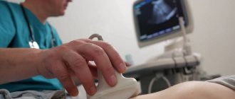
- Echocardiography (ultrasound of the heart) is the most informative method for diagnosing effusion in the pericardial cavity. Thanks to ultrasound, the doctor determines the approximate amount of fluid.
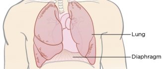
- Chest X-ray is recommended for all patients. An enlarged heart shadow is detected on an x-ray only if the effusion is more than 300 ml. The study allows us to detect signs of involvement of the pleura (the outer lining of the lungs) in the inflammatory process.
- Magnetic resonance imaging is prescribed in the absence of information content, the impossibility of echocardiography, or if a certain localization of the encysted process is suspected.
- Pericardial puncture with drainage and subsequent analysis of the contents of the cavity can alleviate the symptoms of the disease and determine the cause of the development of effusion.
If there is no information about the cause of effusion pericarditis, the doctor may order some of the following tests:
- intradermal tuberculin test (Mantoux test, Diaskintest);
- blood culture for suspected infective endocarditis;
- virological studies using ELISA and PCR methods;
- analysis for HIV, hemophilus influenzae infection;
- exclusion of chlamydial and mycoplasma infections using ELISA and PCR methods;
- determination of antinuclear factor, rheumatoid factor, antibodies to cardiolipins (for systemic lupus erythematosus, rheumatoid arthritis and others);
- antistreptolysin-O titer (for rheumatism);
- determination of antibody titers to myolemma and actomyosin in blood serum (if perimyocardial tuberculosis is suspected);
- determination of the level of thyroid hormones (for hypothyroidism).
Determining the cause of the disease is of paramount importance for successful treatment.
Treatment
Treatment of pericardial effusion should be aimed primarily at eliminating the cause of its occurrence. Often it is the underlying disease (tuberculosis, tumors, renal failure) of the patient. If the cause is not treated properly, then the chance of a full recovery is reduced, and the likelihood of relapse of the disease increases many times over.
If there is a significant amount of effusion in the pericardial cavity, there are no symptoms of pericarditis and signs of inflammation in blood tests, or the ineffectiveness of anti-inflammatory therapy, the main treatment option should be drainage of the pericardial cavity (puncture of the pericardium to evacuate fluid).
There is no specific therapy to reduce effusion alone. In the absence of an inflammatory process in the pericardium, taking NSAIDs, colchicine and corticosteroids does not provide the desired effect. If inflammatory changes are confirmed by a blood test (increased leukocytes, ESR, C-reactive protein), or analysis of fluid evacuated from the pericardial cavity, then the listed groups of drugs are prescribed without fail.
If the etiology of the process is unclear, invasive treatment techniques should be considered, including pericardial windowing (surgery to connect the pericardial and pleural cavity by creating an opening in the pericardium for permanent drainage) and pericardectomy (surgery to excise the pericardium).