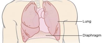
Heart failure is a condition in which the heart is unable to provide sufficient blood flow. As a result, organs and tissues suffer from oxygen starvation and nutritional deficiency. In severe cases, the pathology leads to death. Depending on the speed of development of symptoms, heart failure in cats is divided into acute and chronic.
Acute heart failure in cats, symptoms
Symptoms appear suddenly and the condition worsens quickly, over several hours and sometimes minutes. At the first sign of acute heart failure in a cat, it should be taken to a veterinarian immediately.
Symptoms:
- Shortness of breath, breathing is frequent and shallow, the cat breathes mainly from the stomach.
- The animal may become restless and meow loudly; in severe cases, severe depression develops, even to the point of loss of consciousness.
- Blue discoloration (cyanosis) of the mucous membranes.
- The limbs are cold.
- With the development of pulmonary edema, breathing becomes heavy, with moist rales.
- Due to the detachment of a blood clot, paralysis of the pelvic limbs may develop.
Diagnosis
Radiography
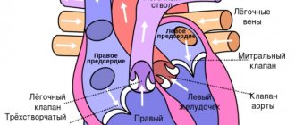
Radiographic features of HCM include enlargement of the left atrium and varying degrees of enlargement of the left ventricle. The classic valentine-shaped heart appearance in dorsoventral and ventrodorsal views is not always present, although the position of the left ventricular apex is usually preserved. The cardiac silhouette appears normal in most cats with mild HCM. Dilated and tortuous pulmonary veins may be seen in cats with chronic increases in pulmonary vein and left atrium pressure. Left-sided congestive heart failure causes patchy infiltrates expressed to varying degrees with interstitial or alveolar pulmonary edema. Radiologically, the distribution of pulmonary edema is variable; A diffuse or localized distribution within lung fields is usually found, as opposed to the characteristic hilar distribution of cardiogenic pulmonary edema in dogs. Pleural effusion is common in cats with advanced or biventricular congestive heart failure.
Electrocardiography
The majority of cats with HCM (up to 70%) have electrocardiographic abnormalities. These include abnormalities characterized by left atrial and left ventricular enlargement, ventricular and/or (less commonly) supraventricular tachyarrhythmias, and signs of left bundle branch block. Atrioventricular conduction delay, complete atrioventricular block, or sinus bradycardia sometimes occur.
Echocardiography
Echocardiography is the best method for diagnosing and differentiating HCM from other diseases. The extent of hypertrophy and its distribution within the free wall of the left ventricle, interventricular septum and papillary muscles is revealed in M-mode and B-mode echo studies. Doppler ultrasound may demonstrate left ventricular systolic and diastolic abnormalities.
Widespread myocardial thickening is common, and hypertrophy is often asymmetrically detected in the left ventricular free wall, interventricular septum, and papillary muscles. Focal zones of hypertrophy also occur. Using B-mode helps ensure that the scanning direction is correct. Standard M-Mode measurements should be taken, but areas of thickening outside these standard positions should also be measured. Diagnosis at an early stage of the disease may be presumptive in cats with mild or only focal thickening. False-positive thickening (pseudohypertrophy) can occur with dehydration and sometimes with tachycardia. False diastole thickness measurements also occur when the ultrasound beam does not cross the wall/septum perpendicularly and when measurements are not taken at the end of diastole, which can occur without a simultaneous ECG, or when the use of B-mode is insufficient for a good measurement. A thickness of the free wall of the left ventricle or interventricular septum (correctly measured) of more than 5.5 mm is considered pathological. Cats with advanced HCM have a diastolic left ventricular septal or left ventricular free wall thickness of 8 mm or more, although the degree of hypertrophy does not necessarily correlate with the severity of clinical signs. Doppler assessments of diastolic function, such as isovolumic relaxation time, mitral inflow, and pulmonary venous velocity, as well as Doppler tissue imaging techniques, are increasingly being used to characterize disease.
Hypertrophy of the papillary muscles may be severe and obliteration of the left ventricle during systole has been observed in some cats. Increased echogenicity (brightness) of the papillary muscles and subendocardial areas is usually a marker of chronic myocardial ischemia with resultant fibrosis. Left ventricular shortening fraction is usually normal or increased. However, some cats have mild to moderate left ventricular dilatation and reduced contractility (contraction fraction 23-29%; normal contractility fraction 35-65%). Sometimes there is enlargement of the right ventricle and pleural or pericardial effusion.
Cats with dynamic left ventricular outflow tract obstruction also often have mitral valve SAM or early closure of the aortic valve leaflets on M-mode imaging. Doppler ultrasonography can demonstrate mitral regurgitation and turbulence in the left ventricular outflow tract, although positioning the ultrasound beam along the blood stream with maximum ventricular ejection velocity is often difficult and it is easy to underestimate the systolic gradient.
Enlargement of the left atrium can range from mild to severe. Spontaneous enhancement (rotation, smoke echo) is seen within the enlarged left atrium in some cats. It is assumed that this is the result of blood stasis with cell aggregation and is a precursor to thromboembolism. Thrombosis is sometimes visualized within the left atrium, usually in its appendage.
Other causes of myocardial hypertrophy must be excluded before a diagnosis of idiopathic HCM is made. Myocardial thickening may also occur due to infiltrative disease. Variations in myocardial echogenicity or wall irregularity may be detected in such cases.
Excess connective tissue appears as bright, linear echoes within the left ventricular cavity.
Clinicopathological features
Cats with moderate to severe HCM have high circulating concentrations of natriuretic peptides and cardiac troponins. In cats with congestive heart failure, plasma concentrations of tumor necrosis factor (TNF) have been found to be increased to varying degrees.
Figure 1 Radiographic findings in feline HCM. Lateral (A) and dorsoventral (B) views demonstrating left atrial enlargement and mild ventricular enlargement in a male domestic shorthair cat. Lateral © view in a cat with HCM and severe pulmonary edema
Figure 2 Electrocardiogram in a cat with HCM showing infrequent ventricular premature beats and left axis deviation. Leads 1,2,3, speed 2.5 mm/sec. 1cm=1 mV
Figure 3 Echocardiographic findings in feline HCM. M-mode image (A) at the level of the left ventricle in a 7-year-old male domestic shorthair cat. The thickness of the free wall of the left ventricle and the interventricular septum in diastole is approximately 8 mm. B-mode image (B) in the right parasternal short-axis view of the left ventricle during diastole (B) and systole© in a male Maine Coon with hypertrophic obstructive cardiopathy. In (B), note the hypertrophied and brightly colored papillary muscles. In ©, note the almost complete obliteration of the left ventricular chamber during systole. IVS, interventricular septum; LV, left ventricle; LVW, left ventricular free wall; RV, right ventricle
Figure 4 A, Mid-systole M-mode echo image in the cat from Figure 3 (B and C). An echo from the anterior mitral valve leaflet is visible in the left ventricular outflow tract (arrow) due to abnormal systolic movement of the anterior mitral valve leaflet toward the interventricular septum (SAM). B, M-mode echocardiogram at the level of the mitral valve, also demonstrating SAM (arrows).
Ao, aorta; LA, left atrium; LV, left ventricle.
Figure 5 Color Doppler image during systole in a male domestic longhaired cat with hypertrophic obstructive cardiopathy. Note the turbulent flow above the protrusion of the thickened interventricular septum into the lumen of the left ventricular outflow tract and the mild mitral regurgitation of the mitral valve that often accompanies SAM. Right parasternal position along the long axis of the left ventricle. Ao, aorta; LA, left atrium; LV, left ventricle.
Figure 6 Echocardiogram obtained from a right parasternal short-axis view of the left ventricle at the level of the aorta and left atrium in an older male domestic shorthair cat with restrictive cardiomyopathy. Note marked enlargement of the left atrium and thrombus (arrows) within the atrial appendage. A, aorta; LA, left atrium; RVOT, right ventricular outflow tract
Causes
The main causes of heart failure in cats:
- Hypertrophic cardiomyopathy - the myocardium grows inward, resulting in a decrease in the volume of the heart chambers. A very common pathology in some cat breeds.
- Severe stress.
- High blood pressure.
- Inflammatory and degenerative pathologies of the myocardium.
- Hyperthyroidism.
- Infectious and invasive diseases.
- Long-term lack of taurine in the diet.
- Oncological diseases.
- Congenital heart defects – patent ductus botallus, pathologies of valves, cardiac septa.
Types of disease
Like almost any disease, this too can have an acute or chronic form. There are several manifestations of pathology by type:
- Congenital.
- Acquired.
- Primary.
- Secondary.
The last one on the list can develop against the background of a previous infection. In addition, there are some cat breeds that have a tendency to develop heart and vascular diseases. Basically, these are large animals, Maine Coons, for example.
Heart failure in cats treatment
For acute heart failure in cats, treatment includes:
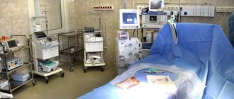
- Oxygen therapy - the animal is placed in an oxygen chamber, if the clinic equipment allows - oxygenation of the blood is carried out (its direct saturation with oxygen using a special device).
- Providing the animal with maximum rest.
- Intravenous administration of cardiac glycosides, diuretics, steroids, energy substrates, soda solution, electrolytes, antibiotics.
- Symptomatic therapy - the prescription of manipulations and drugs that eliminate the cause of heart failure.
- Maintenance therapy - the use of vitamins, immunostimulants, nutrient solutions.
- If diuretics are insufficiently effective and fluid accumulates in the body cavities, it is pumped out through a puncture.
By eliminating the cause of the pathology and providing timely assistance, it is possible to avoid irreversible changes in the animal’s body. In some cases, the acute process becomes chronic.
For chronic heart failure, treatment is often lifelong and includes:
- A therapeutic diet.
- Normalizing body weight, minimizing situations that can cause stress.
- Prescribing medications to thin the blood, normalize blood pressure, increase heart contractility, diuretics, potassium and magnesium supplements, and vitamins.
How to provide first aid? Memo for owners
Acute attacks of heart failure are very dangerous not only for the health, but also for the life of the pet. In case of fainting, you need to act quickly, so the specialists of the Berloga veterinary clinic have prepared a step-by-step guide:
- Place your pet on a horizontal surface (table, bed).
- Turn your head to the side.
- Remove your tongue from your mouth.
- Apply a cool compress (wet the cloth in cold water, preferably with ice, wring it out and apply it for a few minutes, then change the compress to a new one).
- Try to revive your pet with a cotton ball soaked in ammonia.
- Keep your paws higher than your head to increase blood flow.
- Seek veterinary help immediately.
Add this article to your bookmarks so that in an emergency you do not hesitate and provide timely assistance to your pet.
Prevention
It is impossible to completely eliminate the risk of developing pathology. Significantly reduce the likelihood of its occurrence:
- Proper feeding.
- Absence of factors that cause stress in the cat.
- Examination by a veterinarian every year, if the breed of the animal predisposes to the disease - every 6 months.
- Exclusion from breeding of animals with a history of congenital heart pathologies.
Pet owners need to know that a number of heart pathologies, such as feline hypertrophic cardiomyopathy and some others, may not manifest themselves for a long time. But if the animal’s body is subjected to stress, including anesthesia, cardiac pathology can become threatening to the normal existence and even life of the cat. Therefore, before carrying out planned surgical interventions, it is very important to undergo a cardiac examination (Ultrasound of the heart), during which hidden cardiac pathologies of cats, if any, will be identified.
You can read more about the importance of cardiac examination of animals in the section Heart ultrasound and Cardiology.
In our clinic, cardiology appointments are conducted by doctors Lidiya Mikhailovna Biryukova and Olga Vladimirovna Evstifeeva.
Diagnostics
- Blood and urine tests.
- X-ray examination of the chest.
- Ultrasound.
- ECG.
If a cat is diagnosed with heart failure, it is recommended to protect it from breeding offspring, since the disease is often inherited.
Among other things, the development of this anomaly in an animal is determined using the following methods:
- Collecting a complete medical history (data about the pet from the owner). It is important to prepare and provide the veterinarian with detailed, comprehensive information; it is best to bring it to the appointment in writing, so as not to miss important details about the health and vital functions of your pet.
- Listening, palpation and other methods of visual and physical examination. Even by measuring an animal’s blood pressure, you can come across a sign of disease: low readings usually indicate the presence of serious heart problems.
Caring for a pet with heart failure
The pet will need the most comfortable living conditions that eliminate stress. It is recommended to place the sleeping area in a quiet place away from noisy household appliances and TV.
You will have to have a conversation with the children, explaining the nuances of handling a sick cat. They must understand that hugging too tightly can have very unpleasant consequences.
You should not refuse loads. They should be moderate so that the cat can maintain a normal weight. Changes should also affect nutrition:
- Reduce the amount of salt. It retains water and impairs natural blood circulation.
- Add more proteins and taurine. Eating low-calorie protein foods helps maintain muscle mass and prevents the development of obesity. And thanks to taurine, it is possible to normalize blood pressure, stimulate blood flow and improve immunity. This beneficial element can be found in sea fish.
If your pet eats dry food, choose granules from a special veterinary line. If you have a poor appetite, it is better to replace “crackers” with wet canned food. Sick animals eat them much more willingly due to their pleasant aroma and texture.