Heart failure is a disease that is accompanied by insufficient contractility of the heart, which does not meet the metabolic needs of the body
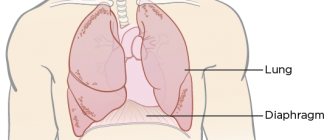
A healthy heart easily pumps blood from the left ventricle through the body to the organs. This allows you to provide them with oxygen and essential nutrients. After fulfilling its saturation function, the blood returns to the heart, but to the right side. From there, the blood is sent to the lungs, where it is saturated with oxygen and again begins its path through the left ventricle.
In other words, the disease is a weakening of the very pumping function of the heart. The lesion occurs either on the right side of the heart or on the left (right and left ventricular heart failure, respectively). Progressive disease may involve damage to both sides of the heart. In addition, deficiency can be chronic or acute.
Causes of right ventricular failure
Acute right ventricular failure develops when:
- massive pulmonary embolism;
- serious attacks of bronchial asthma;
- rupture of the septum between the ventricles;
- myocardial infarction;
- acute pneumonia.
The chronic form of the pathology is provoked by:
- congenital heart defects;
- constructive pericarditis;
- pathologies of the respiratory system;
- severe obesity;
- disorders of the pulmonary circulatory system;
- tumors;
- aortic aneurysm.
Classification of the disease
There are acute and chronic forms of pathology.
- The acute condition is dangerous because it develops in just a few hours or days. It is characterized by the patient's serious condition. This condition leads to hemodynamic disturbances. To save life, it is necessary to carry out emergency resuscitation measures.
- The chronic condition develops over a long period of time, usually several years, against the background of chronic pathologies of the bronchi and lungs, and major heart diseases.
Also distinguished:
- “Pure” (primary) failure. This pathology is very rare.
- Secondary failure. This pathology is usually provoked by a wide range of diseases of cardiac and non-cardiac nature.
Both forms of the condition are treatable.
Symptoms of right ventricular failure
The following symptoms are characteristic of acute failure:
- decreased skin temperature;
- pulsation and swelling of the jugular veins;
- increased heart rate;
- dyspnea;
- pulmonary edema.
Chronic failure is characterized by:
- pain in the right hypochondrium;
- impaired blood circulation in the kidneys;
- enlarged liver;
- accumulation of fluid in the abdominal and chest cavities;
- swelling of the lower extremities.
Patients often get tired quickly, suffer from a reduction in mental and physical activity, and insomnia. Some patients are depressed. This is due to the fact that with low cardiac output, blood circulation to the brain is reduced and the functioning of the central nervous system changes. Liver disorders and congestive gastritis that occur against the background of the condition also make themselves felt. Patients often suffer from constipation, nausea, bloating and even vomiting.
Any symptom of right ventricular failure can easily be confused with signs of many other diseases. That is why, if you suspect any pathology, you should immediately contact a professional.
The prognosis largely depends on the severity of the patient’s condition, the duration of the disease and its characteristics. Massive thromboembolism, for example, often causes death. If pathology is detected in time and its treatment is started, you can save not only the life, but also the health of the patient.
Diagnostics
Diagnosis of the condition is carried out using methods such as:
- Electrocardiogram (ECG). During the examination, specialists quickly identify signs of right ventricular overload, complete or incomplete right bundle branch block, and thromboembolism.
- Chest X-ray. This examination makes it possible to determine inflammatory processes, pneumothorax and hydrothorax. In some cases, specialists also detect pulmonary edema. This pathology requires separate therapy.
- Ultrasound of internal organs. This examination is carried out if there is a suspicion of blood stagnation in the liver, changes in the kidneys, etc. Ultrasound is especially important if the doctor thinks that the disease may arise.
All these examinations are usually performed when a patient is admitted to the hospital with acute failure.
After stabilization of the patient’s condition, the following examinations are prescribed:
- EchoCG.
- 24-hour Holter monitoring.
- Blood test for troponins, D-dimers and other indicators.
- FVD (respiratory function test).
The studies are informative for bronchial asthma, chronic obstructive bronchitis and other major pathologies.
The set of diagnostic measures is selected individually, depending on:
- patient's condition;
- existing symptoms;
- previously suffered diseases.
Additionally, vascular studies may be performed.
Age and gender factor
Statistical studies prove the connection between the spread of the disease and the gender and age of the patient.
In the age group up to 65 years, women suffer from congestive heart failure to a lesser extent; above this age, the rates become equal. Death as a result of acute heart failure is recorded in men after 45 years.
In old age, symptoms of congestive heart failure are more common, this is explained by chronic diseases, poor lifestyle, and reduced immunity.
Treatment
Therapy is always selected individually and depends on the underlying pathology.
Patients with bronchopulmonary diseases undergo treatment adjustments. Thanks to this, it is possible to reduce the number of exacerbations and reduce the load on the heart. Improving the functionality of the lungs allows you to provide oxygen to all internal organs. Patients with heart defects often undergo surgical interventions. They allow you to eliminate even severe pathologies.
When right ventricular failure is detected, patients are usually recommended to take certain medications.
Among them:
- Diuretics.
- Calcium channel antagonists.
- Nitrates.
These drugs reduce blood flow to the right atrium. In addition, they dilate blood vessels, which helps reduce pulmonary hypertension. Medicines eliminate not just the signs of pathology, but its causes.
Important! Certain medications are often taken for life. This is due to the fact that without special means, decompensation will quickly occur. The patient can only be removed from it in a hospital.
Advanced deficiency requires:
- compliance with bed rest;
- absolute peace;
- refusal of physical and emotional stress.
In some cases, minor physical activity is allowed (provided you maintain normal health).
Particular attention is paid to diet.
Experts recommend:
- Reduce the amount of liquid consumed. Thanks to this, the increase in the volume of edema is prevented.
- Salt-free diet. By adhering to it, you can avoid fluid retention in the body.
- Eat only easily digestible foods with plenty of vitamins and microelements.
We take a comprehensive approach to therapy. Thanks to this, you can count on its maximum efficiency. At the same time, we do not inflate the prices of therapy. The exact cost of services depends on many factors. The doctor can calculate it after consultation and diagnosis with all the diagnoses. You can even find out the approximate price for the services of our clinic in Moscow by phone.
The review presents an analysis of modern literature data concerning the mechanisms of liver damage in various cardiovascular diseases. The role of the liver in fat metabolism, coagulation and anticoagulation functions, and hormonal status is shown. There are difficulties in correcting liver function without affecting the metabolism of the body as a whole. Liver dysfunction can be of a wide variety of types. Correct correction of liver disorders is a task for the near future.
Influence state of the liver in the course of cardiovascular disease
This review presents an analysis of recent literature data concerning the mechanisms of liver injury in various cardiovascular diseases. It was shown the role of the liver in fat metabolism, coagulation and anticoagulation function, hormonal status. There are difficulties in the correction of the liver without affecting the metabolism of the organism as a whole. Liver function abnormalities may be the most diverse character. Proper correction of liver disorders is a problem in the near future.
The close connection between liver damage and cardiovascular diseases is beyond doubt. The relationship between changes in the heart and liver must be considered from two perspectives: on the one hand, changes occur in the liver due to primary myocardial damage and associated central hemodynamic disorders, oxygen starvation and humoral changes [10], and on the other hand, liver pathology may be an important factor for the emergence or aggravation of existing cardiovascular disorders [5] and influence the prognosis. A decrease in the functional capabilities of the liver due to its damage leads to the loss of one of the most important mechanisms of the humoral link of vascular regulation and homeostasis, the circulation of biogenic amines (mediators of blood circulation regulation) is disrupted, the rate of aldosterone inactivation slows down, and the content of the vasodilator substance increases [4].
As is known, liver dysfunction can occur with heart failure, for example with rheumatic heart defects - mitral stenosis and tricuspid insufficiency, with constrictive pericarditis. There are several pathogenetic factors responsible for the development of disturbances in the structural and functional state of the liver in heart failure. One of the main ones is a decrease in left ventricular (LV) cardiac output, leading to a decrease in hepatic blood flow. With a decrease in pressure in the hepatic artery, compensatory dilatation develops, which is apparently mediated by adenosine, produced in constant quantities by unknown cells into the space surrounding the arterioles and venules. In the absence of additional factors (increased physical activity, hypotension, shock, sepsis), the described mechanisms help avoid damage [16].
Ischemic hepatitis has been described in acute left ventricular failure due to shock, hypotension, acute myocardial infarction, especially complicated by cardiogenic shock, and pulmonary embolism. Apparently, the role of venous congestion in the liver in heart failure is also important. An increase in venous pressure leads to hepatocyte atrophy. Stagnation of blood in the sinusoids contributes to perisinusoidal edema and disrupts the oxygenation of hepatocytes. As for the role of arterial hypoxemia in the damaging effect on the liver, there is no clear opinion. Although it has been shown that the overall decrease in tissue oxygen saturation in CHF has a certain significance in aggravating the severity of hepatocyte damage [24], at the same time, most patients with stable CHF do not have significant arterial hypoxemia and the degree of liver damage does not correlate with the severity of hypoxemia [10 ]. It is believed that the development of centrilobular necrosis of hepatocytes is possible only with the development of severe shock, but it is not possible to reliably judge the degree of shock only from absolute blood pressure data.
The liver is a key link in maintaining homeostasis in the body, creating a unified system of metabolic processes. The following syndromes of liver damage are distinguished: cytolytic, cholestatic, hepatodepressive, mesenchymal-inflammatory [9]. The study of liver enzymes is used mainly to indicate cytolytic syndrome. For a “stagnant” liver, a special role in its genesis is played by hydrostatic cytolysis, caused by the development of biliary hypertension and hypertension in
hepatic vein system. With cardiovascular liver damage, the increase in aminotransferases is small and usually does not exceed normal values by 2-3 times [10]. An excess of 5 or more times is typical for ischemic hepatitis, more often observed in acute heart failure, shock, and hypotension [15].
To assess the functional usefulness of the liver and the depth of damage, it is extremely important to analyze its synthetic function. Tests of this group are rarely used in practice and, for the most part, only the protein spectrum and prothrombin index are assessed [25]. It has been noted that a pronounced decrease in prothrombin activity is observed with liver necrosis or secondary intrahepatic cholestasis, leading to secondary deficiency of vitamin K. Other factors of the blood coagulation system also change towards hypocoagulation, explaining the tendency to bleeding in patients with cirrhosis. Thus, many biochemical parameters are subject to external influence and can change during other, extrahepatic pathological processes, especially in patients with myocardial damage and concomitant diseases.
Another feature of the interaction between the functions of the liver and heart can be considered the changes that occur during pathology of the cardiovascular system in individuals with a previously compromised liver. First of all, this applies to situations where fat metabolism in general and the liver in particular is “reconfigured” in a pathological way.
In 1980, when Ludwig first introduced the new concept of “non-alcoholic steatohepatitis”, its definition was first formulated [11]. Non-alcoholic steatohepatitis (NASH) is regarded as an independent nosological entity, which is characterized by increased activity of liver enzymes in the blood and morphological changes in liver biopsies, similar to changes in alcoholic hepatitis, however, patients with NASH do not drink alcohol in quantities that can cause liver damage [6] . In recent years, scientists have attached great importance not only to fatty liver disease in general, but also to its variant, non-alcoholic fatty liver disease (NAFLD). The concept of NAFLD includes fatty degeneration, fatty degeneration with inflammation and damage to hepatocytes - non-alcoholic (metabolic) steatohepatitis and fibrosis [2].
NAFLD should always be considered in close connection with the concept of “metabolic syndrome” and its manifestations such as insulin resistance, obesity, dyslipidemia and concomitant arterial hypertension. According to the definition given in the National Clinical Guidelines of the National Clinical Committee, metabolic syndrome is characterized by an increase in visceral fat mass, a decrease in the sensitivity of peripheral tissues to insulin and hyperinsulinemia, which cause disturbances in carbohydrate, lipid, purine metabolism and hypertension.
Changes in the body observed in metabolic syndrome have stimulated interest in studying their role in the progression of cardiovascular diseases [23]. The study showed that patients with visceral obesity, high leptin levels and low adiponectin levels have a high risk of developing coronary artery restenosis after stenting, as well as a higher risk of developing and progressing atherosclerosis in previously intact areas of the coronary artery [12]. In addition, patients with ultrasonographically diagnosed NAFLD have a higher prevalence of clinically manifested CVD compared with controls without steatosis [22, 17].
In a retrospective cohort study of 420 patients with NAFLD followed for 7.6 years, the incidence of death from any cause (but the most common were CVD or cancer) was higher in patients with AN steatohepatitis or cirrhosis than in the general population [12]. The connection between CVS and liver dysfunction is proven by the fact that 100% of patients with liver cirrhosis were found to have impaired diastolic relaxation of the left ventricle (diastolic dysfunction), and increased NT-proBNP levels [7].
Liver dysfunction during myocardial infarction, apparently, can be of a different nature, from mild forms, manifested by a slight increase in liver enzymes, to severe disorders, manifested by “failures” of all liver functions.
An extreme variant of liver dysfunction in the presence of acute cardiac pathology is hepatic cell failure. It occurs in acute myocardial infarction complicated by prolonged cardiogenic shock. Hepatocellular failure is a true, endogenous, primary liver failure. Mortality in hepatocellular failure reaches 50-85% [20,18]. Hepatic coagulopathy due to liver dysfunction can be due to many reasons [14]. This is the main organ for the synthesis of most coagulation factors, both external and internal coagulation pathways. Along with other organs, the liver is involved in the formation of heparin [19]. The hepatocyte is the main site of synthesis of proteins of the coagulation system, such as vitamin K-dependent factors II, VII, IX, X, labile factor V, contact factors XI, XII, fibrinogen and fibrin-stabilizing factor XIII. The half-lives of all of the above coagulation proteins are quite short. Consequently, acute necrosis of liver cells may lead to a decrease in the levels of these proteins. The concentration of factor VII, whose half-life is 100-300 minutes, decreases most noticeably [1]. It is the first to decrease in liver failure and the first to be restored during correction.
The complexity of many processes of mutual influence of the cardiovascular system and liver requires careful and effective selection of drugs, the action of which is aimed at stabilizing the state of these systems. Correction of liver function, apparently, should be considered in close connection with the correction of metabolism in general and, in particular, diabetes mellitus. One such remedy is biguanides. Metformin is the only drug from this group that is currently the mainstay in the treatment of type 2 diabetes [24]. It has been established that biguanides do not affect insulin secretion, and in the presence of insulin they increase peripheral glucose utilization, reduce gluconeogenesis, increase intestinal glucose utilization, and also reduce elevated serum insulin levels in patients suffering from obesity and T2DM. According to some authors, biguanides have a slight anorectic effect, and their use over a long period has a positive effect on lipid metabolism (lowering cholesterol and triglycerides).
The studies have proven that metformin significantly reduces the area of necrosis after ligation of the left coronary artery in experimental animals, improves post-ischemic reperfusion not only in the experiment, but also in patients after acute transient ischemia, and with its long-term use a positive effect is clearly revealed, confirmed by improved hemodynamics in small blood and lymphatic vessels [21].
The literature data presented above allows us to conclude that liver dysfunction can be of a very diverse nature; they mutually affect the cardiovascular system. On the other hand, recent data suggest that these influences can be very complex, due to the fact that the liver is an important link in many metabolic processes in the body. It seems difficult to find an effective drug that affects only the liver without having a positive effect on metabolism as a whole. And correct, from this point of view, correction of liver function is a task for the near future.
I.F. Yakupov
Interregional Clinical Diagnostic Center, Kazan
Yakupov Iskander Fairuzovich - Candidate of Medical Sciences, Head of the Department of Anesthesiology and Reanimation No. 2
Literature:
1. Barkagan Z.S., Momot A.P. Diagnosis and controlled therapy of hemostasis disorders. - M.: Newdiamed; 2001.
2. Bogomolov P.O., Pavlova T.V. Non-alcoholic steatohepatitis: pathophysiology, pathomorphology, clinical picture and approaches to treatment. — Pharmateka. - 2003. - T. 73, No. 10. - P. 12-15.
3. Bueverov A.O., Bogomolov P.O. Multifactorial genesis of fatty liver disease. - Hepatology Forum, 2006. - pp. 6-12.
4. Vorobyov L.P., Maev I.V. The state of hemodynamics of the pulmonary circulation in liver diseases // Doctor. case. - 1990. - No. 7. - P. 16-19.
5. Garbuzenko D.V. Multiorgan hemodynamic disorders in liver cirrhosis // Therapist. arch. - 2007. - No. 2. - P. 73-76.
6. Carneiro de Mura M. Clinical perspectives in gastroenterology, hepatology. - 2001. - No. 3. - P. 12-15.
7. Komissarenko I.A., Golovanova E.V., Khanina E.A., Guseinadze M.G. Predictors of chronic heart failure in patients with alcoholic cirrhosis of the liver. // Cardiovascular therapy and prevention, 2010; 9 (6), Appendix 1.
8. Loginov A.S., Zvenigorodskaya L.A., Speranskaya I.E. and others. On the issue of cardiac fibrosis of the liver // Cardiology. - 1984. - No. 9. - P. 111-113.
9. Podymova S.D. Liver diseases / S.D. Podymova. - M.: Medicine, 1998. - 704 p.
10. Storozhakov G.I., Ettiner O.A.. Liver damage in chronic heart failure // Journal. "Heart failure". - 2005. - No. 6. - P. 28-31.
11. Liver steatosis and non-alcoholic steatohepatitis: modern view on pathogenesis, diagnosis and treatment. Yu.M. Stepanov, Doctor of Medical Sciences, Professor, A.Yu. Filippova, PhD; Department of Gastroenterology and Therapy, Faculty of Postgraduate Education, Dnepropetrovsk State Medical Academy, 2004.
12. Chumakova G.A., Veselovskaya N.G., Kozarenko A.A. Activity of local fat depots in severe angina before and after stenting of the coronary arteries. — Cardiovascular therapy and prevention, 2010; 9 (6), Appendix 1.
13. Adams LA, Lymp JF, St. Sauver J. et al. The natural history of nonalcoholic fatty liver disease: a population based cohort study. Gastroenterology, 2005; 129: 113-21.
14. Amitrano L., Guardascione MA, Brancaccio V., Balzano A. Cogulatiom disorders in liver disease. Semin. Live Dis. 2002; 22 (1): 83-96.
15. De la Monte SM, Arcidi JM, Moore GN et al. Midzonal necrosis as a pattern of hepatocellular injury after schok // Gastroenterology. - 1984. - Vol. 86. - P. 627-631.
16. Ezzat WR, Lautt WW Hepatic arterial pressure-flow autoregulation is adenosine mediated // Am. J. Physiol. - 1987. - Vol. 252, No. 4. - P. 836-845.
17. Lin YC, Lo HM, Chen JD Sonografic fatty liver, overweight and ischemic heart disease. World J Gastroenterol 2005; 11: 4838-42.
18. Marudanayagam R., Shanmugam V., Gunson B. et al. Aetiology and outcome of acute liver failure. HPB (Oxford) 2009; 11 (5): 429-434.
19. Panasiuk A., Prokopowicz D., Zak J., Panasiuk B. Reticulated platelets as a marker of megakaryopoiesis in liver cirrhosis. Relation to thrombopoietin and hepatocyte growth factor serum concentration. Hepatogastroenterology 2004; 51(58): 1124-1128.
20. Rusu EE, Voiculescu M., Zilisteanu DS, Ismail G. Molecular adsorbents recirculating system in patients with severe liver failure. Experience of a single Romanian Centre. J. Gastrointestin. Live Dis. 2009; 18 (3): 311-316.
21. Sirtori CR, Pasik C., Re-evaluation of a biguanide, metformin: mechanism of action and tolerability // Pharmacol Res. - 1994. - Vol. 30. - P. 197-228.
22. Targher G., Bertolini L., Padovani R. et al. Prevalence of nonalcoholic fatty liver disease and its association with cardiovascular disease in patients with type 1 diabetes. J Hepatol 2010; 53: 713-8.
23. Targher G., Marra F., Marchesini G. Increased risk of cardiovascular disease in nonalcoholic fatty liver disease: causal effect or epiphenomenon? Diabetologia 2008; 51: 1947-53.
24. Wiernsperger N., Membrane physiology as a basis for the cellular effects of metformin in insulin resistance and diabetes // Diabetes Metab. - 1999. - Vol. 25. - P. 110-127.
25. Wolke AM, Brooks KM, Schaffner F. The liver in congestive heart failure // Primery cardiology. - 1982. - No. 12. - P. 130-140.
Prevention
Prevention of failure (as well as any other pathologies of the cardiovascular system) comes down to:
- giving up bad habits;
- proper nutrition;
- maintaining emotional calm;
- weight control;
- active lifestyle.
Of course, in some cases it is simply impossible to prevent the disease (especially if we are talking about a genetic predisposition to it). However, you can always reduce risks. It is also important to understand that usually right ventricular failure occurs against the background of an underlying disease. It is also important to carry out its prevention. Your doctor will tell you about this.
To prevent the condition from getting worse:
- regular consultations with a specialist;
- taking prescribed medications;
- moderate physical activity.
Acute form
Along with chronic, there is an acute form of heart failure. The reasons for its occurrence are:
- myocardial infarction;
- major stroke;
- structural defects of the heart, congenital and acquired;
- hypertension.
A distinctive feature of acute cardiovascular failure is the suddenness of the attack and the absence of the listed stages of development. The person's condition quickly deteriorates and death is possible within a few minutes. The occurrence of pathology is indicated by a sharp deterioration in condition, suffocation, severe cough with foam or red sputum, blue discoloration of the skin, and cold sweat.
In such cases, first aid is to call an ambulance. Then the victim should be taken out into the open air, the collar and other tight clothing should be unbuttoned. The best pose is considered to be sitting, with legs down. A nitroglycerin tablet should be given under the tongue. Repeat the dose every 10 minutes until the ambulance arrives.
Important! In acute heart failure, you should not take a supine position.