Leukocytes, or white blood cells, are divided into three groups:
- Tissue macrophages or phagocytes. Macrophages are found inside organs and provide tissue protection. The mechanism of their work is that they capture the infectious agent and destroy it. These cells do not circulate throughout the body, so they cannot be detected in a blood test. But their predecessors, monocytes, are recorded in the bloodstream.
- Granular leukocytes. These cells contain granules or inclusions - vesicles with active substances, which are released from the cell in the event of invasion of infectious agents. Granular leukocytes take over the fight against bacteria. These cells include: neutrophils, basophils and eosinophils.
- Agranular leukocytes (agranulocytes). There are no inclusions in the structure of such leukocytes. These include lymphocytes. There are three types of agranular white blood cells: natural killer (NK) cells, T lymphocytes, and B lymphocytes. Each group of cells has its own characteristics and performs different functions.
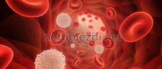
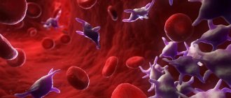
Killer cells
Figure 1. Lymphocytes attack a cancer cell.
Image: animaxx3d / Depositphotos Natural killer (NK) cells are an important component of the immune system. The function of these cells is to destroy tumor cells and virus-infected cells of the body.
Tumor cells are formed in the human body every day, and it is the normal functioning of NK cells that is necessary to prevent tumors from developing. Natural killer cells get their name because they find and destroy damaged cells themselves.¹
T lymphocytes provide the body with a cellular immune response. This means that they form a chain of cells that act on the causative agent of the disease in order to destroy it. T-lymphocytes are different, they include:
- T-helpers. These cells find the infectious agent and present it to killer T cells. They are able to secrete special substances: cytokines and growth factors that activate the immune response.
T-helpers are not capable of destroying pathogens, but without them the functioning of cellular immunity is impossible. The human immunodeficiency virus (HIV) infects these cells. As a result of prolonged carriage of HIV without treatment, acquired immunodeficiency syndrome (AIDS) develops. AIDS is a deadly disease in which a person’s immune system does not work and the body becomes unable to resist the bacteria, viruses and fungi around it. Patients with AIDS experience severe, combined forms of infections that often do not respond to therapy.
At the same time, too strong activity of T-helper cells is also not the norm. In this case, they can react to normal cells of the body and lead to their destruction. Such conditions are called autoimmune diseases. The most common of them are autoimmune thyroiditis (Hashimoto's disease), type 1 diabetes mellitus, multiple sclerosis, rheumatoid arthritis, and systemic lupus erythematosus.
- Killer T cells. They are otherwise called T-cytotoxic lymphocytes. The function of killer T cells is to destroy cells infected with viruses and tumor cells. These lymphocytes differ from natural killer cells in that they react to specific antigens that T helper cells “indicate” to them.
- T-suppressors or T-regulators. Such cells act on T-helper and T-killer cells and reduce their activity in order to avoid an excessive immune response.
B lymphocytes are cells that provide the humoral immune response. This type of response involves antibodies. The body needs this immunity to neutralize toxins and pathogens that pathogenic bacteria produce. Antibodies are able to bind to pathogens or toxins, neutralize them and attract tissue macrophages to them. Antibodies are distributed throughout the body, they are found in the blood, intercellular fluid, and on mucous membranes.
B lymphocytes are of the following types:
- Actually B cells. These are lymphocytes that have not yet come into contact with antigens. After meeting a foreign agent, they are activated and transform into another type of B lymphocyte.
- Memory B cells. They have already encountered the antigen and “remembered” it. If the same pathogen is re-introduced, memory cells are able to quickly react to it. These are long-lived cells. Acquired immunity, which appears after an illness or vaccination, is based on the work of these lymphocytes.
- Plasma cells. Such lymphocytes do not live long and are formed in large numbers during the introduction of infectious agents. Their function is to produce antibodies.²
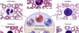
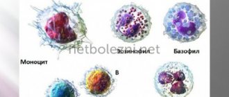
Figure 2. Types of lymphocytes. Image: mikrostoker/Depositphotos
Types of lymphocytosis
An increase in the number of lymphocytes in women is associated with a major source of dysfunction. Experts identify several types of pathology:
- Reactive – provoked by diseases that cause disruption of the functionality of the immune system. The list is presented: infectious mononucleosis, tuberculosis, syphilis, HIV infection, severe depletion of the body. To normalize test results, it is necessary to carry out therapy for the infectious disease.
- Malignant – cancerous tumors provoke the formation of acute or chronic lymphocytosis. The phase of the disease directly depends on the stage of development of the neoplasm.
- Post-infectious – deviations in tests are caused by previous infections. Lymphocytosis is accompanied by influenza, chickenpox, rubella, hepatitis, scarlet fever, and measles.
A sharp increase in the number of lymphocytes in the blood should alert a woman. Sometimes deviations make it possible to timely detect latent pathological processes or the initial stages of malignant neoplasms. Small changes should not be ignored; patients need to reconsider their usual diet, eliminate bad habits, and do not forget about seasonal vitamin therapy.
Blood test for lymphocytes
A blood test for lymphocytes is taken as part of an extended general blood test with a leukocyte formula. The formula prescribes the absolute number and relative content of all types of white blood cells. Blood can be donated from a finger or a vein.
In test forms you can find different designations for lymphocytes: LYMPH, LYM, LYM%. The unit of measurement is the number of cells per nanoliter of blood, as well as their percentage among all leukocytes.
Indications for analysis
A general blood test is the most accessible method for diagnosing various diseases and conditions. This analysis is indicated for undergoing a routine medical examination, before hospitalization, before and after surgery, for diagnosing diseases when first seeking medical help. Therefore, if the patient comes to the doctor in a timely manner, changes in the content of lymphocytes in the blood are detected quickly.
A doctor may suspect disturbances in the formation of lymphocytes and prescribe an analysis in the case of frequent and/or long-term infectious diseases. Sometimes these diseases can be difficult, difficult to treat and lead to complications.
Preparing for analysis
In order for a blood test to show an accurate result, proper preparation for it is necessary:
- Blood is taken for analysis on an empty stomach. The day before you should not eat fatty, spicy or sweet foods. It is better if the dinner is light and at least 8 hours pass before the analysis.
- Before the study, you need to get enough sleep and eliminate physical and emotional stress.
- You should avoid overload on the day before donating blood: do not exercise too much, do not go to the bathhouse or sauna, and maintain a drinking regime.
- It is necessary to refrain from smoking at least 2 hours before donating blood for analysis.
- If you are taking any medications, you must tell the medical staff when your blood is drawn.
- Norms of lymphocytes in the blood - tables with norms for women, men, children
- The normal number and percentage of lymphocytes differ depending on gender and age.
Table 1. Norm of lymphocytes in the blood of women and men.
| Absolute quantity | Percentage | |
| Among women | 1 - 3.4 x 10⁹/l | 17-38% |
| In men | 1 - 4.8 x 10⁹/l | 17-38% |
Table 2. Norm of lymphocytes in the blood of children.
| In children | Up to 1 month | 1-6 months | 6-12 months | 1-6 years | 7-12 years | 13-15 years old |
| Absolute quantity | 1.3-4.4 x 10⁹/l | 2.7-12.3 x 10⁹/l | 2.4-10.2 x 10⁹/l | 1.6-9.5 x 10⁹/l | 1.3-6.7 x 10⁹/l | 1.26-5.8 x 10⁹/l |
| Percentage | 22-25% | 45-70% | 44-66% | 30-61% | 29-50% | 28-45% |
Leukocyte crossover
In children, there is a condition called leukocyte crossover. A child is born with approximately the same ratio of neutrophils and lymphocytes as an adult: neutrophils 60-65%, lymphocytes 16-34%. After birth, the number of neutrophils gradually decreases, and the number of lymphocytes increases. By the fifth or sixth day of life, their content is equalized and is approximately 45%. This is the cross of the leukocyte formula. Further, up to 4-5 years, lymphocytes in the blood test will dominate and make up up to 70% of all white blood cells. At the age of 5-6 years, a second crossover occurs: the proportion of cells equalizes again, after which the composition of the blood gradually approaches the picture of an adult, when there are more neutrophils than other leukocytes.
Classification
There are no clear numerical criteria for dividing lymphocytosis according to severity. Conventionally, moderate (up to 60%) and high lymphocytosis (more than 60%) are distinguished. The main cause of high lymphocytosis is considered to be malignant diseases of the hematopoietic and lymphatic tissue. Lymphocytosis, like other types of leukocytosis, is divided into:
- Absolute
. Absolute lymphocytosis means an increase in the number of lymphocytes along with the total number of leukocytes (in adults more than 4000, in children under 5 years of age more than 6000). - Relative
. Relative lymphocytosis is much more common and means a percentage increase in lymphocytes (more than 40%) against the background of a normal or even reduced total number of leukocytes.
Causes of elevated lymphocytes
An increase in the level of lymphocytes in the blood is called lymphocytosis. Lymphocytosis can be:
1. Absolute: when the number of lymphocytes in the blood is increased - more than 4.8 x 10⁹/l in adults.
The causes of absolute lymphocytosis may be:
- Various viral infections (respiratory infections, herpetic diseases, chicken pox, mononucleosis, cytomegalovirus, viral hepatitis).
- The initial stage of HIV.
- Toxoplasmosis.
- Damage to the thyroid gland with a decrease in its function (hypothyroidism).
- Malignant tumors (lymphomas, leukemias).
- Vasculitis.
- Whooping cough.
- Tuberculosis.
- Strict diet, fasting.
- Eating more carbohydrates than proteins and fats.
Lymphocytosis can also occur during menstruation and in the days before it, after heavy physical labor, during severe stress, after vaccination.
2. Relative: in case of an increase in the percentage of lymphocytes among other white blood cells. In this case, their share in adults is more than 38%. In this case, the number of lymphocytes may be within normal limits, and their content may be higher due to a decrease in the number of other leukocytes. The most numerous in the leukocyte formula of adults are neutrophils; normally they contain 47-72%. Accordingly, with a decrease in their number in the blood, the proportion of lymphocytes can significantly increase. And relative lymphocytosis will be the first thing that catches your eye when you receive the result of a blood test. The following conditions can lead to this:
- Viral infections (chickenpox, influenza, measles, hepatitis).
- Toxoplasmosis.
- Chronic bacterial infections.
- Fungal diseases.²
Diagnostics
The level of lymphocytes is measured during a clinical blood test. Due to the fact that lymphocytosis has a fairly wide etiological spectrum, if it is detected, you should contact a generalist (general practitioner or pediatrician) so that, based on the patient’s complaints, anamnestic data, and physical examination, he will prescribe an additional examination, which may include:
- Blood tests
. The leukocyte formula is calculated to determine the percentage of all forms of leukocytes. A blood smear is studied using microscopy to identify atypical mononuclear cells, Botkin-Gumprecht shadows (remnants of destroyed lymphocytes). Inflammatory markers are determined - increased ESR, CRP. To detect tumor antigens, immunophenotyping and immunohistochemical studies are performed. - Pathogen identification
. In order to identify an infectious agent, tests are performed for the presence of antibodies to pathogens and their DNA (by ELISA, PCR). Bacteriological studies are carried out - culture, sputum microscopy (tuberculosis, whooping cough), serological diagnostics - Wright, Heddelson reaction (brucellosis), microprecipitation reaction (syphilis). - Instrumental Research
. With tuberculosis, an X-ray of the lungs shows an increase in the hilar and mediastinal lymph nodes, infiltration of the upper lobes of the lungs, and sometimes effusion into the pleural cavity. In case of mononucleosis and hemoblastoses, ultrasound of the abdominal cavity reveals pronounced splenomegaly, less often hepatomegaly. - Histological studies
. In chronic lymphocytic leukemia, a large number of lymphoblasts are found in the bone marrow punctate. In lymphomas, a lymph node biopsy obtained by fine-needle aspiration reveals diffuse proliferation of lymphoid cells with blast morphology. A specific sign of lymphogranulomatosis is Berezovsky-Sternberg giant cells.
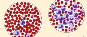
Leukocyte count
Consequences of lymphocyte deviation from the norm
A decrease in the number of lymphocytes in the blood leads to dysfunction of the immune system. In this condition, the body has poor resistance to viral infections, and a person can suffer from various diseases with difficulty and for a long time.
An increase in lymphocytes in the blood indicates the presence of an infection in the body, which may not manifest itself clinically.
Relative lymphocytosis indicates a violation of the formation of other leukocytes, including neutrophils. In this case, it will be difficult for a person to tolerate bacterial infections.
How to reduce the level of lymphocytes in the blood
The most common cause of high lymphocyte counts is infection, but sometimes leukemia may be the cause. Therefore, in the case of lymphocytosis, it is important to find its cause and prescribe treatment. Various medications are usually used: steroid hormones, immunosuppressants. Nutritional supplements can also be used: echinacea, fish oil.
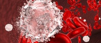
Figure 3. Blood cells in a healthy person and in leukemia. Image: mikrostoker/Depositphotos
Forecast
In some cases, the level of lymphocytes can be a guideline for predicting the development of the disease. For example, lymphocytosis in tuberculosis, both in children and adults, indicates a favorable course of the disease and a speedy recovery. Conversely, if the cause of an excessive increase in lymphocytes is malignant hematological diseases, this may indirectly indicate a high probability of death. However, first of all, it is necessary to focus on the underlying pathology and its severity. Therefore, any degree of lymphocytosis requires a thorough examination to determine the cause and timely treatment.
How to increase the number of lymphocytes in the blood
Depending on the condition, the doctor may prescribe the following treatment methods:
- Combination antiretroviral therapy for HIV.
- Antibiotics, antiviral, antifungal or antiparasitic drugs to treat chronic diseases.
- Gamma globulin for preventing infections in people with B-cell lymphocytopenia
- Bone marrow stem cell transplantation for some forms of cancer (such as leukemia, multiple myeloma, and some types of lymphoma)
If a decrease in lymphocytes in the blood is a side effect of taking a medication (such as immunosuppressants and steroids), your doctor may reduce your dose or recommend stopping it.
People with low lymphocyte counts should strengthen their immune system by eating a nutrient-rich diet that provides adequate amounts of protein, vitamins and minerals.³
These foods will help strengthen your immune system:
- Citrus fruit. Almost all citrus fruits contain large amounts of vitamin C, which is important for the functioning of the immune system.
- Red bell pepper. It is a rich source of vitamin C and beta-carotene. Beta-carotene is converted into vitamin A in the human body, which helps maintain healthy eyes and skin.
- Broccoli. Broccoli is rich in vitamins A, C and E, as well as fiber, various minerals and antioxidants.
- Garlic. Garlic contains many sulfur-containing compounds such as allicin.
- Ginger. Ginger reduces inflammation and helps relieve sore throat.
- Spinach. Rich in vitamin C, as well as numerous antioxidants and beta-carotene, which may boost the immune system's ability to fight infections.
- Yogurt. Natural yogurt contains live cultures of bacteria that can stimulate the immune system.
- Almond. Nuts contain a lot of fats, which help vitamin E to be absorbed. Almonds contain large amounts of vitamin E and healthy fats.
- Sunflower seeds. Sunflower seeds are rich in phosphorus, magnesium and vitamins B6 and E, selenium.
- Green tea. Tea contains flavonoids, which are good antioxidants. Green tea is a source of the amino acid L-theanine, which helps the functioning of T-lymphocytes.
- Papaya, kiwi. These fruits are also rich in vitamin C. They contain a lot of potassium, magnesium, and folic acid.
- Bird. Chicken and turkey are rich in vitamin B6. Broth made from chicken bones contains gelatin, chondroitin and other nutrients beneficial for stimulating the immune system.
- Seafood. Some seafood contains high amounts of zinc. Lymphocytes need zinc to function effectively. Shellfish varieties high in zinc include: oysters, crabs, lobsters, and mussels.³
Certain foods help strengthen your immune system. Photo: NewAfrica/Depositphotos
What is the danger?
An increase in lymphocytes in itself is not dangerous: the diseases that caused such a change may pose a danger. Let's consider the most common diseases in which a laboratory sign occurs:
- Infectious mononucleosis occurs when infected with the Epstein-Barr virus (EBV), a member of the herpes virus family.
. Typical manifestations: difficulty breathing through the nose without discharge, sore throat, sudden enlargement of lymph nodes, weakness, rise in temperature. As the disease progresses, symptoms of damage to internal organs appear: rashes on the body, yellowing of the skin and sclera, darkening of urine and discolored feces. - Herpes virus infection begins with severe weakness, drowsiness, weakness, enlarged lymph nodes, and vesicular rashes appear on days 2-3
. The localization of the bubbles is different: lips, nasal mucosa, genitals, torso. Shingles is one of the manifestations of herpes infection and is a very serious disease. - Lymphocytosis may be the first sign of HIV infection, so doctors, when lymphocytes are elevated, first prescribe a blood test for HIV and hepatitis B and C
. The immunodeficiency virus can circulate in the body for up to 10 years after infection without causing clinical symptoms. Frequent herpetic rashes, colds, weakness and drowsiness may cause slight concern. During the period of the first manifestations, the temperature rises, the lymph nodes enlarge, a sore throat develops, diarrhea and headache appear. - Blood cancer - leukemia, lymphoma - the most terrible diagnosis when the number of lymphocytes increases
. The main symptoms are identical for any leukemia: pronounced enlargement of lymph nodes, night sweats, weakness, fever, fatigue, shortness of breath and cough.
Stages of chronic lymphocytic leukemia
There are two staging systems for CLL, which are based on a single principle: taking into account the mass of the tumor (lymphocytosis), the size of the lymph nodes, the degree of enlargement of the liver and spleen, the presence or absence of anemia and thrombocytopenia. The combination of these factors influences life expectancy and predetermines the prognosis.
Stages of CLL according to the European Binet classification
| Stage | Characteristics | Forecast | ||
HGB anemia | Platelets PLT | Enlarged lymph nodes | ||
| A | >100 g/l | >100×109 /l | in 1-2 regions | good |
| B | >100 g/l | >100×109 /l | in 3 or more areas | intermediate 7+ years |
| C | >100 g/l | >100×109 /l | any number of zones | bad |
Stages of CLL according to the American RAI classification
| Stage | Characteristics | Forecast | |||
| Lymphocytosis | Anemia | Platelets PLT | Lymph nodes are enlarged | ||
| 0 | >15×109/l in bone marrow: >40% | No | norm ≥150 x109 /l | No | good |
| I | There is | No | normal/about normal | Yes | relatively good |
| II | There is | No | normal/about normal | yes/no + spleno/hematomegaly | intermediate 6+ years |
| III | There is | Yes | normal/about normal | Not really | bad |
| IV | There is | Not really | <110 x109 /l | Not really | bad |
Role of leukocytes
The content of the article
During physiological tissue renewal, the lymphocyte is right there; restoration of damaged organs, damaged skin occurs - it is here too; in any inflamed place we again encounter a lymphocyte... This representative of white blood cells - leukocytes - is almost omnipresent, and it sometimes behaves inexplicably.
For example, its “relatives” neutrophils, eosinophils, basophils and monocytes work hard at the site of inflammation; absorb bacteria, cell debris, and secrete proteolytic enzymes that help digest dead tissue. And the lymphocyte seems to stand on the sidelines, showing no visible activity. Is it so? And what explains his presence in this and other situations?
These questions were answered only in the middle of our century, almost seven decades after the discovery of lymphocytes. Subtle studies have shown that the lymphocyte is not an outside observer at all. He is the leading organizer and active participant in all immune and protective reactions occurring in the body.
The role of leukocytes in cell repair and division
Scientists conducted an interesting experiment. Lymphocytes from an animal in whose body an active process such as liver regeneration was taking place were injected into a healthy animal. And the cells of his normal, undamaged liver began to actively divide.
The experiment was repeated again and again and the same amazing effect was obtained. Scientists have suggested that the participation of lymphocytes in the processes of cell division is due not only to their duty to carry out immune surveillance, but that they also have a construction, morphogenetic function.
It was known before that leukocytes (including lymphocytes) influence cell division. In the experiment, serum in which leukocytes were cultured was applied to the wound, and the wound healed much faster.
This was explained by the so-called trophic function of leukocytes, believing that they enrich the serum with nutrients. It was believed that, in addition to these substances, tissue cells use “fragments” of dying leukocytes for regeneration.
Today we can say with confidence that the matter is not only in nutrients (this factor cannot be completely denied), but mainly in a specific biologically active substrate that promotes cell reproduction. It is unclear what substance (or maybe substances?) is part of this substrate; it has not yet been possible to isolate it. But it has been reliably established that it is again the lymphocyte that produces it and transfers the “regeneration stimulus” to the wife.
The most amazing thing is that lymphocytes carry more than just a stimulus to division as such. This “curious” cell is always well aware of which organ is undergoing regeneration. Lymphocytes from an animal with a regenerating liver cause increased division of mainly liver cells in healthy animals; lymphocytes of an animal with a recovering kidney - cell division of the recipient's kidney.
The lymphocyte also carries the signal for cell proliferation in cases where increased cell division occurs in the donor’s body, not associated with damage to any organ.
For example, hypoxia (lack of oxygen), as is known, leads to increased formation of red blood cells - carriers of the respiratory pigment hemoglobin. So, if you take lymphocytes from an animal in a state of hypoxia and inject them into an animal that does not experience oxygen starvation, then after some time you can observe stimulation of the production of red blood cells.
What structures of the lymphocyte play the role of a storage and reproducing device? Perhaps this is one of the most interesting and complex riddles that a lymphocyte has ever asked researchers. It is simply necessary to solve it, because there is reason to believe that this will open the way to the treatment of many diseases.
How often in clinical practice doctors are faced with the need to influence regenerative processes. And lymphocytes could provide invaluable help here. After all, they have high mobility, have wide access to almost all tissues and organs and are able to transfer their “skills” to the body, in which it is necessary to stimulate the regeneration of a certain tissue.
Perhaps lymphocytes will help to some extent solve the complex problem of treating hereditary diseases.
While studying hereditary pathology - osteopetrosis in mice and rats, which is expressed in abnormal bone formation and premature compaction, experts discovered the following fact. Injecting sick mice and rats with lymphocytes from their healthy “brothers and sisters” led to the normalization of bone formation.
True, provided that the activity of the sick animals’ own lymphocytes is suppressed. Isn’t it amazing that a hereditary malformation, such as osteopetrosis, could be corrected, albeit in an experiment, with the help of ordinary lymphocytes!