October 15, 2012
What is blood made of? We talk about its main components - plasma, red blood cells, leukocytes and platelets.
Blood is a type of connective tissue, a distant relative of bones, cartilage and even fat. It circulates throughout the body with the help of the heart and blood vessels and has two main functions: transportation
nutrients and oxygen to cells and
protection
against infectious agents, including bacteria and viruses. What is blood made of? Let's talk about its main components - plasma, red blood cells, white blood cells and platelets.
All about plasma
Plasma is a liquid formed by water and dry substances. It makes up the bulk of the blood - about 60%. Thanks to plasma, blood has a liquid state. Although according to physical indicators (density) plasma is heavier than water.
Macroscopically, plasma is a transparent (sometimes cloudy) homogeneous liquid of light yellow color. It collects in the upper part of the vessels when the formed elements settle. Histological analysis shows that plasma is the intercellular substance of the liquid part of the blood.
Plasma becomes cloudy after a person consumes fatty foods.
Composition and objectives of non-protein compounds in plasma
Plasma contains:
- Organic compounds based on nitrogen. Representatives: uric acid, bilirubin, creatine. An increase in the amount of nitrogen signals the development of azotomy. This condition occurs due to problems with the excretion of metabolic products in the urine or due to the active destruction of protein and the entry of large amounts of nitrogenous substances into the body. The latter case is typical for diabetes, fasting, and burns.
- Organic compounds that do not contain nitrogen. This includes cholesterol, glucose, lactic acid. Lipids also keep them company. All these components must be monitored, as they are necessary to maintain full functioning.
- Inorganic substances (Ca, Mg). Na and Cl ions are responsible for maintaining a constant pH of the blood. They also monitor osmotic pressure. Ca ions take part in muscle contraction and stimulate the sensitivity of nerve cells.
Blood plasma composition
Albumen
Albumin in plasma blood is the main component (more than 50%). It has a small molecular weight. The place of formation of this protein is the liver.
Purpose of albumin:
- Transports fatty acids, bilirubin, drugs, hormones.
- Takes part in metabolism and protein formation.
- Reserves amino acids.
- Forms oncotic pressure.
Doctors judge the condition of the liver by the amount of albumin. If the albumin content in plasma is reduced, this indicates the development of pathology. Low levels of this plasma protein in children increase the risk of developing jaundice.
Globulins
Globulins are represented by large molecular compounds. They are produced by the liver, spleen, and thymus.
There are several types of globulins:
- α – globulins. They interact with thyroxine and bilirubin, binding them. Catalyze the formation of proteins. Responsible for transporting hormones, vitamins, lipids.
- β – globulins. These proteins bind vitamins, Fe, and cholesterol. They transport Fe and Zn cations, steroid hormones, sterols, and phospholipids.
- γ – globulins. Antibodies or immunoglobulins bind histamine and take part in protective immune reactions. They are produced by the liver, lymph tissue, bone marrow and spleen.
There are 5 classes of γ-globulins:
- IgG (about 80% of all antibodies). It is characterized by high avidity (antibody to antigen ratio). Can penetrate the placental barrier.
- IgM is the first immunoglobulin that is formed in the unborn baby. The protein has high avidity. It is the first to be detected in the blood after vaccination.
- IgA.
- IgD.
- IgE.
Fibrinogen is a soluble plasma protein. It is synthesized by the liver. Under the influence of thrombin, the protein is converted into fibrin, an insoluble form of fibrinogen. Thanks to fibrin, a blood clot forms in places where the integrity of the vessels has been compromised.
Other proteins and functions
Minor fractions of plasma proteins after globulins and albumins:
- Prothrombin,
- Transferrin,
- immune proteins,
- C-reactive protein,
- Thyroxine binding globulin,
- Haptoglobin.
The tasks of these and other plasma proteins boil down to:
- Maintaining homeostasis and aggregative state of blood,
- Controlling immune reactions
- Transport of nutrients
- Activation of the blood clotting process.
Functions and tasks of plasma
Why does the human body need plasma?
Its functions are varied, but basically they come down to 3 main ones:
- Transporting blood cells and nutrients.
- Establishing communication between all body fluids that are located outside the circulatory system. This function is possible due to the ability of plasma to penetrate the vascular walls.
- Providing hemostasis. This involves controlling the fluid that stops bleeding and removing the resulting blood clot.
Electrolyte composition of blood plasma
It is known that the total water content in the human body is 60–65% of body weight, i.e. approximately 40–45 l (if body weight is 70 kg); 2/3 of the total amount of water is intracellular fluid, 1/3 is extracellular. Part of the extracellular water is in the vascular bed (5% of body weight), most of it is outside the vascular bed - this is interstitial, or tissue, fluid (15% of body weight). In addition, a distinction is made between “free water”, which forms the basis of intra- and extracellular fluid, and water associated with various compounds (“bound water”).
The distribution of electrolytes in body fluids is very specific in its quantitative and qualitative composition.
Of the plasma cations, sodium occupies a leading place and makes up 93% of their total quantity. Among the anions, chlorine and bicarbonate should be highlighted first. The sum of anions and cations is almost the same, i.e. the entire system is electrically neutral.
Sodium . It is the main osmotically active ion in the extracellular space. In blood plasma, the concentration of Na+ ions is approximately 8 times higher (132–150 mmol/l) than in erythrocytes.
With hypernatremia, as a rule, a syndrome develops due to overhydration of the body. The accumulation of sodium in the blood plasma is observed in a special kidney disease, the so-called parenchymal nephritis, in patients with congenital heart failure, in primary and secondary hyperaldosteronism.
Hyponatremia is accompanied by dehydration of the body. Correction of sodium metabolism is achieved by introducing solutions of sodium chloride with the calculation of its deficiency in the extracellular space and cell.
Potassium . The concentration of K+ ions in plasma ranges from 3.8 to 5.4 mmol/l; in erythrocytes it is approximately 20 times more. The level of potassium in cells is much higher than in the extracellular space, therefore, in diseases accompanied by increased cellular breakdown or hemolysis, the potassium content in the blood serum increases.
Hyperkalemia is observed in acute renal failure and hypofunction of the adrenal cortex. Lack of aldosterone leads to increased urinary excretion of sodium and water and retention of potassium in the body.
With increased production of aldosterone by the adrenal cortex, hypokalemia occurs, and the excretion of potassium in the urine increases, which is combined with sodium retention in the tissues. Developing hypokalemia causes severe disturbances in the functioning of the heart, as evidenced by ECG data. A decrease in serum potassium content is sometimes observed when large doses of adrenal cortical hormones are administered for therapeutic purposes.
Calcium . Traces of calcium are found in erythrocytes, while in plasma its content is 2.25–2.80 mmol/l.
There are several fractions of calcium: ionized calcium, non-ionized calcium, but capable of dialysis, and non-dialyzable (non-diffusing) protein-bound calcium.
Calcium takes an active part in the processes of neuromuscular excitability (as an antagonist of K+ ions), muscle contraction, blood clotting, forms the structural basis of the bone skeleton, affects the permeability of cell membranes, etc.
A distinct increase in the level of calcium in the blood plasma is observed with the development of tumors in the bones, hyperplasia or adenoma of the parathyroid glands. In such cases, calcium enters the plasma from the bones, which become brittle.
Determining the calcium level during hypocalcemia is of great diagnostic importance. The state of hypocalcemia is observed in hypoparathyroidism. Dysfunction of the parathyroid glands leads to a sharp decrease in the content of ionized calcium in the blood, which may be accompanied by convulsive attacks (tetany). A decrease in plasma calcium concentration is also noted in rickets, sprue, obstructive jaundice, nephrosis and glomerulonephritis.
Magnesium . In the body, magnesium is localized mainly inside the cell - 15 mmol/per 1 kg of body weight; magnesium concentration in plasma is 0.8–1.5 mmol/l, in erythrocytes – 2.4–2.8 mmol/l. Muscle tissue contains 10 times more magnesium than blood plasma. The level of magnesium in plasma, even with significant losses, can remain stable for a long time, replenished from the muscle depot.
Phosphorus . In the clinic, when testing blood, the following fractions of phosphorus are distinguished: total phosphate, acid-soluble phosphate, lipoid phosphate and inorganic phosphate. For clinical purposes, the content of inorganic phosphate in blood plasma (serum) is often determined.
The level of inorganic phosphate in the blood plasma increases with hypoparathyroidism, hypervitaminosis D, thyroxine intake, UV irradiation of the body, yellow liver degeneration, myeloma, leukemia, etc.
Hypophosphatemia (decreased plasma phosphorus levels) is especially characteristic of rickets. It is very important that a decrease in the level of inorganic phosphate in the blood plasma is observed in the early stages of the development of rickets, when clinical symptoms are not sufficiently pronounced. Hypophosphatemia is also observed with insulin administration, hyperparathyroidism, osteomalacia, sprue and some other diseases.
Iron . In whole blood, iron is contained mainly in erythrocytes (about 18.5 mmol/l); in plasma its concentration averages 0.02 mmol/l. Every day, during the breakdown of hemoglobin in erythrocytes in the spleen and liver, about 25 mg of iron is released and the same amount is consumed during the synthesis of hemoglobin in the cells of hematopoietic tissues. The bone marrow (the main erythropoietic tissue of humans) contains a labile supply of iron that exceeds 5 times the daily requirement for iron. The supply of iron in the liver and spleen is significantly greater (about 1000 mg, i.e. a 40-day supply). An increase in iron content in blood plasma is observed with weakened hemoglobin synthesis or increased breakdown of red blood cells.
With anemia of various origins, the need for iron and its absorption in the intestine increase sharply. It is known that in the duodenum, iron is absorbed in the form of ferrous iron. In the cells of the intestinal mucosa, iron combines with the protein apoferritin and ferritin is formed. It is assumed that the amount of iron entering the blood from the intestines depends on the content of apoferritin in the intestinal walls. Further transport of iron from the intestine to the hematopoietic organs occurs in the form of a complex with the blood plasma protein transferrin. The iron in this complex is trivalent. In the bone marrow, liver and spleen, iron is deposited in the form of ferritin - a kind of reserve of easily mobilized iron. In addition, excess iron can be deposited in tissues in the form of metabolically inert hemosiderin, well known to morphologists.
Lack of iron in the body can cause disruption of the last stage of heme synthesis - the conversion of protoporphyrin IX into heme. As a result of this, anemia develops, accompanied by an increase in the content of porphyrins, in particular protoporphyrin IX, in erythrocytes.
Microelements . Mineral substances found in tissues, including blood, in very small quantities (10–6–10–12%) are called microelements. These include iodine, copper, zinc, cobalt, selenium, etc. Most trace elements in the blood are in a protein-bound state. Thus, plasma copper is part of cerruloplasmin, erythrocyte zinc is entirely associated with carbonic anhydrase (carbonate dehydratase), 65–70% of blood iodine is in organically bound form - in the form of thyroxine. Thyroxine is found in the blood mainly in protein-bound form. It forms a complex predominantly with a specific globulin that binds it, which is located during electrophoresis of serum proteins between two fractions of α-globulin. Therefore, thyroxine-binding protein is called interalphaglobulin.
Cobalt found in the blood is also found in protein-bound form and only partially as a structural component of vitamin B12. A significant portion of selenium in the blood is part of the active site of the enzyme glutathione peroxidase and is also associated with other proteins.
Previous page | Next page
CONTENT
How to get plasma?
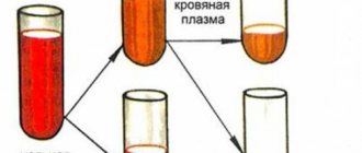
Plasma is obtained from the blood using centrifugation. The method allows you to separate plasma from cellular elements using a special apparatus without damaging them . The blood cells are returned to the donor.
The plasma donation procedure has a number of advantages over simple blood donation:
- The volume of blood loss is less, which means less harm is caused to health.
- Blood can be donated for plasma again after 2 weeks.
There are restrictions on plasma donation. Thus, a donor can donate plasma no more than 12 times per year.
Plasma donation takes no more than 40 minutes.
Plasma is the source of such important material as blood serum. Serum is the same plasma, but without fibrinogen, but with the same set of antibodies. They are the ones who fight pathogens of various diseases. Immunoglobulins contribute to the rapid development of passive immunity.
To obtain blood serum, sterile blood is placed in an incubator for 1 hour. Next, the resulting blood clot is peeled off the walls of the test tube and placed in the refrigerator for 24 hours. The resulting liquid is added to a sterile vessel using a Pasteur pipette.
Red blood cells
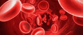
They are also called red blood cells. Red blood cells are the most common type of blood cell
and are what gives blood its red color.
One cubic milliliter of blood contains about five million red blood cells. These cells do not have a nucleus, but they contain millions of hemoglobin molecules. This iron-containing protein binds oxygen molecules obtained in the lungs and transports it throughout the body. After oxygen is delivered to the body's cells, red blood cells take carbon dioxide from them and transport it to the lungs to be removed from the body using the lungs. Red blood cells are produced in the red bone marrow of the bones of the skull, ribs and spine. More than two and a half million red blood cells are produced in the body every day, and approximately the same number of these cells die. The lifespan of one red blood cell is about three months. If there are not enough red blood cells produced, which carry oxygen and remove carbon dioxide from tissues, anemia
.
It is characterized by a feeling of weakness, fatigue and lack of attention. In more severe cases, shortness of breath, rapid heartbeat and sleep disturbances may occur. Attention!
If you experience these symptoms for a long time, be sure to consult a doctor.
Blood pathologies affecting the nature of plasma
In medicine, there are several diseases that can affect the composition of plasma. All of them pose a threat to human health and life.
The main ones are:
- Hemophilia. This is a hereditary pathology when there is a lack of protein, which is responsible for coagulation.
- Blood poisoning or sepsis. A phenomenon that occurs due to infection entering directly into the bloodstream.
- DIC syndrome. A pathological condition caused by shock, sepsis, severe injuries. It is characterized by blood clotting disorders, which simultaneously lead to bleeding and the formation of blood clots in small vessels.
- Deep venous thrombosis. With the disease, the formation of blood clots in the deep veins (mainly in the lower extremities) is observed.
- Hypercoagulation. Patients are diagnosed with excessive blood clotting. The viscosity of the latter increases.
Plasma test or Wasserman reaction is a study that detects the presence of antibodies in plasma to Treponema pallidum. Based on this reaction, syphilis is calculated, as well as the effectiveness of its treatment.
Plasma, a liquid with a complex composition, plays an important role in human life. It is responsible for immunity, blood clotting, homeostasis.
Leukocytes
White blood cells - leukocytes - play an important role in the body's immune system, protecting it from infection. They detect, destroy and remove pathogenic microorganisms, harmful and toxic substances from the body. In addition, white blood cells are able to fight cancer cells. There are several types of white blood cells, each with their own unique functions. Monocytes
are produced in the bone marrow and make up three to eight percent of the total volume of all white blood cells.
Monocytes can transform into other blood cells - macrophages and dendritic cells. Monocytes, which have turned into macrophages
, destroy “invaders” found in the body, including bacteria and parasites.
In addition, macrophages ingest cellular debris and dead cells infected with viruses. Having absorbed them, macrophages are able to “raise the alarm” by transmitting information about infection to other cells of the immune system. Dendritic cells
help the adaptive immune system, also called “acquired immunity.”
They do not destroy foreign elements on their own, but help identify them as hostile and “teach” other blood cells to do so. This important information is saved - in the future, when encountering a familiar “invader,” the cells react faster and more efficiently. Neutrophils
are produced in the bone marrow and make up 50 to 70 percent of all blood cells.
These cells are the first to respond to infection - within an hour after infection. Neutrophils fight not only infections in internal organs, but also on the surface of the skin. For example, pus, which is one of the visible signs of infection, consists mainly of dead neutrophils and bacteria. Eosinophils
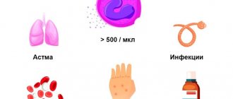
are also produced in the bone marrow and make up one to three percent of all white blood cells. Eosinophils circulate not only in blood vessels - they can be found in various organs. For example, the gastrointestinal tract contains the largest number of eosinophils. These cells kill bacteria and parasites. Unfortunately, they can mistake even beneficial substances for an “invader,” causing an allergic reaction and inflammation.
This is important to know
Viruses such as hepatitis C and HIV are transmitted through blood.
Find out more about how to avoid infection. B cells
are also produced in the bone marrow but leave it immature.
They develop into fully functional B cells only after reaching the organs of the lymphatic system - the spleen and lymph nodes. After this, B cells circulate in the blood, performing “immune surveillance” functions. One type of B cell is able to attach to the invader and act as a marker so that other cells of the immune system recognize them correctly. The second type of B cells is responsible for producing antibodies. B cells live long and have a kind of “memory”. They recognize infections that the body has encountered before and help the immune system recognize and destroy them much faster. T cells
are also capable of “learning.”
They can recognize bacteria, viruses and even cancer cells if they have previously appeared in the body. T cells come in two main types: helper T cells and killer T cells. Helper T cells
respond to information about infection received from macrophages and dendritic cells.
They begin to actively divide and produce substances to activate B cells and T killer cells. Killer T cells
attack body cells that are infected with bacteria or viruses. Killers “scan” each cell and, having detected the presence of infection, kill it.
