1.What is blood osmolarity?
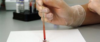
Blood osmolarity testing shows the amount of chemicals dissolved in the blood serum, the liquid part of the blood. Chemicals that affect blood osmotic concentration include sodium, chloride, bicarbonate, protein, and glucose. Blood osmolarity analysis is performed on venous blood material.
The osmotic concentration of the blood is partially controlled by a special antidiuretic hormone. Water constantly leaves our bodies - through sweat, urine and even breathing. If you do not drink enough water, then the osmolarity of the blood plasma, i.e. the concentration of chemicals is increasing. When the osmotic concentration of the blood increases, antidiuretic hormone is released into the blood. Antidiuretic hormone prevents fluid loss and increases its content in the blood. This helps blood plasma osmolarity return to normal.
On the contrary, if you drink too much water, your blood plasma osmolarity drops. When blood osmolarity drops, antidiuretic hormone is not produced. This increases the volume of urination, which prevents overhydration and fluid accumulation in the body.
Osmotic dilution and concentration of urine
Only the kidneys of warm-blooded animals have the ability to form urine with a higher osmotic concentration than blood. Many researchers tried to unravel the physiological mechanism of this process, but only in the early 50s of the 20th century was the hypothesis substantiated according to which the formation of osmotically concentrated urine is associated with the mechanism of the countercurrent-rotary multiplying system of some parts of the nephron.
The principle of countercurrent exchange is quite widespread in nature and is used in technology. Let us consider the mechanism of operation of such a system using the example of blood vessels in the limbs of Arctic animals. To avoid large heat losses, blood in parallel arteries and veins of the extremities flows in such a way that warm arterial blood warms cooled venous blood moving towards the heart (Fig. 204).
Arterial blood flows into the foot at a low temperature, which sharply reduces heat transfer. Here such a system functions only as a countercurrent exchanger: in the kidney it has a multiplying effect. To better understand its operation, consider a system consisting of three parallel tubes. Tubes I and II are arcuately connected at one end (Fig. 204, B).
The wall common to both tubes has the ability to transport salts, but it does not allow water to pass through. When a liquid with a concentration of 300 mOsmol/L is poured into such a system through inlet 1 and it does not flow, then after some time, as a result of the transport of salts, the liquid in tube I will become hypotonic, and in tube II it will become hypertonic. In the case when the liquid flows continuously through the tubes, the concentration of salts begins.
At each horizontal level, the difference in their concentrations due to a single effect of salt transport cannot exceed 200 mOsmol/l, however, along the length of the tube, single effects multiply and the system begins to work as a countercurrent multiplying system. Since, as the liquid moves, not only salt, but also a certain amount of water is extracted from it, the concentration of the solution increases increasingly as it approaches the bend of the loop.
In tube III, the permeability of the walls to water is adjusted; when the wall begins to let water through, the volume of liquid in it decreases. In this case, the water moves towards a higher osmotic concentration. As a result, the concentration of liquid in tube III increases and the volume of liquid contained in it decreases. The concentration of substances in it will depend on a number of conditions, including the operation of the countercurrent multiplying system of tubes I and II. As will be clear from the following discussion, the work of the renal tubules in the process of osmotic concentration of urine is similar to the described model.
Depending on the state of the body's water balance, the kidneys secrete diluted or concentrated urine. All sections of the tubules, medulla vessels, and interstitial tissue take part in the process of osmotic concentration of urine in the kidney. Of the 100 ml of filtrate formed in the glomeruli, ²/3 of it is reabsorbed by the end of the proximal segment. The fluid remaining in the tubules contains osmotically active substances in the same concentration as the ultrafiltrate of blood plasma, although it differs from it in composition due to the reabsorption of a number of substances in the previous parts of the nephron.
Next, the tubular fluid passes from the renal cortex into the medulla - into the descending (thin) section of the nephron loop (loop of Henle) and moves to the top of the renal papilla, where the tubule bends 180°, and the urine passes into the ascending section of the loop, located parallel to its descending department.
The functional significance of the various parts of the loop is ambiguous. When fluid from the proximal tubule enters the thin descending nephron loop, it enters the area of the kidney, in the interstitial tissue of which the concentration of osmotically active substances is higher than in the renal cortex. This increase in osmolar concentration in the outer zone of the medulla is due to the activity of the thick ascending limb of the nephron loop. Its wall is impermeable to water, and the cells transport C1⁻ and Na⁺ ions into the interstitial tissue. The wall of the descending part of the loop is permeable to water, and therefore water is absorbed from the lumen of the tubule into the surrounding interstitial tissue of the kidney along an osmotic gradient, and osmotically active substances remain in the lumen of this part of the tubule.
The farther from the cortex along the longitudinal axis the fluid is located in the descending limb of the loop, the higher its osmolar concentration. In each adjacent sections of the descending limb of the loop there is only a slight increase in osmotic pressure, but along the length of the loop the osmolar concentration gradually increases from 300 mOsmol/l to almost 1450 mosmol/l. In other words, at the top of the nephron loop, the osmolar concentration of fluid increases several times and at the same time its volume decreases. With further movement of fluid along the ascending section of the nephron loop, reabsorption of C1 ⁻ and Na ⁺ ions occurs, water remains in the lumen of the tubule, therefore, the initial parts of the distal convoluted tubule always receive hypotonic fluid, the concentration of osmotically active substances in which is less than 200 mOsmol/l.
Water is reabsorbed from the hypotonic fluid along the osmotic gradient, the osmolar concentration of the fluid in this section increases, i.e., the fluid in the lumen of the tubule becomes isosmotic. The final concentration of urine occurs in the collecting ducts; they are located parallel to the tubules of the nephron loop, in the renal medulla.
As noted above, the osmolar concentration increases in the interstitial fluid of the renal medulla. As a result, water is reabsorbed from the fluid of the collecting ducts and the concentration of urine in them increases, balancing with the ever-increasing osmolar concentration of the inner medulla of the kidney. Ultimately, hyperosmotic urine is released, in which the maximum concentration of osmotically active substances can be equal to the osmolar concentration of the interstitial fluid at the apex of the renal papilla (Fig. 205).
Under conditions of water deficiency in the body, the secretion of antidiuretic hormone from the pituitary gland (ADH) increases, which increases the permeability of the walls of the final parts of the distal segment and collecting ducts for water.
Unlike the outer renal medulla, where the increase in osmolarity is based primarily on chloride transport, the increase in osmolarity in the inner renal medulla depends on several mechanisms. The accumulation of urea plays a special role in osmotic concentration. The walls of the proximal tubule are permeable to urea. In this section of the nephron, up to 50% of filtered urea is reabsorbed.
However, when extracting liquid from the convoluted distal tubule, it turned out that the urea content was even slightly higher than the amount that came with the filtrate, amounting to about 110%. It has been shown that there is an intrarenal urea circulation system that is involved in the osmotic concentration of urine. In the lumen of the collecting ducts, due to the reabsorption of water, the concentration of urea increases; ADH increases the permeability of the collecting ducts in the medulla not only for water, but also for urea.
When the permeability of the tubular wall to urea increases, it diffuses into the renal medulla. The constant supply of urea, C1⁻ and Na⁺ ions, reabsorbed by the cells of the thin ascending limb of the nephron loop and collecting ducts, into the inner medulla ensures an increase in osmotic concentration in the renal medulla. Following an increase in the osmolarity of the interstitial tissue surrounding the collecting ducts, the reabsorption of water from them also increases and the efficiency of the osmoregulatory function of the kidney increases. Changes in the permeability of the tubular wall to urea provide insight into why urea clearance decreases as urine output decreases.
The direct blood vessels of the renal medulla, like the tubules of the nephron loop, also form a countercurrent system that plays a very important role in osmotic concentration. Due to the peculiarities of the location of the vasa recta, effective blood supply to the renal medulla is ensured, but osmotically active substances are not washed out, since the same changes in osmotic concentration are observed in the blood of the vasa recta as in the thin descending section of the nephron loop.
As the blood moves through it, the osmotic concentration gradually increases, and during its return movement to the renal cortex, salts and other solutes that diffuse through the vascular wall pass into the interstitial tissue. Thus, the concentration gradient of osmotically active substances is maintained, i.e. The vasa recta function as a countercurrent system. The speed of blood movement through the straight vessels affects the amount of Na⁺, Cl⁻ and urea ions removed from the medulla, which participate in the creation of the osmotic gradient, and the outflow of reabsorbed water.
With a water load, the relative proximal reabsorption of ions and water does not change, and the same amount of fluid enters the distal nephron as without a load. At the same time, the wall of the distal sections of the renal tubules remains waterproof, and the cells continue to reabsorb sodium salts from the flowing urine; in this case, hypotonic urine is released, the concentration of osmotically active substances in which is below 50 mOsmol/l. The permeability of the tubules to urea is low, and it is excreted in the urine without accumulating in the renal medulla.
The collecting ducts also allow the reabsorption of sodium, chloride, and other ions. Their main functional feature is that the reabsorption of substances occurs in small quantities, but against the most significant gradient, which causes significant differences in the concentration of a number of inorganic substances in the urine compared to blood.
Thus, the activity of the nephron loop, the terminal parts of the distal collecting ducts determines the ability of the human kidneys to excrete large volumes (up to 900 ml/h) of diluted, hypotonic urine during water stress, and to excrete only 10-12 ml/h of urine when there is a lack of water in the body. , 4'/g times osmotically more concentrated than blood. The ability of the kidney to osmotically concentrate urine is exceptionally developed in some desert rodents, which allows them to go without drinking water for a long time.
4.What are the risks and what could interfere with the analysis?
What are the risks of an osmolarity blood test?
Possible risks can only be associated with the blood collection itself. In particular, the appearance of bruises at the puncture site and inflammation of a vein or artery (phlebitis). Warm compresses several times a day will relieve phlebitis. If you are taking blood thinning medications, you may bleed at the puncture site.
What can change blood osmolarity?
Plasma osmolarity may change due to:
- Drinking alcohol before the test;
- Recent blood transfusion.