Thrombocrit - what is it?
Thrombocrit is a blood test indicator that indicates what proportion of whole blood consists of platelets.
The value of this general analysis parameter is determined only by a hematology analyzer and is calculated using the formula: platelet count * average platelet volume (MPV) * 10-4.
The value of the PCT indicator is directly affected by the average platelet volume MPV, as well as factors such as smoking, lack of vitamins B12, B9, B6, and depressive disorders.
Knowledge of the study results allows us to assess the effectiveness of the blood coagulation system. The test indicator reflects the human body’s tendency to bleed or form blood clots.
A thrombocrit test is prescribed as part of a comprehensive examination:
- heart and blood vessels;
- kidney;
- reproductive organs;
- endocrine system;
- respiratory organs;
- immune system.
Based on the test results, the risk of possible complications during organ transplantation is assessed and the course of diseases during treatment with immunosuppressive drugs is predicted.
Knowing the PCT value allows one to assess the risk of thrombosis when prescribing medications that affect the parameters of the coagulation system. Such an assessment is necessary for drawing up a treatment regimen and predicting the outcome of the disease in bedridden patients, with artificial ventilation, and before surgery.
Significance in diagnosis
First of all, by determining thrombocrit indicators, the following aspects are established:
- Determination of the risk of bleeding, increased thrombosis (thrombocytosis condition).
- Assessment of the benefit/risk ratio when prescribing certain medications.
The most well-known reasons for determining this indicator in the blood are:
- The patient has a high temperature of unknown origin.
- Evaluation of treatment with immunosuppressive drugs.
- Severe viral, bacterial, fungal infection of the body.
- The patient is on artificial ventilation.
- Complications after organ transplantation.
Important. It is necessary to take into account that any change in the number of platelet germ cells - an increase (thrombocytosis), a decrease (thrombocytopenia), a change in shape, aggregation - immediately affects the thrombocrit numbers.
- Thrombocrit is increased in the blood of an adult and a child. What does this mean, treatment
To assess platelet distribution in peripheral blood, it is necessary to know its normal values.
When thrombocrit is low
A decrease in PCT occurs when the production of platelets or platelet precursors, which are megakaryocytes, is blocked. With a decreased PCT, there is a tendency to bleed.
This indicator is reduced during pregnancy. The drop in thrombocrit in women can be 2 times lower than normal, but during pregnancy this reduces the risk of thrombosis, and means that blood circulation and fetal nutrition will not be affected.
PCT decreases in conditions caused by:
- anemia - folate deficiency, aplastic, megaloblastic;
- autoimmune diseases – systemic lupus erythematosus, collagenosis;
- chronic diseases of the liver, kidneys;
- poisoning by poisons, drugs - diuretics, cytostatics, antibiotics, corticosteroids;
- chemotherapy;
- oncological diseases - hemoblastosis, leukemia.
When the thrombocrit is lowered and is less than 0.11%, then in adults this indicates a disorder of hematopoiesis in the bone marrow or an acceleration of the breakdown of platelets in the spleen.
A low PCT thrombocrit in a child’s blood test may be explained by:
- in infants - prematurity, low birth weight, hypoxia;
- in a child of an older age group – parasitic infection.
Normal indicators
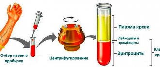
The PCT indicator, which is nothing more than thrombocrit, cannot be determined without special modern equipment, since platelets, being outside the bloodstream, are able to “overgrow” with pseudo-similarities.
At the same time, they still manage to increase in size at least ten times. It is for this reason that a laboratory technician cannot do without an automatic analyzer when determining the thrombocrit index. Determination of thrombocrit level in blood
The norm for this indicator ranges from 0.15 to 0.4. In some medical publications you can find information in which the thrombocrit norm differs slightly from that indicated, but only slightly. The norm for this indicator in an adult, an infant or in children of different ages is proportionally different from each other. At the same time, a proportional change in the indicator is not observed due to physiological fluctuations.
In particular:
- during the day or at different times of the year, the thrombocrit indicator may deviate by 10% (in particular, when depression occurs at night or in early spring);
- in women, thrombocrit is reduced by 50% during menstruation, as well as during pregnancy (this is a kind of protective reaction of the body that prevents the formation of blood clots);
- Thrombocrit indicators will be doubled if the patient was subjected to significant physical activity before the analysis.
Lack of platelets
But what to do if, on the contrary, the thrombocrit is low? There are a lot of cases when there is not enough thrombocrit in the blood and its amount drops below normal. A case where the thrombocrit is below normal is otherwise called thrombocytopenia. The most common cause of thrombocytopenia is taking medications that are inappropriate for the body. That is, in this case, thrombocytopenia manifests itself as a side effect of the drug. A decrease in platelet count appears from cirrhosis, hypothyroidism, leukemia, hepatitis, bone marrow dysfunction, alcohol abuse and anemia.
In addition, thrombocytopenia may occur if the patient has recently had a tooth pulled out. The appearance of thrombocytopenia leads to a loss of elasticity of blood vessels, as a result of which they become more fragile.
To determine the causes of a decrease in platelets, the doctor prescribes the following tests:
- Ultrasound of the liver and spleen. This is necessary in order to identify tumors, if any. In addition, ultrasound helps to study in detail the density and size of internal organs.
- Genetic research. It is carried out to determine hereditary thrombocytopenia, again if there is one.
- Magnetic resonance imaging.
- Blood test for various antibodies.
The reasons described above are the most common, and only after all these studies can the doctor prescribe pharmacological drugs to the patient that will help get rid of thrombocytopenia.
- What does a deviation from the norm indicate?
RDW and PDW in general blood test22779
table 2
| Index | x 109/l | % |
| Band neutrophils | 0,04-0,3 | 1-6 |
| Segmented neutrophils | 2-5,5 | 45-72 |
| Basophils | up to 0.065 | up to 1 |
| Eosinophils | 0,02-0,3 | 0,5-5 |
| Lymphocytes | 1,2-3 | 19-37 |
| Monocytes | 0,09-0,6 | 3-11 |
The results of a general blood test (Table 1) show many indicators. Let's consider the main ones:
- RBC – total number of red blood cells (erythrocytes). The pathological increase in these cells is associated with impaired hematopoiesis. A decrease in red blood cells is usually a consequence of anemia, hemolysis and blood loss.
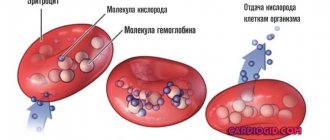
- HGB stands for hemoglobin, which is a protein containing iron. It transports oxygen to tissues, and carbon dioxide from them, and also maintains acid-base balance. A decrease in hemoglobin most often occurs due to anemia.
- HCT – hematocrit. It is defined as the ratio between the red blood cells that have settled to the bottom after taking the test and the total blood volume. An increase in this indicator indicates polyuria, erythrocytosis or erythremia. A decrease in hematocrit level occurs with anemia and an increase in circulating blood volume.
- PLT – platelets. These cells are responsible for blood clotting. If their number decreases, then the cause may be viral diseases, bone marrow lesions, bacterial infections and other pathologies. An increase in the number of platelets is caused by a wide variety of ailments: from joint diseases to cancer.
- CPU – color indicator. It determines the saturation of red blood cells with hemoglobin. If it is insufficient, this may indicate iron deficiency anemia, anemia or lead poisoning. When CP rises above normal, the cause is oncology, gastric polyposis and deficiency of vitamins B9 and B12.
- Erythrocyte indices: MCV - average volume of erythrocytes, used to determine water-salt balance and type of anemia;
- RDW is the degree of red blood cell diversity, which determines how different cells differ from each other in volume;
- MCH – average hemoglobin content in erythrocyte; this criterion is considered analogous to the color indicator;
- MCHC – average concentration and content of hemoglobin in red blood cells; this indicator is calculated taking into account the level of hematocrit and hemoglobin.
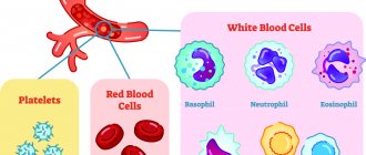
Now let's move on to the leukocyte formula (Table 2). It determines the percentage of different types of white blood cells in the blood, that is, the relative content of each type of white cell. What is this formula for? It is very important because with any changes in the body, the percentage of certain types of white cells in the blood decreases or increases. This is associated with a decrease or increase in other types. Based on the information obtained from the leukocyte formula, one can judge the course of a particular pathology, the occurrence of complications, and also more accurately predict the outcome of the disease.
Elevated platelet levels: what to do?
What to do if there is an excess of thrombocrit, and it is above normal, and what could this mean? It must be remembered that if the thrombocrit is elevated, then this is dangerous in any case, and you should not delay treatment. Absolutely always, the presence of an excess of blood platelets indicates one of the diseases that are described below:
- Arthritis.
- Enteritis.
- Leukemia.
- Erythrocytosis.
- Lymphogranulomatosis.
- Chronic myeloid leukemia.
- Anemia.
- Hemolysis.
- Kidney/liver cancer.
In addition, increased thrombocrit can appear from an acute infection in the body, large blood loss, severe stress and poisoning. A case where the thrombocrit is higher than normal is also called thrombocytosis.
There is also so-called “secondary” thrombocytosis. It appears with too much weight, alcohol abuse and various injuries. In this case, the culprit is thrombopoietin, which is responsible for cell maintenance, division and maturation, which is found in the bone marrow. In the presence of secondary thrombocytosis, the amount of thrombopoietin in the blood increases.
In any case, doctors recommend paying special attention to these cells, and if they are damaged, consult a doctor. If the patient is confirmed to have a high platelet level, he is prescribed an ultrasound of the abdominal cavity of the pelvic organs, blood and urine tests, and a test to detect C-reactive protein. And only after all these studies, the attending physician will be able to find out the real causes of thrombocytosis.
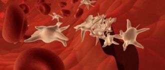
Platelets
Platelets are the third and last formed elements of blood studied during a general blood test, but in terms of their importance for human health and vital activity they are far from in last place. Platelets, or platelets, are tiny (2-4 microns), flattened, anucleate cells with an uneven surface. They are produced by the bone marrow and perform important functions: they form the primary plug at sites of damaged blood vessels, provide their surface for the plasma coagulation reaction, and then release growth factors that promote wound healing and tissue regeneration.
Total platelet count (PLT)
Platelet rate:
- Women and men – 180-320 109/l
- Children – 160-400 109/l
A pronounced decrease in the content of platelets in the blood when deciphering the results of a general analysis indicates a high risk of prolonged bleeding and extensive blood loss if a person receives a serious injury. And a pathological increase in their number can lead to the formation of blood clots (thrombi) blocking blood vessels, which is also very dangerous.
Platelet deficiency is collectively called thrombocytopathy. It is of three types: a decrease in the number of cells (thrombocytopenia), an abnormal increase (thrombocytosis) and a violation of their functional activity (thrombasthenia).
Platelets are elevated – reasons:
- Blood loss due to injury, childbirth or surgery;
- Iron-deficiency anemia;
- Acute inflammatory process or exacerbation of a chronic disease, for example, rheumatism;
- Splenectomy;
- Oncological diseases;
- Erythremia;
- Exhaustion or extreme fatigue.
Platelets are low – reasons:
- Hemophilia (congenital bleeding disorder);
- Aplastic anemia;
- Systemic lupus erythematosus;
- Autoimmune thrombocytopenic purpura;
- Some viral, bacterial and parasitic infections, such as malaria or toxoplasmosis;
- Heart failure;
- Paroxysmal nocturnal hemoglobinuria;
- Evans syndrome and DIC syndrome;
- Renal vein thrombosis;
- The period after blood transfusion;
- Prematurity in infants;
- Taking blood thinning medications such as aspirin.
Platelet indices (MPV, PDW, PCT)
The automatic analyzer calculates three platelet indices based on information about the total content of blood platelets, their sizes and volumes. These indicators in the decoding of a general blood test are designated by abbreviations consisting of several Latin letters.
MPV (mean platelet volume)
This index characterizes the average volume of one platelet and is expressed in femtoliters. It is known that very young platelets are large in size and do not work efficiently enough, while old ones shrink and gradually lose their functionality. This means that if a person has an increased MPV, his blood clotting is impaired, and if it is low, the bone marrow produces too few new platelets.
MPV rate:
- Women and men – 7.0-10.0 fl
- Children – 7.4-10.4 fl
PDW (platelet distribution width)
This index reflects the degree to which platelets differ from each other in volume, or their anisocytosis, and is measured as a percentage. We have already considered a similar indicator when we talked about red blood cells. In the case of platelets, it is also very important to take into account the previous index, MPV, when assessing the PDW value, because this is the only way to objectively judge the condition and functionality of the blood platelets.
PDW standard:
- Women and men – 15-17%
- Children – 10-17%
PCT (platelet crit)
This index is otherwise called thrombocrit, is an analogue of hematocrit, is also expressed as a percentage and describes the ratio of platelet volume to total blood volume. If the reading is significantly lower than normal, this may indicate temporary clotting problems or even hemophilia. If the thrombocrit is higher than normal, a person is at risk of forming clots and clogging blood vessels.
PCT Standard:
- Women and men – 0.1-0.4%
- Children – 0.15-0.4%
Head of Clinical Diagnostic Laboratory
Institution "6th Central Regional Communal Center of the Leninsky District of Minsk" - Veysaga Chungara Umberto