At the age of 30-40, small red moles begin to appear on the body. However, they are not moles in the true sense as they are made up of many small blood vessels. These formations are called angiomas.
Rice. 1. Angioma on the body
The exact cause of the appearance of angiomas on the body is unknown, however, bad heredity plays a large role. That. if your fathers and mothers had similar formations on their bodies, then the likelihood of them appearing on you is very high.
Rice. 2. Multiple angiomas - vascular tumors
Angiomas on the body usually do not develop into malignant tumors. However, if they appear, it is worth consulting with a dermatologist and dermatosurgeon to rule out the presence of melanoma or another type of skin cancer. This is because melanoma and Merkel carcinoma (neuroendocrine skin cancer) can appear as small red papules that can be easily confused with angiomas.
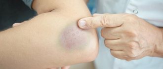
Rice. 3. Red mole - angioma
During the consultation, a specialist doctor will help you determine the diagnosis and advise whether it is worth removing your existing skin lesions.
What if you have an angioma?
If you have an angioma, it should still be removed. Removal can be done with a coagulator or radio knife, but in most cases they leave a noticeable scar on the skin. This is explained by the fact that angioma consists of vessels, and the coagulator and radio knife cannot separate the vessels from other tissues and cauterize everything. It is better to remove angiomas on the body using a laser, since a laser beam of a certain wavelength is capable of destroying only tumor vessels without affecting neighboring tissues.
Symptoms of angioma
Symptoms of angioma depend on the type of vascular tumor, its size and location. In newborns, it is diagnosed immediately or several months after birth, and can grow very quickly, covering large areas of the skin.
Vascular angiomas can affect:
- Integumentary tissues - skin, mucous membranes of the genitals, subcutaneous tissue. Angiomas are also found in the mouth.
- Musculoskeletal system - muscles, bones. A severe form of the disease includes hemangioma of the vertebral body.
- Internal organs.
If integumentary hemangiomas cause cosmetic defects, then hemangiomas of internal organs provoke the occurrence of disturbances of important functions - urination, breathing, vision, defecation.
Angiomas of muscles and bones are accompanied by:
- skeletal deformation;
- severe pain in the joints;
- frequent fractures;
- radicular syndrome (compression of the spinal nerves).
Venous angiomas of the brain (cerebellum, left/right frontal lobe, etc.) are very dangerous. They lead to subarachnoid hemorrhage and epilepsy. Also, due to compression of the brain vessels by the tumor, the following symptoms may occur:
- convulsions;
- headache;
- nausea, vomiting;
- dizziness;
- noise in the head;
- taste disturbances;
- speech problems.
During the growth process, angiomas can become inflamed, causing phlebitis and thrombosis. A common complication is bleeding due to trauma.
If you notice similar symptoms, consult a doctor immediately. It is easier to prevent a disease than to deal with the consequences.
Advantages of laser removal of angiomas on the body:
- Selectivity of the laser beam - only the vessels that make up the tumor are destroyed;
- Bloodlessness - the procedure for laser vaporization of a hemangioma takes place without a drop of blood, even if the formation is large;
- High cosmetic result - after laser removal of angioma there are no traces left on the skin;
- Single use - removal of angioma is carried out at one time;
- No anesthesia required.
Rice. 4. Angioma under microscopy consists of vessels
Rice. 5. Removal of angioma on the body with a laser
Treatment
The main goal of treatment is to stop the growth of the tumor, eliminate it and resume normal function of the arteries and veins.
In some cases with small congenital angiomas that do not pose a danger to the patient, observation of such angioma may be recommended and intervention is not required.
Various methods are used for therapy.
Among them are laser treatment, radiation therapy when localized in the orbital region, retrobulbar space. For bleeding or punctate lesions, as well as for angiofibromas, diathermocoagulation is indicated. The sclerosing method is carried out in the case of a deep-lying structure of small size. In this case, alcohol is introduced into the cavity of the object. Hormone therapy is used for the progressive development of the pathological process, simultaneous damage to several anatomical areas. Cryotherapy is also used, especially this method is used in pediatric surgery. The surgical method is also used when the object is located deep.
With complete and timely treatment, the prognosis is favorable. For large neoplasms in difficult-to-reach locations (large vessels, internal organs), the prognosis is not so optimistic.
Establishing a high-quality and accurate diagnosis with the choice of optimal treatment is the task of a specialist, therefore, if these manifestations are detected, consultations with a therapist, surgeon, or angiologist are recommended.
Removal of angioma on the body at the Laser Surgery Center “ATLANTiK”
Our center performs removal of angiomas on the body using two types of lasers:
- Nd:YAG
- Diode-laser.
Removal of angiomas is carried out on the day of treatment, the removal session lasts no more than 5-10 minutes, anesthesia is not required for small formations up to 5 mm.
During laser removal, angioma on the skin disappears before our eyes as a result of sealing of blood vessels; large angiomas larger than 5 mm first darken and then peel off from the surface of the skin within 7-14 days, depending on the treatment area.
There are no traces left on the skin after laser removal of angiomas.
Cavernous angioma of the brain
Cavernous angiomas (cavernomas) are most often congenital vascular anomalies, represented by single, much less often multiple cavities, separated inside by septa (septa) and filled with blood. The blood supply to the cavity is carried out from small arterioles and capillaries, and the outflow of blood through venules is of the same order. Due to the small caliber of the feeding vessels, the blood pressure in cavernomas is low, so the draining veins do not hypertrophy and are not visible during SCT/MRI/Angiography. A special feature of cavernomas is a very thin, defective vascular wall, so thin that the cellular elements of the blood - red blood cells - “sweat” through it and settle in the adjacent medulla. This process is called “diapedetic hemorrhage.” The products of the natural breakdown of hemoglobin (hemosiderin), contained in red blood cells, form a zone of chronic, specific and very recognizable changes on MRI around the cavernoma.
Cavernomas can be localized in any part of the brain, mainly in the cerebral hemispheres, as well as in the brain stem, cerebellum, subcortical ganglia, corpus callosum and lateral ventricles. In approximately 30-40% of cases, cavernous angiomas are combined with another type of vascular malformation - venous angiomas. Venous angiomas are an anomaly in the development of venous vessels in the form of a “bundle” of small veins (“Gorgon’s head”) gathering into one large drainage vein.
The incidence of cavernous angiomas is several cases per million population.
What is the danger of having a cavernous angioma?
Cavernomas can remain asymptomatic throughout life. However, in a number of patients, clinical manifestations can be of two types, both individually and in combination:
- Hemorrhage. Sometimes, an increase in blood pressure inside a cavernoma leads to local destruction of the vascular wall and the formation of intracerebral hemorrhage. Unlike AVMs, hemorrhages from cavernomas are never massive and do not pose a threat to the patient’s life, with the exception of extremely rare cases of cavernomas located in the lower parts of the medulla oblongata, where the centers of cardiovascular and respiratory regulation are located. However, when the focus of hemorrhage is located in any functional area, even a small volume of hemorrhage can lead to the appearance of neurological symptoms (for example, the development of contralateral hemiparesis when the focus is located in the precentral gyrus of the frontal lobe).
- Epileptic syndrome . In some cases, due to the chronic presence of hemosiderin in the medulla or with the development of acute hemorrhage, a focus of pathological brain bioactivity may form, clinically manifested by epileptic seizures of various structures (convulsive, non-convulsive, absence seizures, vegetative, polymorphic and others).
Thus, cavernomas extremely rarely pose a threat to the patient’s life, but can have a significant impact on the quality of life
How to diagnose a cavernoma?
Magnetic resonance imaging (MRI) is the most informative method for diagnosing cavernomas. In T1 and T2 scanning modes, a cavity surrounded by a black rim of hemosiderin may be visible, with signs of acute and subacute hemorrhages. The most sensitive mode is T2* (“T2 with star”), which allows you to diagnose even small (1-2 mm) cavernomas that are not visible in other scanning modes
Computed tomography - its diagnostic value is limited, as a rule, to the acute period of hemorrhage (several days), when the image shows a focus of bleeding
Angiography is not informative in diagnosing cavernomas
How to get rid of a cavernoma?
In case of episyndrome formation and/or hemorrhage, two treatment methods are possible:
Surgical removal of cavernoma is a highly effective method that allows you to once and for all relieve the patient from the risk of repeated hemorrhages. Surgery is also effective if the patient has epileptic seizures, but its effectiveness is reduced in the case of a long history of epileptic syndrome
Radiosurgery is carried out according to the same principle as for arteriovenous malformations - the higher the dose, the better the effect. Therefore, cavernomas up to 1 cm in diameter are best treated, when the maximum permissible doses of radiation can be safely used. Morphological changes in this case are similar to those in arteriovenous malformations - the walls of the cavernoma and the mouths of the feeding vessels undergo hyalinosis, as a result of which the blood flow decreases, diapedetic hemorrhages stop, and the risk of repeated spontaneous intracerebral hemorrhages is reduced by 50-70%. Episodic attacks also become less frequent or disappear completely, but as in the situation with surgery, the longer the history of the disease, the less likely it is to completely recover from them
The generally accepted tactic for asymptomatic, incidentally detected cavernomas is observation.
Is it necessary to irradiate multiple cavernomas?
As in the case of single cavernomas, radiosurgical treatment is carried out only on the lesion that manifests itself clinically - hemorrhage or epileptic seizures
What to do with venous angioma?
The question often arises: how to get rid of venous angioma? Firstly, you should know that venous angiomas never manifest themselves as hemorrhages or epileptic seizures. Secondly, although they represent a venous network that is abnormal in structure, they nevertheless take full part in the local venous circulation of the brain. The only option for the appearance of clinical symptoms associated with venous angioma is the spontaneous development of thrombosis of the draining vein. This situation is more hypothetical than real. In this case, the patient will experience cerebral venous infarction with severe swelling at the site of impaired venous outflow. And only one way can help cope with such a situation - thrombolytic and anticoagulant therapy. Attempts to surgically or radiosurgically influence venous angiomas not only do not make any sense, but in a similar manner to thrombosis will lead to gross local disturbances of venous blood flow. Thus, the mere presence of venous angioma is not a reason for any treatment.
Which cavernoma treatment method is best?
From the point of view of preventing recurrent hemorrhages, the effectiveness of surgical treatment is higher. However, when cavernomas are localized in functionally important or hard-to-reach areas of the brain (for example, in the subcortical nuclei, brain stem), surgery itself has a high risk of residual neurological deficit, and radiosurgery can largely avoid these complications. Therefore, the decision on the optimal method of influence is made individually, taking into account all possible factors, incl. and taking into account the informed choice of the patient himself
Fig. 1 Result of radiosurgical treatment of brainstem cavernoma. On the left is a cavernous angioma of the lateral surface of the brain stem at the time of Gamma Knife radiosurgery. On the right - a significant reduction in the size of the cavernoma 3.5 years after radiosurgery. No recurrent hemorrhages
Diagnostics of education
In exceptionally extreme cases, only by chance, liver hemangioma in a child or adult can be detected at an early stage. In the vast majority of cases, it is found when the size reaches serious volumes.
The following diagnostic methods are used:
- Ultrasound of the gallbladder and liver (may show a round tumor with clear boundaries);
- MRI of the biliary tract and liver (in addition to the outlines of the formation, it will also show its contents);
- MSCT of the abdominal cavity (layer-by-layer will demonstrate the boundaries and contents of the tumor).
When a tumor is detected, it is necessary to determine its qualitative characteristics. To confirm hemangioma, angiography of the celiac trunk is performed; static liver scintigraphy may also be needed. To determine the degree of benignity (malignancy) of atypical cells, hepatoscintigraphy is prescribed.
Along with this, clinical tests are carried out:
- liver tests;
- complete blood count, blood biochemistry;
- blood test for genetic markers.