What it is?
The body is a mechanism in which complex processes occur every day. The patient does not notice this and continues with his daily routine. These processes are responsible for the proper functioning of organs and internal systems and maintaining health. One of the important processes for the body is pressure.
When the internal balance is disturbed, a person begins to experience unpleasant feelings. Therefore, to maintain health, it is never suitable to pay due attention to all types of internal pressure. First of all, this relates to intracortical pressure. The increase in pressure inside the abdominal cavity is called intrazonal hypertension (IAH). The disease develops as a result of abnormal functioning of the lungs, hearts, kidneys and intestines.
If a person is healthy, indebile pressure varies from 0 to 5 mm Hg. - This is the norm. In an adult patient in critical condition, this figure can rise to 7 mmHg. The increase is observed in several other concerns about the body's concerns - obesity and childbirth. In this case, the pressure indicator does not go beyond 10-15 mmHg.
The patient’s body has time to adapt to new conditions, and this does not affect his health. As a result of surgery, that is, the abdominal cavity, the device will display 13 mmHg. Such values do not threaten the patient's life in such conditions. It is impossible to talk about a forcible increase in pressure without any important reasons.
NSICU.RU neurosurgical intensive care unit website of the intensive care unit of the N.N. Research Institute Burdenko
Introduction
The normal level of intra-abdominal pressure (IAP) for patients in critical condition is 5-7 mmHg.
Sustained or repeated increase in IAP above 12 mmHg. is intra-abdominal hypertension (IAH) [1]. There are 4 degrees of IAH depending on the size of IAP: I degree – 12-15 mmHg; II degree – 16-20 mmHg; III degree – 21-25 mmHg; IV degree > 25 mmHg. [1]. IAH III, IV degrees in combination with signs of newly developed organ dysfunction is abdominal compartment syndrome (ACS) [1]. With ACS, multiple organ failure develops—respiratory, cardiovascular, renal, and liver failure [2, 3]. The causes of organ dysfunction are either an increase in pressure in the abdominal cavity itself, leading to an increase in pleural pressure, a decrease in cardiac output and a decrease in respiratory compliance, or the translocation of bacteria during the formation of ischemia of the intestinal wall [4, 5]. If ACS develops in patients with pathology of the abdominal cavity, retroperitoneal space or pelvis, then this is primary ACS; if it develops in patients with other pathologies, then this is secondary ACS. Abdominal perfusion pressure (APP) is the difference between mean arterial pressure and IAP.
IAH and, in particular, ACS significantly worsen the course of critical illness and disease outcomes, and can also be a direct cause of death [2, 6]. This is the basis for considering VAH and, in particular, ACS to be urgent conditions that require immediate correction. The treatment algorithm for IAH recommended by the International Society for the Study of ACS is presented in Figure 1 [1].
The relationship between intra-abdominal pressure and the central nervous system (CNS) is poorly understood and is of considerable interest. This literature review is devoted to this problem.
Intra-abdominal pressure and the central nervous system
Intra-abdominal and intracranial hypertension
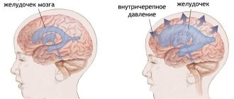
Works devoted to the problem of IAP appeared at the beginning of the 20th century [7], however, the relationship between IAP and the central nervous system began to be studied only at the end of the last century. The first were researchers from Boston University - Josephs LG et al., who in 1994 studied in laboratory conditions the effect of pneumoperetoneum on the dynamics of ICP [8]. Carbon dioxide was injected into the abdominal cavity until the IAP reached 15 mmHg. and parallel ICP measurement. The study included two groups of animals. In the first group, the brain was intact, in the second, brain damage was simulated by inflating an epidurally inserted balloon. ICP in the first group increased from 13.46 ± 1.01 mmHg to 18.72 ± 1.5 mmHg, and in the second - from 22 ± 1.75 mmHg to 27. 4 ± 0.93 mmHg The increase in ICP in both groups was statistically significant. The authors hypothesized that IAH first increases pleural pressure (PP) and then central venous pressure (CVP). This leads to difficulty in venous outflow from the cranial cavity. The deterioration of blood outflow, according to the authors, was the direct cause of the increase in ICP. This assumption was based on the Monroe-Kelly doctrine, which states that ICP is caused by three components - brain parenchyma, cerebrospinal fluid and blood vessels with blood in their cavity [9, 10]. For ICP to remain within normal limits when the volume of any of these components increases, the volume of the other two components must decrease because the cranium is rigid. This mechanism determines both the brain’s compliance and its compensatory abilities aimed at maintaining a normal ICP level [11]. Based on this concept, in animals of the first group with an intact brain, the deterioration of venous outflow was compensated by reducing the volume of other components of the cranial cavity. As a result, ICP increased, but did not reach the level of ICH. In the animals of the second group, even before the formation of VAH, ICH already existed, so they did not have the opportunity to compensate for the increased ICP due to the deterioration of venous outflow. This was the reason for the significant increase in ICH.
In 1996, Bloomfield GL et al. conducted a study that clarified the mechanisms of increased ICP during IAH and examined the relationships between IAP, PP, CVP, cardiac output (CO), and ICP [12]. In laboratory animals, IAP was increased to 25 mmHg. using a balloon inserted into the abdominal cavity. At the same time, dynamic measurements of PP, CVP, CO and ICP were carried out with cerebral perfusion pressure (CPP). The authors showed that when creating an IAH, there was an increase in ICP from 7.6 ± 1.2 mmHg. up to 21.4 ± 1.0 mmHg PP also increased along with CVP, while CO and CPP significantly decreased. When the animals underwent decompressive laparotomy, the IAP actually returned to the initial level – 11.2 ± 1.8 mmHg. At the same time, normalization of PP, CVP, CI and CPP occurred. This work, in fact, confirmed the hypothesis of Josephs LG et al that the cause of ICH in IAH is a violation of venous outflow with a decrease in thoracoabdominal compliance. Another important result of this work was the demonstration that simply increasing IAP to 25 mmHg. can lead to the development of ICH even in animals with an intact brain.
The first clinical works devoted to the study of the relationship between IAP and ICP, as well as laboratory studies, appeared at the end of the last century, when two clinical observations were published in 1995. In the first of them, Bloomfield GL et al described the effective reduction of high ICP, resistant to intensive therapy, immediately after decompressive laparotomy in a patient with severe concomitant trauma [13]. In the second clinical observation, Irgau I et al described an acutely developed pronounced increase in ICP in a patient with severe concomitant trauma in response to the formation of pneumoperetoneum to perform cholecystectomy [14].
In 2001, Citerio G et al conducted a study on 15 patients with severe TBI [15]. A 15-liter bag of water was placed on the patients' stomachs. The criteria for inclusion of patients in the study were the subacute period of TBI and stabilization of their condition, which meant at least a 24-hour period with normal values of ICP and CPP. During the study, multiparametric cerebral monitoring, monitoring of central hemodynamics and, of course, measurement of IAP were performed. All parameters were recorded before, during and 20 minutes after the artificial increase in IAP. The authors found that an increase in IAP from 4.7 ± 9.9 mmHg. up to 15.4 ± 4.1 mmHg. (p<0.001) led to an immediate increase in CVP from 6.2 ± 2.4 mmHg. up to 10.4 ± 2.9 (p<0.001) mmHg. and ICP - from 12.0 ± 4.2 mmHg. up to 15.5 ± 4.4 (p<0.001) mmHg. It took only a few seconds for the pressures to increase, which remained at a plateau until the weight was removed from the abdomen. Then there was an immediate return of all pressures to the original level.
In addition to the monitoring options described above, the authors measured respiratory system compliance and thoracic transmural pressure. A decrease in compliance of the respiratory system was detected from 58.9 ± 9.8 ml/cm H2O to 44.9 ± 9.4 ml/cm H2O. At the same time, thoracic transmural pressure, which is the difference between central venous pressure and intraesophageal pressure, remained stable, and chest compliance significantly decreased - from 204.7 ± 37.1 ml/cm H2O to 123.6 ± 38.0 ml/cm H2O. Interpreting these results, the authors concluded that an increase in IAP leads to an upward displacement of the diaphragm and, accordingly, a decrease in chest compliance. In other words, increased abdominal pressure leads to increased thoracic pressure, increased central venous pressure, decreased blood flow from the cranial cavity, and ultimately increased ICP.
The mechanism of increased ICP due to obstruction of venous outflow from the cranial cavity was described by Huseby JS et al back in 1981 [16]. The authors in their study showed that increasing pressure in the internal jugular vein increases pressure in the superior sagittal sinus and cortical bridging veins. As a result, there is an increase in pressure in all parts of the venous system of the brain and an increase in intracranial blood volume. As a result, ICP increases.
Work carried out in the late 90s - early 2000s made it possible to recommend for routine clinical practice, firstly, to carry out IAP measurements for the timely identification of potentially curable causes of increased ICP in patients with TBI, and, secondly, to take special care when the need to use laparoscopic techniques in patients with combined traumatic brain and abdominal trauma [11]. These recommendations are relevant because the combination of traumatic brain and abdominal trauma is common. According to various authors, the frequency of such a combination reaches 30–40% [17, 18].
In 2004, Joseph DK et al published a large series of observations consisting of 17 patients with severe TBI, resistant to therapy, ICH (30.0 ± 8.1 mmHg) and IAH (27.5 ± 5. 2 mmHg) [19]. The authors showed that decompressive laparotomy is an effective method not only for normalizing IAP, but also ICP. Laparotomy reduced IAP to 21.4 ± 1.0 mmHg. 6 patients with a transient decrease in ICP had a fatal outcome, while 11 patients with sustained normalization of ICP survived.
In 2004–2005, Belgian scientists Malbrain M et al and Deeren DH et al conducted two studies that examined the relationship between IAP and ICP in patients with non-traumatic brain damage [20, 21]. As with patients with TBI, patients with ischemic, hemorrhagic strokes and metabolic encephalopathy were characterized by a direct correlation between the level of IAP and the ICP value. In addition, the authors of these studies actively discuss intracranial relationships, in particular, the pressure-volume relationship. This relationship is nonlinear. In other words, if a patient has increased ICP, even a slight increase in volume in the cranial cavity leads to the development of severe ICH, often resistant to intensive therapy. The role of such a factor, which slightly increases the intracranial volume in a patient in critical condition, may well be played by an increase in intracranial blood volume due to deterioration of venous outflow from the cranial cavity due to IAH. This clinical scenario is more than likely given that IAH develops in 54–65% of critically ill patients [22].
Confirmation that the interest of clinicians in the problem of IAH in neurosurgical patients remains extremely high is a clinical observation published in 2011 in the journal Neurocritical Care [23]. The authors describe a clinical situation that has already become classic, when decompressive laparotomy in a patient with severe concomitant trauma allowed to normalize therapy-resistant ICH.
Central nervous system and intra-abdominal pressure.
The hypothetical probability of a secondary increase in IAP in patients with neurological or neurosurgical pathology and primary damage to various parts of the brain is obvious. However, we were unable to find studies that would examine the relationship between the central nervous system and IAP in this way. Over the past year, we have published 2 papers devoted to this problem. The first work is a clinical observation that describes the development of secondary ACS in a patient with intra-extraventricular craniopharyngioma and a complicated course of the postoperative period [24]. This patient experienced increased neurological symptoms after surgery and then developed ACS. IAP was normalized using prolonged epidural anesthesia (EA) at the lower thoracic level. Gradually, regression of neurological symptoms was achieved. We came to the conclusion that with a complicated course of the postoperative period in patients with tumors of the chiasmatic-sellar region (CHS), the development of IAH is possible, which, in turn, aggravates the course of the postoperative period and the patient’s condition and can worsen the outcome of the disease.
Analysis of this clinical observation allowed us to create a prospective study design [25]. The subjects of the study were patients with chronic tumors and a complicated course of the postoperative period. The purpose of the study was to determine the effectiveness of conservative therapy and EA for the correction of IAH in patients with CSO tumors and a complicated course of the postoperative period. At the same time, the causes and mechanism of development of IAH in this category of patients were studied. As a result, it was revealed that IAH developed in 68.3% of patients, while ACS developed in 22% of patients. This corresponds to the frequency of development of IAH declared by other authors in patients in critical condition [17]. This correspondence is unlikely to be accidental. There is still no generally accepted opinion about the reasons for such frequent development of IAH in critically ill patients. At the same time, it is known that the frequency of cerebral dysfunction that develops as a result of critical illness in patients with a primarily intact nervous system reaches 60–80% [26, 27, 28]. It is possible that cerebral dysfunction developing as a result of a critical condition causes or aggravates the course of IAH.
The mechanism of development of IAP in all observations of our study was the formation of various variants of gastrointestinal motility disorders (GIT), which manifested simultaneously with an increase in IAP and persisted longer than IAP. The cause of the development of gastrointestinal motility disorders is most likely the loss of control of the diencephalic structures over the function of the gastrointestinal tract. Another possible cause is the formation of dishormonal disorders, primarily hypothyroidism, or, most likely, a combination of these causes.
Conservative therapy for IAH that did not reach the ACS level was effective in the vast majority of cases. However, with the development of ACS, which was recorded in 9 patients, conservative therapy, as a rule, was ineffective. It was effective in only two cases. If conservative therapy was ineffective, EA was performed. A contraindication for EA was sepsis, which developed in 2 patients with ACS. Thus, out of 9 patients with ACS, EA was performed in 4 cases, which effectively corrected IAH in all these patients. In addition, the outcomes of patients with ACS who underwent EA were significantly better than the outcomes of patients with ACS who did not undergo EA for various reasons. These results make it possible to include EA in the international algorithm for the correction of IAH, which is shown in Figure 1. However, the encouraging results obtained in the study in patients with CSO tumors are the rationale for further work, the object of which should be patients with other neurosurgical and neurological pathologies.
Conclusion
Intra-abdominal hypertension leads to the development of intracranial hypertension due to increased intrapleural pressure, central venous pressure and deterioration of venous outflow from the cranial cavity. The relationship between IBG and ICH is nonlinear and is described by pressure-volume relationships that are classic for the intracranial situation.
IAP should be measured in patients with ICH because IAP is one of the curative factors, the correction of which can dramatically normalize refractory ICH.
Damage to the chiasmatic-sellar region can lead to the development of IAH of varying severity and ACS, among others. Extended epidural anesthesia at the lower thoracic level may be a promising direction for correcting IAH in this category of patients. Conducting further research on the problem of IAH in patients with primary CNS damage is justified and advisable.
Literature
- Malbrain ML, Cheatham ML, Kirkpatrick A, et al. Results from the International Conference of Experts on Intra-abdominal Hypertension and Abdominal Compartment Syndrome. I. Definitions.// Intensive Care Med. 2006; No. 32: p1722–1732
- Malbrain ML, Cheatham ML, Kirkpatrick A, et al. Results from the International Conference of Experts on Intra-abdominal Hypertension and Abdominal Compartment Syndrome. II. Recommendations.// Intensive Care Med. 2007; No. 33: p 951–962.
- Cheatham ML Intra-abdominal hypertension and abdominal compartment syndrome.// New Horiz.; 1999: No. 7 p 96–115.
- Rosin D, Rosenthal RJ. Adverse hemodynamic effects of intraabdominal pressure-all in the head? // Int J Surg Investig. 2001; No. 2: p335-345.
- Malbrain ML, Chiumello D, Pelosi P, et al. Prevalence of intra-abdominal hypertension in critically ill patients: a multicentre epidemiological study. Intensive Care Med. 2004; No. 30: p 822–829.
- Hagiwara A, Fukushima H, Inoue T, et al. Brain death due to abdominal compartment syndrome cause massive venous bleeding in a patient with a stable pelvic fracture: report of a case.// Surg Today. 2004; No. 34: p82-85.
- Emerson H. Intra-abdominal pressures. Arch Intern Med. 1911;7:754-84.
- Josephs LG, Este-McDonald JR, Birkett DH, et al. Diagnostic laparoscopy increases intracranial pressure. J Trauma 1994; 36(6):815-818. 9. Monro A. Observations on the structure and function of the nervous system. Edinburgh: Creech & Johnson, 1783.
- Mokri B. The Monro-Kellie hypothesis: applications in CSF volume depletion. Neurology 2001;56(12):1746–1748.
- Citerio G, Berra L. Intra-abdominal hypertension and the central nervous system. From Abdominal compartment syndrome, edited by Ivatury RR, Cheatham ML, Malbrain M, Sugrue.// Landes Bioscience. 2006;Texas.
- Bloomfield GL, Ridings PC, Blocher CR, et al. Effects of increased intra-abdominal pressure upon intracranial and cerebral perfusion pressure before and after volume expansion. J Trauma 1996; 40(6):936-941.
- Bloomfield GL, Dalton JM, Sugerman HJ, et al. Treatment of increasing intracranial pressure secondary to the acute abdominal compartment syndrome in a patient with combined abdominal and head trauma. J Trauma 1995; 39(6):1168-1170.
- Irgau I, Koyfman Y, Tikellis JI. Elective intraoperative intracranial pressure monitoring during laparoscopic cholecystectomy. Arch Surg 1995; 130(9):1011-1013.
- Citerio G, Vascotto E, Villa F, et al. Induced abdominal compartment syndrome increases intracranial pressure in neurotrauma patients: A prospective study. Crit Care Med 2001; 29(7):1466-1471.
- Huseby JS, Luce JM, Cary JM, et al. Effects of positive end-expiratory pressure on intracranial pressure in dogs with intracranial hypertension. J Neurosurg 1981; 55)5):704-705.
- Gennarelli TA, Champion HR, copes WS, et al. Comparison of mortality, morbidity, and severity of 59,713 head injured patients with 114,447 patients with extracranial injuries. J Trauma 1994; 37(6):962-968.
- Citerio G, Stocchetti N, Cormio M, et al. Neuro-Link, a computer-assisted database for head injury in intensive care. Acta Neurochir (Wien) 2000; 142(7):769-776.
- Joseph DK, Dutton RP, Aarabi B, et al. Decompressive laparotomy to treat intractable intracranial hypertension after traumatic brain injury. J Trauma 2004; 57(4):687-695.
- Malbrain ML, Chiumbello D, Pelosi P, et al. Prevalence of intra-abdominal hypertension in critically ill patients: A multicenter epidemiological study. Intensive Care Med 2004; 30(5):822-829.
- Deeren DH, Dits H, Malbrain ML. Correlation between intra-abdominal and intracranial pressure in nontraumatic brain injury. Intensive Care Med. 2005:31(11), 1577-1581.
- Andrews PJ, Citerio G. Intracranial pressure. Part one: Historical overview and basic concepts. Intensive Care Med 2004; 30(9):1730–1733.
- Dorfman JD, Burns JD, Green DM, et al. Decompressive laparotomy for refractory intracranial hypertension after traumatic brain injury. Neurocrit Care, 2011, ahead of print.
- Popugaev KA, Savin IA, Goryachev AS et al. Intra-abdominal hypertension and secondary abdominal compartment syndrome in a patient with craniopharyngioma in the postoperative period. Problems of Neurosurgery, 2011, 1:66-71.
- Popugaev KA, Savin IA, Goryachev AS et al. Secondary abdominal compartment syndrome in the complicated course of the postoperative period in patients with tumors of the chiasmal-sellar localization. Anesthesiology and Reanimatology, 2011, No. 4, accepted for publication.
- Ely EW, Inouye SK, Bernard GR, et al. Delirium in mechanically ventilated patients: validity and reliability of the confusion assessment method for the intensive care unit (CAM-ICU). JAMA 2001, 286:2703–2710
- Ely EW, Margolin R, Francis J, et al. Evaluation of delirium in critically ill patients: validation of the Confusion Assessment Method for the Intensive Care Unit (CAM-ICU). Crit Care Med 2001, 29:1370–1379.
- Thomason JW, Shintani A, Peterson JF, et al. Intensive care unit delirium is an independent predictor of longer hospital stay: a prospective analysis of 261 non-ventilated patients. Crit Care 2005, 9:R375–R381.
Measurement methods
To measure intrabacal pressure, one of two existing methods is used:
- catheter;
- Surgical method.
In the first case, a catheter is used, which is introduced into the patient through the bladder. It is considered less informative than the surgical method. This applies when the second is not possible. In this case there should be no complications.
Measuring internal pressure using a surgical method is possible during surgery. The doctor places a special sensor in the abdominal gown, which after a while shows the pressure level. The measuring device can also be placed on the liquid colon. This has good information value. Decreased or increased values indicate the development of a pathological process in the human body.
If you begin to notice characteristic symptoms, you should ask your doctor.
Clinical picture
Indicators are observed frequently. At the same time, irrelevant changes are not disclosed in any way. This is not a sign of a serious illness.
Significant changes in the previous pressure value look like this:
- feeling of gravity in the stomach area;
- weakness and dizziness;
- High blood pressure;
- Nausea, often leading to vomiting;
- Rattling and shaking in the stomach;
- incomplete defecation files;
- dull or painful pain;
- bloating;
- Bloody abdominal cavity.
If there is a block of Peter's hypertension, it is difficult to determine. Nonspecific symptoms are not enough. To make an accurate diagnosis, the patient must be subjected to a complete examination. Based on the data obtained, the doctor makes a diagnosis and prescribes treatment.
The patient may have specific symptoms, in addition to general symptoms. The strength of their manifestation and nature depends on the reason for which the disease developed. Regardless, it is contraindicated to have the patient perform self-healing. This can not only lead to complications, but also cause death if emergency medical care is not provided.
Infections
Some infectious diseases occur with virtually no symptoms, and their manifestations can easily be attributed to poor health associated with blood pressure. For example, if you are infected with the herpes simplex virus, cytomegalovirus, or Epstein-Barr virus, you may experience weakness, chills (due to a slightly elevated temperature), and pain in the head and muscles.
If it really was the herpes simplex virus, then within a day or two you will notice a rash on your lips (or on your genitals, in the case of genital herpes). With other infections, there may be no external symptoms at all, so the cause of the lethargic state will remain a mystery for a long time.
So those who have been tormented by symptoms of “pressure” for a long time, but motherwort and coffee do not help, should get tested for hidden infections.
Treatment
It is difficult to make a valid diagnosis based on the symptoms listed. Other diseases may also have the same set of warning symptoms. To make an accurate diagnosis, you need to see a doctor who will conduct all the necessary diagnostic examinations and tests. First of all, two factors are considered that determine the cause and severity of the disease.
To confirm the diagnosis, the patient is given an umbrella, which is inserted rectally. During the procedure, the patient does not feel pain, only slight discomfort is possible. This method is used to measure performance. It cannot be used to reduce intra-abdominal pressure.
If the patient does not ignore the symptoms and begins treatment as soon as possible, this will stop the first stage of the disease. In this case, the patient will avoid complications in the form of multiple organ failure. The duration of therapy depends on the stage of the disease. In this case, treatment is carried out using several methods:
- Conservative treatment - medications and physiotherapeutic procedures.
- Radical - surgical intervention.
If the patient complains of being unwell and has serious complaints, an immediate diagnosis is carried out. In this case, IAP is measured. If it exceeds 25 mmHg. St., urgent surgery is required. Abdominal surgery is used.
Drug therapy and physiotherapeutic methods
It contains the following drugs:
- sedatives;
- painkillers;
- muscle relaxants.
Vitamins and mineral complexes are prescribed to maintain the function of the digestive tract.
Physiotherapeutic procedures are carried out after the main treatment. They help stabilize water and electrolyte balance in the body. This may be the installation of a drainage tube or an enema with medicinal decoctions. Diuresis stimulation is often used.
Factors that increase intra-abdominal pressure should be avoided. The patient should not wear tight-fitting clothing; it is better to choose a loose fit. To protect your health, avoid over-tightening your trouser belt. The lying position should be at least 20 degrees.
Avoid exercises that require you to lift heavy weights (more than 10 kg). The abdominal muscles should also be protected from excessive tension. The patient is advised to avoid exercise. A varied diet will help keep the body in a stable state.
Are women the stronger or weaker sex? And how does physical activity affect women's health?
Useful information from the gynecologist of the Dixion clinic - Voronkova T.V.
Have you noticed
signs of urinary incontinence
during physical exertion, coughing or sneezing? Is it often impossible to keep gases in the intestines or constipation? Do you experience discomfort during sexual intercourse, a feeling as if something is interfering with the vagina, or a more pronounced opening of the genital opening, or perhaps prolapse of the vaginal walls or genitals?
Patients often do not talk about these problems even to doctors.
.
Isn't this a disease? That's not fatal? Women are the weaker sex, and weak not because they are unable to lift weights, but because such loads contradict the female anatomy, causing weakness of the “pelvic floor”
(i.e., the system of muscles and ligaments that hold the internal genital organs in the correct position ).
This causes prolapse of the genital organs, and “sagging” of the bladder and (or) rectum along with the walls of the vagina. The pelvic floor resembles a hammock, and with stress and increased intra-abdominal pressure, this “hammock” can weaken, dragging with it the walls of the vagina, the cervix and the uterus itself.
There are many stresses and factors predisposing to weakening of the pelvic floor in a woman’s life!
This:
1. long and traumatic childbirth;
2. systemic pathology:
- persistent “stretch marks” on the skin after childbirth or significant weight changes,
- phlebeurysm,
- separation of the muscles of the anterior abdominal wall,
- inguinal and umbilical hernias,
- “hypermobility” of the joints - increased flexibility of the joints.
3. excess weight (especially due to the growth of adipose tissue around the internal organs (female “apple” figure);
4. physical activity
, and ATTENTION!
fitness loads
, especially with a weight
of more than 5-10 kg
and it doesn’t matter whether we lift a sack of potatoes, carry children in our arms or perform “deadlifts”
- systematic work with heavy weights slowly but surely squeezes out the pelvic floor muscles and stretches the ligaments on which the organs are attached.
5. regular constipation, the presence of diseases such as bronchial asthma or chronic bronchitis also increases intra-abdominal pressure
6. physical exercise and sports involving jumping
(athletics, basketball, volleyball, currently popular trampolining, etc.)
7. decrease in the level of female sex hormones (during menopause, pelvic floor prolapse is observed in more than 50% of women).
Of course, hyperextension (prolapse) of the pelvic floor is not an instant process
However, patients often turn to doctors when their symptoms are severe.
And if it is practically impossible to influence the pelvic floor ligaments, then it is possible to strengthen the muscles and ensure the prevention of pelvic prolapse and correction in the initial stages; for this there are sets of exercises for strengthening the muscles of the perineum
(“Kegel exercises”), some types of yoga, etc.
Corrective options for women with symptomatic prolapse include observation, pelvic muscle training, mechanical supports (pessaries), and surgery.
The use of pessaries should be considered preoperatively in women who have symptomatic prolapse.
The radical and most effective method for correcting pelvic organ prolapse in advanced stages is surgical treatment - a combination of pelvic floor reconstruction with the use of a special mesh implant.
You can assess the condition of the pelvic floor, conduct the necessary diagnostic studies to clarify the severity of the process and choose the optimal method of correction by making a gynecological appointment at the Dixion Clinic
.
Nutrition for the patient
The diet is selected individually for each case. In this case, general recommendations are taken into account:
- During the day you should drink at least 2 liters of liquid in any form, including first courses.
- Meals should be shared. Take in small portions every 2-3 hours.
- The consistency of cooked food should be either puree or liquid.
- Completely eliminate foods that cause increased gas formation from your diet.
High abdominal pressure is a result of obesity. Uncontrolled consumption of harmful foods poisons the body. When IAP is detected in patients with this problem, a diet is prescribed. A set of physical exercises has been developed to normalize weight.
In case of increased or decreased IAP, the first step is to promptly seek medical help.
In this case, you can not only cure the disease at an early stage, but also avoid complications. Preventative measures aimed at strengthening the entire body play an important role. If you have chronic diseases, attend physiotherapeutic procedures from time to time.
Hypothyroidism
The manifestations of hypothyroidism - decreased thyroid function - are in many ways similar to the symptoms of hyper- and hypotension. And basically we are talking about lethargic health and constant weakness. Also, people with hypothyroidism, like those who suffer from low blood pressure, have pale skin and cold hands and feet.
But those whose thyroid gland is not working well have additional symptoms. For example, they gain excess weight and cannot lose it. Their skin dries out and they lose their hair. Many people experience constipation, women's menstrual cycles are disrupted, and periods become more painful. Also, patients with hypothyroidism may experience severe irritability and develop depression. If lethargy and weakness have been present for a long time, perhaps it is not the pressure, but the level of hormones. It is worth taking tests to assess the function of the thyroid gland and, if necessary, starting hormonal therapy.