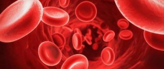
General information
Normal lactate levels are low. It increases with a lack of oxygen and disruption of the energy production mechanism. The main amount of energy is produced in mitochondria. This process uses oxygen and glucose. This generation of energy is called aerobic. If this process is disrupted (for example, there is not enough oxygen), energy production occurs anaerobically due to the breakdown of glucose. This produces lactate. If the production of lactic acid exceeds the liver's ability to utilize it, this substance accumulates. In this case, the development of lactic acidosis is possible. In many cases, the body’s compensatory capabilities are sufficient to avoid severe consequences. Sometimes lactic acidosis is not compensated for. In this case, the acid-base balance is disturbed, nausea, weakness, sweating, rapid breathing appear, and coma is possible.
The causes of excess lactate are divided into two groups. The most common is lactic acidosis A. It occurs when blood circulation is slow or oxygen is not being taken up sufficiently by the lungs. This condition can develop with a heart attack, shock from severe blood loss, sepsis, pulmonary edema and in a number of other cases. With type B lactic acidosis, metabolism is impaired and the need for oxygen is increased. It may be caused by kidney or liver disease, leukemia and other diseases.
BLOOD LACTATE LEVEL AS AN INDICATOR OF STAT ANALYSIS
Home \ Publications \ BLOOD LACTATE LEVEL AS AN INDICATOR OF STAT ANALYSIS
BLOOD LACTATE LEVEL AS AN INDICATOR OF STAT ANALYSIS V.A. Torshin, Ph.D., Moscow
The biochemical and clinical interpretation of blood lactate levels appears quite simple at first glance: only one reaction occurs when pyruvate is converted to lactate and vice versa [1]. However, the discovery of understanding the production of lactate from glucose by glycolysis, as well as the conversion of lactate to glucose by gluconeogenesis, is one of the greatest achievements of biochemistry in the 20th century. The energy required for normal functioning and maintenance of cell homeostasis is in the form of adenosine triphosphate (ATP), formed during the oxidation of glucose. The complete oxidation of 1 mole of glucose produces 36 moles of ATP. The aerobic pathway for glucose conversion occurs in two stages. The 1st stage is a chain of sequential reactions, as a result of which in the cytosol of the cell glucose is converted into pyruvate with the formation of 2 moles of ATP. The 2nd stage occurs inside the mitochondrion and includes the Krebs cycle (tricarboxylic acid cycle), while pyruvate is completely metabolized to CO2 and H2O. At the same time, oxidative phosphorylation occurs, resulting in the formation of another 34 moles of ATP. Oxygen is required for the intramitochondrial part of the process to function. Therefore, under anaerobic conditions, pyruvate accumulates in cells. Excess pyruvate is converted to lactate, which diffuses from the cells into the bloodstream. In addition, glycolysis is accompanied by the production of hydrogen ions, which leads to acidosis, first intracellular, then tissue and finally systemic.
The lactate concentration in the extracellular fluid is about 1 mmol/L, reflecting the dynamic process of formation and consumption of 1200 to 1500 mmol lactate during the day [1]. Lactate, as a product of anaerobic glycolysis, is formed in tissues such as skeletal muscles, intestines, brain, skin and red blood cells. Intense exercise can lead to an increase in lactate production in muscles 10 times compared to the resting state. Resynthesis of glucose from lactate occurs in the liver through gluconeogenesis. The processes of gluconeogenesis and oxidative phosphorylation, respectively, consume from 1200 to 1500 mmol of hydrogen per day. That is, the liver plays a more significant role in maintaining the acid-base balance compared to the kidneys, which normally excrete only 60-75 mmol of hydrogen per day.
The ability to determine the level of lactate in the blood of mammals was first demonstrated by Gaglio in 1886. The determination took several days and required about 200 ml of animal blood. In 1940, Barker and Summerson significantly simplified the technique, which allowed Broder and Weil to conclude in 1964 that there was a correlation between blood lactate levels and the severity of shock. Further development led to the creation of even more simplified methods, which were nevertheless recognized as reference.
The importance of blood lactate level in the assessment of patients in critical condition required the use of the method in the STAT analysis mode from a small sample of whole blood in combination with indicators of CBS, electrolyte balance and indicators of oxygen status. These problems became solvable with the development of the amperometric, enzymatic method using a substrate-specific electrode. In work [3], using large clinical material (1218 intensive care patients), a high degree of correlation was proven between reference methods (determining lactate in plasma and whole blood) and a new method that allows monitoring blood lactate levels in patients in critical condition.
The works of [1,2,3,4] and other authors have proven the role of assessing blood lactate levels in patients in critical condition as:
- tissue oxygen debt indicator
- indicator of the effectiveness of therapy
- a prognostic sign of an unfavorable outcome.
The role of lactate as an indicator of tissue oxygen debt has been proven in [3]:
- intense exercise
- circulatory shock (hemorrhagic, cardiogenic, septic)
- cardiac arrest
- severe hypoxemia
- severe anemia
- grand mal seizures
- status asthmaticus
- carbon monoxide poisoning
- sepsis
- vitamin B1 deficiency
- certain types of tumors
- a number of liver diseases
- congenital metabolic disorders
- poisoning with a number of substances (ethanol, methanol, metformin, ethylene glycol, etc.).
The most interesting is the accumulating data on the importance of lactate as a prognostic sign of an unfavorable outcome. A number of authors have proven [1,2,3], firstly, an earlier increase in lactate levels compared to other indicators of developing shock (hypotension, oliguria, decreased pH, etc.). Secondly, there was a clear correlation between blood lactate levels in critically ill patients and mortality rates. The work [4] noted that when lactate increased to 2.7 mmol/l, mortality did not increase, when the level reached 4.0 mmol/l it reached 50%, and when it increased to 8 mmol/l it was 90%. The prognostic role of lactate in newborns is likely to be associated with other reference values. In the group of surviving newborns born with asphyxia, lactate levels of 17-18 mmol/l were recorded [1].
Considering the growing interest among clinicians in such an indicator as the level of blood lactate, we can expect many center studies that can put an end to such problems as determining reference limits, metabolic features of newborns, etc.
Bibliography:
- S.B. Chelnokov, N.A. Pudina
Blood lactate level in newborns born with asphyxia. Materials of the 1st Russian Congress on Pediatric Anesthesiology and Reanimatology. Moscow. 2001; 321-322.
- James A. Kruse
Understanding Blood Lactate Analysis. The Journal for Respiratory Care Practitioners. 1995; 63-69.
- John Toffaletti
Elevations in blood lactate: Overview of use in critical care. Scand J Clin Lab Invest. 1996; 56, Supp224; 107-110.
- Javier Aduen et al.
The Use and Clinical Importance of a Substrate-Specific Electrode for Rapid Determination of Blood Lactate Concentrations. JAMA, December, 7. 1994- Vol 272.No 21.
- John Toffaletti
Blood Lactate: Biochemistry, Laboratory Methods and Clinical Interpretations. Critical Reviews in Clinical Laboratory Sciences. Vol 28, Issue 4; 1991; 253-268.
News all
07/06/2021 X Baltic Forum "Current problems of anesthesiology and resuscitation" June 30 - July 3, 2021
06/03/2021 The 7th Conference “Laboratory Diagnostics of Emergency Conditions 2021” took place
05/11/2021 Conference “Laboratory diagnostics of emergency conditions 2021” June 3, Novotel, St. Petersburg
01/22/2021 Seminar for managers of veterinary clinics
04/09/2020 Manufacturers and suppliers called for simplification of registration of medical devices
When is a lactate test necessary?
The test results enable the doctor to determine the reasons for the disruption in the supply of oxygen to the organs and systems of the body. A blood lactate test is used to identify lactic acidosis and hypoxia and assess their severity. The lactate level makes it possible to differentiate the types of metabolic acidosis. For newborns, analysis is prescribed in case of asphyxia. In sports medicine, the study is used to monitor the condition of athletes during intense training.
The test is prescribed in the following cases:
- suspected lack of oxygen (symptoms such as rapid breathing, sweating, pale skin, shortness of breath, sudden weakness);
- impaired clarity of consciousness, fainting, fever, severe headaches, symptoms of meningitis;
- suspicion of diabetes, sepsis, heart or kidney pathologies, shock.
What is lactic acid
How not to quit fitness
The average visitor to a sports club has enough enthusiasm for about six months. Find out why your annual membership hasn't paid for itself in this article about the most common fitness mistakes.
So, get acquainted. Lactic, or α-hydroxypropionic, or 2-hydroxypropanoic acid. It is used both in the chemical and food industries - as a preservative. Food additive E270 is lactic acid, and E271-279 are its salts, lactates. However, lactic acid is not only a raw material for the food and chemical industries. It is also formed in the human body.
Interpretation of results: norm, reasons for increased performance
Reference values: 0.5-2.2 mmol/liter. The decrease in indicators is not critical. High levels of this substance can indicate various diseases, including life-threatening conditions. For example, an increase in indicators is observed in acute heart failure, leukemia, circulatory disorders, and diabetes. Only a doctor can make an accurate diagnosis and prescribe treatment. Also, an increase in indicators can be caused by emotional or physical stress, alcohol intoxication. Even in the absence of symptoms, if abnormalities are detected, the patient's condition should be monitored by a doctor.
Where does the pain come from?
Then why do muscles hurt the next morning after training? The fact is that under intense loads, the so-called myofibrils are destroyed - thin threads running along the muscle fibers. This is how pieces of dead tissue form in the muscles. The body's immune system finally destroys them and removes them from the body. However, while the destruction process is taking place, free radicals are released in the tissues, and the cells begin to lack water. As a result, pain receptors located on cell membranes are excited, and the person feels pain in the muscles.
How to avoid pain
Determining the load
City dwellers move much less than they think.
Calculate how active you are and find out how much you need to move. First
, dose the loads, increasing them gradually and systematically.
If the load is selected correctly, there will be less pain or no pain at all. Be sure to familiarize yourself with the basic principles of effective training to avoid not only discomfort the next day after exercise, but also sports injuries. Secondly
, keep your workouts regular - this will help your muscles get used to constant stress.
Thirdly
, if it was not possible to avoid overtraining, take time for full recovery. Healthy sleep, as well as foods containing antioxidants, help speed up the process of breaking down substances that cause muscle pain.
Diagnostics
Despite the fact that gouty arthritis has pronounced symptoms, only 10% of patients can be correctly diagnosed during the first attack. In other cases, a diagnosis of other types of arthritis is made. Diagnostic criteria for gout are:
- acute arthritis of the 1st toe;
- the presence of large and small tophi;
- increased levels of uric acid in the blood;
- detection of EOR crystals in joint fluid and tissues.
If at least two criteria are identified, the diagnosis of gouty arthritis is considered reliable.
Laboratory research:
- general blood test
- signs of inflammation; - biochemical blood test
- uric acid content more than 0.32 mmol/l; increased levels of C-reactive protein (a sign of an inflammatory reaction); - general urine analysis
; - examination of synovial fluid using polarization microscopy
- identification of MUN crystals and a large number of leukocytes.
Instrumental studies:
- Ultrasound of joints
- detection of EOR crystals on the surface of cartilage and tophi; - radiography of the joints
- in the early stages there are no changes; later, bone changes are revealed; - computed tomography (CT)
- reveals the presence of changes in the spine.
Total
So, metabolic acidosis - acidification of the muscle cell environment during intense work - is associated with the use of ATP energy, and not with the synthesis and accumulation of lactate. Its production is necessary for the cell to replenish the costs of the coenzyme NAD +, necessary for glycolysis and the production of new “energy” ATP molecules.
This production (as well as the outward transport of lactate) requires the consumption of protons, reducing their concentration in the cell. Therefore, the formation and accumulation of lactate can be a good indicator of acidification of the cellular environment, but they are not related as cause and effect.
Source:
https://ajpregu.physiology.org/
Indications
- Diagnosis of circulatory pathologies resulting in tissue hypoxia (insufficient oxygen consumption by tissues);
- Assessing the degree of acidosis and prescribing resuscitation measures;
- Detection of diseases of the cardiovascular system;
- Suspicion of non-insulin-dependent diabetes mellitus;
- Determining the cause of lactic acidosis;
- Assessment of the acid-base status of the body and blood pH;
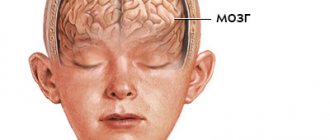
- Diagnosis of asphyxia (severe oxygen deprivation) and enzymopathy (impaired enzyme activity) in newborns;
- Pathological changes in muscles and tissues;
- Differential diagnosis of myopathies (hereditary muscle diseases).
The analysis is deciphered by specialists: endocrinologist, cardiologist, oncologist, traumatologist, surgeon, therapist, pediatrician, etc.
Recounting protons
The first and main source of protons in an actively working muscle cell is considered not to be the synthesis, but the breakdown of ATP, the energy of which is used for contractions and relaxations: ATP + water -> ADP + phosphate + proton. Phosphate itself is capable of serving as a buffer system that softens fluctuations in the acidity of the environment (this is how it works in the body), but it is actively involved in new reactions in the cell, and does not solve this problem very effectively.
Another source of protons is the above-mentioned coenzyme NAD+, which during glycolysis reactions loses a proton, turning into NADH. By the way, a significant part of the protons (as well as phosphate and pyruvate) found in the intracellular environment are transported to mitochondria and used for the processes of oxidative phosphorylation that take place there. Thus, mitochondria can also be called a factor in reducing acidity. But when muscles absorb energy with enormous intensity, converting ATP into ADP, this reaction is stronger than anything acting against it.
Rice. 3.
The balance between the production and use of protons in a working muscle cell. The appearance of protons is associated with ATP hydrolysis and glycolysis reactions. They are consumed in the reactions of creatine phosphate and lactate. In addition, protons bind to inorganic phosphate and buffer compounds in the cytoplasm.
Increased values (lactic acidosis)
- Pathologies of the cardiovascular system: heart failure, cardiogenic shock (acute left ventricular failure), Raynaud's syndrome (severe vascular disease, spasm of small blood vessels);
- Blood flow disorders and diseases of the circulatory system;
- Non-insulin-dependent diabetes mellitus;
- Increased physical activity (usually among professional athletes);
- Tetany (convulsions due to calcium metabolism disorders);
- Tetanus (a bacterial disease affecting the nervous system);
- Epilepsy (pathology of the nervous system, manifested by convulsive seizures with loss of consciousness);
- Hepatitis (viral inflammation of the liver) in acute form;
- Cirrhosis of the liver (an abnormal change in the structure of the organ tissue);
- Oncological processes: lymphoma (cancer of the lymphatic system), leukemia (blood cancer), etc.;
- Poliomyelitis (highly contagious disease of the nervous system, spinal paralysis);
- Tissue hypoxia (oxygen starvation);
- Hypotension (low blood pressure);
- Heavy bleeding;
- Pulmonary insufficiency, hyperventilation (impaired frequency or depth of breathing).
A temporary increase in lactic acid concentration is possible as a result of:
- deficiency of vitamin B1 in the body;
- long-term regular use of alcohol;
- poisoning with chemical elements: ethanol, salicylates, toxins, methanol, etc.;
- dehydration (dehydration of the body);
- pregnancy (in the 3rd trimester the level of lactic acid may increase slightly);
- taking medications: sodium drug, nitroprusside, adrenaline, metformin, fructose or glucose, propylene glycol, methylprednisolone, phenformin, etc.
Cause and mechanism of development (etiology and pathogenesis) of the disease
The cause of gouty arthritis is a disorder of purine metabolism. Purines are chemical compounds that form the basis of nucleic acids necessary for the formation of DNA and RNA molecules. As cells break down, purines are broken down into uric acid (UA). The latter enters the intercellular space and into the blood plasma, where it combines with sodium, forming a salt - monosodium urate (MUN).
An increased level of urate in the blood (hyperuricemia - GU) may be a consequence of a genetic predisposition (the kidneys do not eliminate MUN completely), high blood pressure (BP), consumption of large amounts of animal food, and alcohol.
With an excess of urates, when they can no longer dissolve in the surrounding fluid (EOR concentration more than 0.4 mmol/l), the salts crystallize, deposit in the articular and periarticular tissues and are surrounded by protein rings. This formation is called tophi. The release of MUN from the tophi causes an immunological reaction: a large number of neutrophils (one of the types of leukocytes responsible for cellular immunity) appear in the synovial membrane and joint fluid.
Neutrophils ingest MUN crystals, which causes the release of proinflammatory (inflammation-causing and maintaining inflammation) cytokines and the development of an acute inflammatory response in the synovium. Acute attacks in the form of synovitis are very painful, but do not leave any consequences. The long-term chronic course of the disease with frequent repeated attacks leads to the destruction of articular cartilage, proliferation of bone tissue, deformation and dysfunction of the joint. Deposition of MUN in the kidneys causes a decrease in their function.
Factors contributing to increased urate levels in the blood:
- the presence in the diet of a large number of meat dishes, offal, eggs, alcoholic beverages,
- overweight;
- taking certain medications - diuretics, aspirin, nicotinic acid, medications to lower blood pressure (Concor), etc.;
- lead poisoning;
- increased breakdown of purines in blood diseases, psoriasis, etc.;
- increased formation of purines;
- impaired renal function and uric acid excretion.