Tumor markers are specific substances found in the blood and urine, which can be both waste products of the tumor and substances synthesized by healthy cells in response to the invasion of cancer cells. Detection of tumor markers in the body makes it possible to diagnose the oncological process at an early stage and monitor the dynamics of pathology during treatment.
Who needs to be tested for tumor markers?
People whose relatives have been diagnosed with tumors of various locations should take tests for tumor biomarkers as screening tests 1–2 times a year.
People who have benign tumors (adenoma, myoma, fibroma) or tumor-like formations (kidney cysts, ovarian cysts) need to be examined with the same frequency. The rest can be tested for tumor markers once every 2-3 years.
People who have undergone cancer therapy should be tested for tumor biomarkers: every month during the first year, once every 2 months during the second year, once every 3 months during the third to fifth year. Five years after cancer treatment, it is sufficient to undergo tumor marker testing once every 6–12 months.
Tumor biomarkers can be detected in urine, blood, prostatic juice, fluid taken from the pleura or abdominal cavity. Specific antibodies are added to a sample of biological fluid taken from the patient. They form specific complexes with the protein structure of the tumor marker, which are identified by laboratory methods. Non-protein tumor biomarkers are determined by other methods.
Normal tumor markers in women
Despite the high efficiency of diagnosing the amount of protein compounds in the blood and/or urine, as well as the risk of developing cancer in any person, the analysis is not prescribed to everyone, and it is not preventive for people who are not at risk and do not have any indications for this.
Checking the normal level of tumor markers for women is indicated in the following cases:
- There are cases of cancer in the family history.
- Benign neoplasms were previously diagnosed.
- Congenital pathologies.
- Received injuries, suffered serious illnesses in the pelvic organs and mammary glands.
- Control of metastases and treatment.
- Previously underwent oncology treatment (control of relapse and absence of metastases).
If after treatment the level of markers does not return to normal, it means that the selected program is ineffective.
Most frequently identified tumor markers
75% of cancer patients have elevated alpha-fetoprotein (AFP) levels. Most often it is elevated in liver cancer; also, its high level can indicate testicular and ovarian cancer (in 5% of cases).
The tumor marker beta-2-microglobulin indicates the development of lymphoma and multiple myeloma. Markers CA 27-29, CA 15-3 indicate possible breast cancer, CA 125 - ovarian cancer. Biomarkers LASA-P, CA 72-4 make one suspect the presence of ovarian cancer and gastrointestinal tumors. The CA 19-9 marker is detected in cancer of the pancreas.
Sources
- Inaba S., Shimozono N., Yabuki H., Enomoto M., Morishita M., Hirotsu T., di Luccio E. Accuracy evaluation of the C. elegans cancer test (N-NOSE) using a new combined method. // Cancer Treat Res Commun - 2021 - Vol27 - NNULL - p.100370; PMID:33901923
- Huang Z., Chen L., Wang Y., Fu L., Lv R. Molecular markers, pathology, and ultrasound features of invasive breast cancer. // Clin Imaging - 2021 - Vol79 - NNULL - p.85-93; PMID:33895560
- Qiu X., Zhang H., Zhao Y., Zhao J., Wan Y., Li D., Yao Z., Lin D. Application of circulating genetically abnormal cells in the diagnosis of early-stage lung cancer. // J Cancer Res Clin Oncol - 2021 - Vol - NNULL - p.; PMID:33893839
- Zhang Y., Wu Q., Xu L., Wang H., Liu X., Li S., Hu T., Liu Y., Peng Q., Chen Z., Wu X., Fan JB. Sensitive detection of colorectal cancer in peripheral blood by a novel methylation assay. // Clin Epigenetics - 2021 - Vol13 - N1 - p.90; PMID:33892797
- Koshiaris C., Van den Bruel A., Nicholson BD., Lay-Flurrie S., Hobbs FR., Oke JL. Clinical prediction tools to identify patients at highest risk of myeloma in primary care: a retrospective open cohort study. // Br J Gen Pract - 2021 - Vol - NNULL - p.; PMID:33824161
- Chantzichristos D., Svensson PA., Garner T., Glad CA., Walker BR., Bergthorsdottir R., Ragnarsson O., Trimpou P., Stimson RH., Borresen SW., Feldt-Rasmussen U., Jansson PA., Skrtic S., Stevens A., Johannsson G. Identification of human glucocorticoid response markers using integrated multi-omic analysis from a randomized crossover trial. // Elife - 2021 - Vol10 - NNULL - p.; PMID:33821793
- Lewin J., Kottwitz D., Aoyama J., deVos T., Garces J., Hasinger O., Kasielke S., Knaust F., Rathi P., Rausch S., Weiss G., Zipprich A., Mena E. ., Fong T.L. Plasma cell free DNA methylation markers for hepatocellular carcinoma surveillance in patients with cirrhosis: a case control study. // BMC Gastroenterol - 2021 - Vol21 - N1 - p.136; PMID:33765926
- Chiancone F., Fabiano M., Carrino M., Fedelini M., Meccariello C., Fedelini P. Impact of systemic inflammatory markers on the response to Hyperthermic IntraVEsical Chemotherapy (HIVEC) in patients with non-muscle-invasive bladder cancer after bacillus Calmette-Guérin failure. // Arab J Urol - 2021 - Vol19 - N1 - p.86-91; PMID:33763253
- Ladak Z., Garcia E., Yoon J., Landry T., Armstrong EA., Yager JY., Persad S. Sulforaphane (SFA) protects neuronal cells from oxygen & glucose deprivation (OGD). // PLoS One - 2021 - Vol16 - N3 - p.e0248777; PMID:33735260
- Chen C., Yang H., Cai D., Xiang L., Fang W., Wang R. Preoperative peripheral blood neutrophil-to-lymphocyte ratios (NLR) and platelet-to-lymphocyte ratio (PLR) related nomograms predict the survival of patients with limited-stage small-cell lung cancer. // Transl Lung Cancer Res - 2021 - Vol10 - N2 - p.866-877; PMID:33718028
Tumor marker monitoring
Monitoring the dynamics of tumor markers plays an important role in cancer therapy. This allows you to determine the effectiveness of treatment and timely adjust its regimen. For example, during chemotherapy and radiation therapy, it is possible to increase the level of markers, which does not indicate an exacerbation of the process, but rather lysis of the tumor. Together with other indicators, biomarkers make it possible to monitor the patient’s health status during therapy and at its end.
You can get tested for tumor markers at the Medical Genetics Center.
When to donate blood for markers and how to prepare
Let us immediately note that what a blood test for tumor markers shows is not a diagnosis. This is only the first step, which can carry up to 95% of what is happening in the body. Therefore, checking the oncoprotein norm is always accompanied by other examination methods, in particular, magnetic resonance and computed tomography, ultrasound, and radiography.
The main indications for analysis are:
| Diagnosis of early detection of cancerous compaction. |
| Checking for the presence of metastases in the absence of any symptoms. |
| Drawing up the most suitable and effective treatment program for cancer, selecting medications and their dosages. |
| Monitoring and evaluating the effectiveness of the chosen treatment program and the effectiveness of the drugs used. |
Testing the normal level of tumor markers is prescribed for risk groups and people over 50 years of age, since the production of these substances increases with age.
Please note that a blood test for tumor markers in women and men is taken only for each type separately, as prescribed by the doctor. Depending on the suspected location of the tumor, testing only one marker may be sufficient to identify it. But more often than not, the complete picture is provided by the group that best fits together.
Preparation in most cases does not require special actions, and it is almost similar to the requirements for a blood biochemistry test:
| Refusal to eat 4-12 hours before the procedure. You should also exclude coffee, tea and juices. Only non-carbonated water is acceptable, including on the day of blood collection. |
| Stop using medications in consultation with your doctor. If this is not possible, you should definitely notify the laboratory assistant about this. |
| Avoid alcohol and smoking 12 hours before the start (this also applies to hookah, smoking various aromatic tobaccos and electronic cigarettes). |
| Try not to visit a massage therapist or undergo diagnostic manipulations several hours before testing. |
| Reduce physical activity. Enough leisurely walks. |
It is also necessary, if possible, to prevent stressful situations and not to be nervous. Remember that compliance with these requirements will allow you to obtain high-quality and reliable results.
Women need to plan the analysis based on the period of the menstrual cycle. An appropriate specialist will help you choose the right day when your hormonal levels are optimal.
What is analysis and preparation?
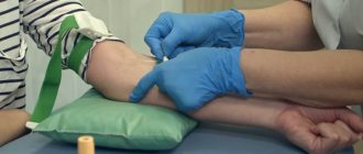
This is a simple and virtually painless procedure; all you need to do is come to the laboratory and donate blood. Blood for tumor markers is taken from a vein and immediately sent by a specialist to the laboratory.
One of the most important stages of analysis is preparation. Strict adherence to the doctor’s recommendations guarantees an accurate result. The general requirements are as follows:
- Blood from a vein is taken on an empty stomach in the morning .
- You are required to stop drinking alcohol the day before donating blood.
- For 3 days, refrain from excessive exercise and heavy food .
- You may have to stop taking some medications for a while . The doctor will tell you at your appointment what medications you should not take, or you can call the clinic administrator.
Since tumor markers increase during inflammatory processes, the procedure is not performed for inflammatory diseases.
Tumor markers (tumor markers) – what are they?
03/21/2018 Tumor markers (tumor markers) are substances of various natures that, when present in liquid media (urine, blood, etc.) of the human body in certain concentrations, indicate the presence of a tumor in it. The value of tumor markers for diagnosis depends on their specificity and sensitivity. Specificity is the percentage of negative tumor marker results in tests in healthy individuals. Sensitivity is the percentage of positive tumor markers in tests in cancer patients. However, to date, there is not a single marker of tumor growth that is 100% found only in patients with cancer. Tumor markers can also increase in inflammatory diseases of various origins, benign processes in the body, etc. Tumor marker CA 125 - early diagnosis of ovarian cancer. Tumor marker CA 125 is a protein that is synthesized by the mesothelium of the serous membranes: pleura, pericardium, peritoneum. In women, this protein is secreted by the endometrium of the uterus, so its concentration in the blood changes during the menstrual cycle. The upper limit of normal CA 125 for 94% of healthy individuals is a level of less than 35 U/ml. In menopausal women, the discriminatory level is less than 20 U/ml. CA 125 levels in patients with ovarian cancer after treatment should be less than 10 U/ml. The CA 125 tumor marker is mainly used for the early diagnosis of ovarian cancer in women of menopausal age and in women with a family history of ovarian cancer. And for young women it has low specificity and sensitivity. They may have other reasons for the increase in this tumor marker:
1. Cancers (uterine endometrial cancer, breast cancer, pancreatic cancer, lung cancer, colorectal cancer, stomach cancer, primary liver cancer, liver metastases. 2. Other diseases. ● Diseases involving the serous membranes (pleurisy, peritonitis , pericarditis). ● Pelvic inflammatory diseases, pneumonia, hepatitis, pancreatitis, cirrhosis, renal failure. In this case, the tumor marker CA 125, like many other tumor markers, acts as an inflammatory protein. ● Ovarian cysts and benign tumors. ● Endometriosis of various localizations. 3. Pregnancy, menstruation. The tumor marker CA 125 is also used to determine the relapse of ovarian cancer: a persistent increase in its level in patients in remission means a relapse of the disease. CA 125 is also used to assess the effectiveness of treatment for ovarian cancer. In the clinical diagnostic laboratory You can undergo testing for the tumor marker CA 125 at the Central Regional Hospital of the Volgodonsk region.
Tumor marker CEA (carcinoembryonic antigen) Tumor marker CEA refers to the antigens of the fetus, in which it is produced in the mucous membranes of the intestines and stomach. After birth, its synthesis decreases. The discriminatory level of CEA is less than 3–5 ng/ml; in smokers a level of less than 10 ng/ml is possible. The CEA tumor marker is primarily a marker for colon and rectal cancer. It reflects the volume of the cancer tumor before surgery, as well as the prognosis and five-year survival of patients after treatment. CEA also increases in adenogenic tumors (tumors from glandular tissue): stomach cancer, lung adenocarcinoma, breast cancer, pancreatic cancer, ovarian cancer, prostate cancer, endometrial cancer. And other diseases of the liver, lungs, and gastrointestinal tract. The tumor markers CEA, CA 19-9 and CA 72-4 together have high sensitivity for gastrointestinal tumors. CEA is a sensitive marker of metastases of adenogenic tumors in the bones, lungs, and liver.
The CEA tumor marker is also used to assess the effectiveness of treatment for patients with adenogenic tumors. Tumor marker SCC (antigen for squamous cell carcinoma) Tumor marker SCC - a marker for squamous cell carcinoma, is a protein that is synthesized by epithelial cells of the skin, bronchi, cervix, esophagus, and anal canal. The upper limit of normal (discriminatory level) is 1.5 ng/ml. The SCC tumor marker increases in the presence of squamous epithelial tumors: cervical cancer, tongue cancer, head and neck cancer, esophageal cancer, lung cancer, laryngeal cancer, vaginal cancer, vulvar cancer and other diseases: skin diseases (eczema, psoriasis, lichen planus ), chronic liver failure, tuberculosis, chronic renal failure. Therefore, the indications for its determination are as follows:
1. Assessing the effectiveness of therapy for patients with squamous cell carcinoma with initially elevated SCC. 2. Monitor patients with squamous cell carcinoma to diagnose relapses.
Tumor marker AFP (alpha fetoprotein) Tumor marker AFP is a fetal antigen, a transport protein of the fetal liver. The upper limit of normal is less than 15 ng/ml. The AFP tumor marker is used to diagnose hepatocellular cancer, as well as to monitor patients. AFP also increases in the presence of metastases of other tumors in the liver. In these patients, it is used to evaluate the effectiveness of treatment. Elevated AFP levels also occur in patients with ovarian tumors (chorionepitheliomas, teratomas, endodermal sinus tumors, dysgerminomas) and testicles. In these patients, AFP is used to determine relapse. The AFP tumor marker is also used to diagnose hepatocellular cancer in patients with liver cirrhosis and chronic hepatitis. Tumor marker hCG (human chorionic gonadotropin) Tumor marker hCG is a hormone synthesized by the placenta and chorion. The discriminatory level of hCG in the blood of men and non-pregnant women is less than 5 IU/ml, the borderline values are 5–10 IU/ml. The hCG tumor marker is used to diagnose pregnancy, hydatidiform mole (hCG levels reach high values, sometimes more than 1,000,000 IU/ml), choriocarcinoma (also a strong increase in hCG levels). And also for assessing the effectiveness of therapy and for the purpose of diagnosing relapses in patients with trophoblastic disease.
Tumor marker CA 19-9 Tumor marker CA 19-9 is a fetal protein synthesized by epithelial cells of the gastrointestinal tract. The discriminatory level of CA 19-9 is 37 U/ml. In adults, it is found in meager concentrations in the glandular epithelium of internal organs, so its increase means the presence in the human body of adenogenic tumors of various localizations: pancreatic cancer, stomach cancer, gall bladder cancer, ovarian cancer, colorectal cancer, esophageal cancer, liver cancer, metastatic liver cancer. Or other diseases: liver cirrhosis, hepatitis, cholelithiasis, cholecystitis, pancreatitis, cholestasis, endometriosis, cystic fibrosis, uterine fibroids. Tumor marker CA 19-9 is more often elevated in pancreatic cancer (76-81% of cases), for which it is the tumor marker of choice. For liver cancer (51-74% of cases), stomach cancer, colorectal cancer. CA 19-9 is elevated in 4179% of ovarian cancer cases. Therefore, this tumor marker is used to diagnose these diseases, assess the effectiveness of treatment and determine the progression of the disease. The CA 19-9 tumor marker is also widely used to monitor patients with endometriosis to evaluate treatment and determine disease relapse. Tumor marker CA 72-4 Tumor marker CA 72-4 is a fetal protein synthesized by the epithelium of the gastrointestinal tract (GIT). In adults, it is found in scanty concentrations in the epithelium of the gastrointestinal tract. The upper limit of normal is 5.3 U/ml. Tumor marker CA 72-4 increases more often in gastric cancer (2979% of cases) and for this it is the tumor marker of choice. It is also used in stomach cancer to diagnose relapses. For ovarian cancer, the sensitivity of CA 72-4 is 7178% and is also considered the marker of choice for this disease. The tumor growth marker CA 72-4 also increases in other cancers: pancreatic cancer, breast cancer, colon cancer, lung cancer, endometrial cancer. Tumor marker CA 15-3 Tumor marker CA 15-3 is associated with breast cancer. The upper limit of normal (discriminatory level) in non-pregnant women is 28 U/ml. In pregnant women in the 3rd trimester of pregnancy, it can increase to 50 U/ml. The sensitivity of CA 15-3 depends on the stage of breast cancer; in the early stages of the disease it has low sensitivity (about 20%), therefore it is not used for the early diagnosis of breast cancer. And with a common process, the sensitivity is 84%. Therefore, the CA 15-3 tumor marker is used to assess the effectiveness of breast cancer treatment and diagnose relapses of the disease. Tumor marker UBC (bladder cancer antigen) Tumor marker UBC is a water-soluble fragment of cytokeratins 18 and 8, synthesized by epithelial cells. As bladder cancer grows, their synthesis increases and they can be detected in urine. In urine, the concentration of the tumor marker UBC is adjusted in relation to the concentration of creatinine. The upper limit of normal is 0.00049 µg/µmol creatinine. The UBC tumor marker is used in the diagnosis of bladder cancer, to assess the effectiveness of therapy and detect relapses, since its sensitivity in patients with bladder cancer is 61-86%, and its specificity is 94%.
Tumor marker CYFRA 21-1 Tumor marker CYFRA 21-1 is an epithelial protein that is a marker of cancer. The upper limit of its normal value is 2.3 ng/ml. The CYFRA 21-1 tumor marker is used to diagnose lung cancer, evaluate the effectiveness of treatment, monitor patients with lung cancer and patients with other cancers: cervical cancer, bladder cancer, esophageal cancer, ovarian cancer, breast cancer, rectal cancer.
Tumor marker TU M2-PK Tumor marker Tu M2-PK is a protein-enzyme for ATP synthesis at low oxygen concentrations, which happens in tumor cells, that is, it reflects the metabolism of tumors as a whole. The upper limit of normal is 17 U/ml, the border zone is 17–20 U/ml. The Tu M2-PK tumor marker is increased in kidney cancer, lung cancer, esophageal cancer, colorectal cancer, stomach cancer, breast cancer, and pancreatic cancer. But it has particularly high specificity (89%) and sensitivity (79%) for kidney cancer, so it is used to monitor patients with kidney cancer to determine the effectiveness of anticancer treatment and early detection of relapses.
Tumor marker NSE (neuron-specific enolase) Tumor marker NSE is an enzyme protein synthesized in the lung and nervous tissues. The upper limit of normal is 12.5 ng/ml. The NSE tumor marker has high sensitivity (45–86%) and specificity for lung cancer and is used to diagnose lung cancer and to evaluate the effectiveness of therapy. It is also used for the diagnosis of neuroendocrine tumors, carcinoids, pheochromocytoma, kidney cancer, and seminomas.
Tumor marker TG (thyroglobulin) Tumor marker TG is a precursor protein of thyroid hormones. The upper limit of normal for the tumor marker TG is 60 ng/ml. The TG tumor marker is characteristic of thyroid cancer, but also of other diseases of this organ: thyrotoxicosis, thyroiditis, toxic thyroid adenoma. Therefore, it is used to detect metastases and relapses of the disease in patients with thyroid cancer, as well as to determine the primary focus in the presence of metastases in the lungs and bones of tumors of unknown location.
Tumor marker BONE TRAP Tumor marker Bone TRAP protein-enzyme of osteoclasts (cells that destroy bone tissue). The upper limit of normal for women under 45 years old is 1.1–3.9 U/ml, for women 45–55 years old — 1.1–4.2 U/ml, and for women in menopause — 1.4–4. 2 U/ml, for men - 1.5–4.7 U/ml. The Bone TRAP tumor marker is a tumor marker of bone metastases from breast cancer and prostate cancer, as well as a marker of the presence of multiple myeloma in the bones. Therefore, it is used to diagnose bone metastases. It also evaluates the effectiveness of treatment of bone metastases. The Bone TRAP tumor marker is also used for osteoporosis, hyperparathyroidism, and Paget's disease. Tumor markers according to the localization of the tumor process are presented below:
Breast tumor markers: CA 15-3, TPS, CEA, CA 72-4. Ovarian tumor markers: CA 125, CA 19-9, CA 72-4, AFP, hCG. Uterine tumor markers: SCC, CYFRA 21-1, CEA, TPS, CA 125, CA 19-9, CA 72-4. Stomach tumor markers: CA 72-4, CA 19-9, CEA. Intestinal tumor markers: CA 72-4, CA 19-9, CEA, Tu M2-PK Lung tumor markers: CYFRA 21-1, CEA, NSE, Tu M2-PK, SCC, CA 72-4. Pancreatic tumor markers: CA 19-9, CA 242, Tu M2-PK. Esophageal tumor markers: SCC, Tu M2-PK.
Tumor marker PSA (prostate-specific prostate antigen, total and free)
PSA is present in healthy, overdeveloped and transformed prostate tissue. It is the most specific and sensitive antigen for diagnosing prostate cancer . For research, blood is taken before biopsy, removal or massage of the prostate, because Mechanical irritation of the gland can cause an increase in PSA levels that lasts up to 3 weeks. The PSA norm is 0-4 ng/ml, a level of 10 ng/ml and above indicates a malignant disease.
How to prepare for a PSA blood test?
-no food is allowed within 8 hours before the test; juice, tea, coffee, alcohol are excluded;
- it is recommended to abstain from sexual intercourse for 5-7 days before the study;
— it is advisable to take the test before an examination by a urologist or 10-14 days after it;
- after a prostate massage or digital rectal examination, cystoscopy or catheterization of the bladder, transrectal ultrasound and after any other mechanical effects on the prostate, it is advisable to wait at least 2 weeks before taking a blood test for PSA, and after a biopsy
Increase in PSA :
Prostate cancer (about 80% of cases),
Benign prostatic hyperplasia (slight increase),
Inflammation or infection in the prostate
Ischemia or infarction of the prostate,
Ejaculation on the eve of the study,
Surgery, trauma, or prostate biopsy
glands.