Vascular malformations is a general term that includes congenital vascular anomalies: veins only - venous malformations (VM) lymphatic vessels only - lymphatic defects of both veins and lymphatic vessels: veno-lymphatic malformations anomalies of arteries connected directly to veins without a capillary network between them: arteriovenous malformations (AVMs)
What are arteries
These are vessels that transport oxygen from the heart to the internal organs. By contracting the myocardium, blood circulation is ensured at a speed of 20 cm/s. Purified blood, full of oxygen and nutrients, is necessary for metabolism.
Passing through organ tissue saturates it with carbon dioxide, excreted through venous hematopoiesis.
They are divided into three types:
- diameter;
- structural features;
- topographic principle.
The diameters are:
- large;
- small.
Large in diameter, unlike other components of the vascular system, are: aorta, carotid and subclavian.
The aorta arises from the left ventricle of the heart along the spinal column, dividing into the left and right iliac branches. It begins a large circle of blood circulation, supplying oxygen to the organs and tissues of the body.
General sleep supports the functioning of the brain, providing it with oxygen and microelements necessary for metabolism.
The subclavian vessel supplies blood to the occipital parts of the brain, medulla oblongata, cerebellum and cervical spine. The left arch departs from the aorta, bending around the pleura and, passing through the upper aperture of the chest, exits to the neck and lies in the space of the first rib.
Arterioles are small in diameter. Their task is to regulate blood flow in the SMC.
- Why do the veins in the arms hurt in the elbow bend?
Arteriolar tone determines peripheral resistance, which, along with the stroke volume of the heart, affects blood pressure.
There are three types:
- elastic;
- muscular;
- mixed.
The first type includes mainly the aorta. Its structure is characterized by the predominance of elastic fibers over muscle fibers.
The muscle type contains smooth muscle fibers and is characterized by a weak outer elastic membrane. An example is arterioles.
The muscular-elastic type is distinguished by the presence of muscle and elastic fibers in the structure of the vessel.
Doctor of Medical Sciences, Professor of the Department of Phthisiology and Pulmonology of the Medical Faculty of Moscow State Medical University, Sergey Lvovich Babak
Degree of oxygen deficiency relative to saturation (SpO2) - pulse oximeter readings
| Degree | SpO2,% (pulse oximetry readings) |
| Norm | more than or equal to 95% |
| 1st degree | 90-94% |
| 2nd degree | 75-89% |
| 3rd degree | less than 75% |
| Hypoxemic coma | less than 60% |
*Recommendations, the required oxygen flow, the regimen and duration of oxygen therapy for COPD are prescribed by the attending physician! Oxygen therapy at home is carried out using oxygen concentrators under the control of pulse oximeter readings.
— My name is Babak Sergey Lvovich. I am a professor at the Department of Phthisiology and Pulmonology of the Medical Faculty of Moscow State University of Medicine A.I. Evdokimov. I have several questions that I would like to devote the remaining time to. The role of oxygen in human daily life. The fact is that those mechanisms that we usually evaluate as oxidative are impossible without oxygen. Life is built around oxygen.
It exists in different forms. There are concepts of atomic oxygen, and there are concepts of molecular oxygen. The most curious thing is that molecular oxygen in the air in the lungs turns into atomic oxygen, which penetrates the blood and delivers it to the muscles. And already inside the muscles, it actively participates in the craps chain, allowing the body to receive the necessary proteins, fats, carbohydrates and nutrients by oxidizing products entering the body with food, water, liquids, and so on. Therefore, this delivery of oxygen by the lungs to the blood performs the function of gas exchange.
This is the most important function, and in short, why we breathe. We breathe only to maintain the constancy of atomic oxygen inside our body. The human lungs are adapted to inhale air at a pressure of one atmosphere containing 21% oxygen, almost 80% nitrogen and not containing any additional other impurities in the form of smoke, in the form of solid particles, and so on. But having a humidity of no higher than 60% at a temperature of about 22 degrees.
There are so many conditions necessary for the lungs in order to transform molecular oxygen into atomic oxygen and create a constant saturation of arterial blood with oxygen. If a person, for example, smokes or inhales some dust particles, or some other impurity components in the air, then the lungs react very harshly to this and do not allow such people to have an adequate level of arterial blood oxygen saturation. That is, it seems to be fighting to ensure that we still breathe the freshest air without pathogenic impurities or foreign particles. The second very important component that should be discussed when we talk about the role of oxygen in daily human life concerns environmental humidity and temperature.
The fact is that humans are adapted to live and survive in different climatic conditions. In conditions of very high humidity, in conditions of low humidity, in conditions of cold temperatures, in conditions of very hot temperatures. In fact, this is a unique creature with a high adaptive reserve. Almost all pulmonary diseases can be accompanied by the development of respiratory failure.
The essence of respiratory failure comes down to the fact that there is a discrepancy between the need for oxygen and the ability to deliver oxygen to the arterial blood. The partial tension of arterial blood with oxygen is less than 55 ml of mercury or an increase in the partial tension of carbon dioxide in the arterial blood is above 45 ml of mercury. These two parameters indicate that a person has experienced some degree of respiratory failure.
To our joy, there is an indirect method, but it is quite accurate, by which we can also find out what the degree of respiratory failure is. This method is called pulse oximetry. Pulse oximetry reflects the saturation of arterial blood with oxygen in the degree of saturation. This degree can also suggest the degree of respiratory failure, for example, arterial blood oxygen saturation in the range from 90 to 93% corresponds to a partial blood oxygen tension of 60 to 80 ml of mercury. Which corresponds to zero degree of respiratory failure.
The parameter of reducing blood saturation to 85% will correspond to the first degree of respiratory failure or a reduction to the level of 50 ml of mercury. A parameter of up to 80% blood saturation usually corresponds to the second degree of respiratory failure and 75% below blood oxygen saturation corresponds to the third degree of respiratory failure. It is believed that no matter how the patient feels, the degree of saturation of arterial blood with oxygen
should not be below 90% arterial blood oxygen saturation. The disease will proceed differently in a person if his arterial blood oxygen saturation is below 90%, that is, a certain degree of respiratory failure will occur.
What diseases are usually accompanied by respiratory failure? First of all, obstructive pulmonary diseases. These include bronchial asthma, they include obstructive bronchitis, they include chronic obstructive pulmonary disease, they include bronchiectasis, they include cystic fibrosis. How common is respiratory failure in the population?
It is impossible to give a direct answer here. Because we are talking about the prevalence of the disease, not the prevalence of the syndrome. Respiratory failure is a syndrome and it is quite difficult to separately calculate the prevalence of the syndrome. If we are talking about the comparison of diseases in which respiratory failure can occur, then this is almost 80% of all pulmonary diseases we encounter among the human population.
Therefore, we can say that the data is extrapolated from the given blood. To say that respiratory failure is a common phenomenon in obstructive pulmonary diseases. What underlies the development of respiratory failure? Primarily there are two main mechanisms. The mechanism of narrowing of the bronchi and the impossibility of bleeding out air containing 21% oxygen and the second mechanism is very important, this is the impossibility of oxygen penetrating through the alveolar membranes.
Here are the two main components influencing the development of respiratory failure. Therefore, we divide it into two different types that occur with obstructive pulmonary diseases, which occur with interstitial lesions of the lung tissue. Let's try to decipher the obstructive component of the development of respiratory failure. What is this connected with? First of all, it is due to the fact that in a number of diseases there is a narrowing of the lumen of the bronchial tree, a narrowing of the lumen of the bronchial tubes.
It's caused by bronchospasm, it's caused by swelling, mucus buildup. These three mechanisms lead to a narrowing of the lumen and the impossibility of air entering the respiratory tract. Therefore, even under normal conditions, when there is enough oxygen in the air to ensure the gas exchange function, it physically cannot penetrate the lower part of the respiratory system and saturate the blood with oxygen. Due to the fact that the development of certain respiratory volumes necessary to maintain the gas exchange function is not achieved.
The second situation is completely different; it is associated with intersocial lesions of the lung tissue. When tidal volume is reduced due to compression of the lung. The lung seems to be compressed a little on one side, and on the other side the membranes thicken and oxygen, at a pressure of one atmosphere, cannot penetrate through the membranes and penetrates worse than it should penetrate, cannot adequately saturate the arterial blood with oxygen. In both cases, increasing the concentration of the oxygen mixture supplied to the lungs leads to a very interesting effect.
Oxygen penetrates into the blood at a higher rate and practically the person loses respiratory failure. Therefore, we are talking specifically about devices in this case that are capable of creating an increased concentration of oxygen in the exhaled mixture; they are called an oxygen concentrator. A separate category is respiratory failure caused not by the oxygen component, but by the accumulation of carbon dioxide, it is called hypercapnic respiratory failure.
The first type of respiratory failure that we talked about before is called hypoxemic or hypoxic respiratory failure, where oxygen does not penetrate into the blood, low concentrations. And the second type of respiratory failure is called hypercapnic, associated with the accumulation of carbon dioxide. The culprit of the protogynesis of the development of this type of respiratory failure is precisely the respiratory muscle. A person cannot physically create an excursion adequate to the need for air oxygen to penetrate the respiratory tract.
This is usually associated with neuromuscular diseases, very often associated with obesity or with damage to the skeletal structure of the chest. Also plays an important role in the expansion of the lungs. How does respiratory failure manifest itself clinically? First of all, a person feels a feeling of lack of air, which has an organic name - shortness of breath. Shortness of breath occurs at rest, shortness of breath occurs during physical activity, so we grade this shortness of breath on a certain scale. We assign a point score, the higher the score, the more severe the person’s shortness of breath
In total, the scale provides four points, starting from two points, shortness of breath is chronic and is a reason to seriously think about the causes of such shortness of breath. Clinical shortness of breath manifests itself, if you look at such a patient with shortness of breath, you will see that there is usually bluish skin, blue lips, and often puffs.
True, in some diseases , chronic obstructive pulmonary diseases, in which shortness of breath is very characteristic, we even distinguish two different phenotypes of such a disease. One phenotype is called pink puffing patients, and the other is called blue panting patients. The pink ones that puff are called Pinkpuffers, and the blue ones that pant are called Blue Blowers.
So, Blue Blowers usually have a hypoxemic type of respiratory failure, they are cyanotic, the air supply is very useful for them. Pink-puffing patients more often have a hypercapnic type of respiratory failure with CO2 accumulation and oxygen in this case is not very useful. But on the contrary, it is necessary to have ways to strengthen the disabling part. That is, by changing the ventilation of the lungs in order to flush out CO2 in such patients, since the accumulation of oxygen in the blood causes an increase in the level of CO2 in the blood.
Frequency and seasonality of diseases causing respiratory failure. If we talk about the frequency and seasonality of these diseases, then, in my opinion, these diseases should still be divided into two main categories: obstructive diseases and restrictive diseases with damage to the lungs. If we are talking about the obstructiveness of the disease, then of course, first of all, they are associated with changes in humidity and ambient temperature.
Since this leads to the fact that sputum can swell in the lumen of the bronchus and clog small bronchi, this causes disturbances in the flow of air through the bronchial tree. Therefore, patients usually have chronic obstructive bronchitis twice a year. COPD has this type of exacerbation associated with climate change. A very important component influencing the frequency of exacerbations is continued smoking; such patients have obstructive diseases.
Regular inhalations of toxic gases and fumes support very pronounced inflammation in the respiratory tract and it overlaps with the course of treatment of the disease itself, causing an increase in the frequency of exacerbations. In this case of exacerbation of the disease, there is a sharp increase in shortness of breath, an increase in the secretion of sputum mucus more than usual, this causes the patient to begin to choke and experience varying degrees of respiratory failure.
With which he usually comes to our hospital or is subject to treatment at home. Seasonality in this case is not as important as maintaining those factors that can maintain inflammation of the respiratory tract. The situation is completely different with such an obstructive disease as bronchial asthma. This is a separate category of patients who are usually allergic and have hay fever, and at the moment of flowering of herbs, plants and flora, to which they react very sharply, they experience an exacerbation of bronchial asthma.
Exacerbations are associated specifically with the allergic component, and much attention is paid to the concept of a hypoallergenic regime in patients with asthma, maintaining this and combating hay fever or a reaction to flowering plants, all kinds of herbs, trees, and so on. If we are talking about restrictive diseases, such as pulmonary fibrosis, then they have neither frequency nor seasonality of exacerbation, the process is associated with something else.
The process is often associated with an additional infection, which the patient can get due to a cold or a viral infection. We are essentially talking about pneumonia, pneumonia. Pneumonia in such patients is very severe and very often patients are tormented by destructive diseases; when they get pneumonia, they get a very pronounced degree of respiratory failure. And they literally die from lack of oxygen in the arterial blood.
It must be said that oxygen is a medicine. Like every medicine, it must be considered as a kind of poison, which is given little by little under certain conditions. Since the principle of doing no harm should work in this case too. You can’t just breathe in a certain volume or flow of oxygen. In this way, you can seriously disrupt the humidity of the respiratory tract and disrupt the structure of the respiratory tract, causing yourself serious harm. Oxygen is a powerful oxidizing agent. I would really like our listeners and viewers to remember that the ozone you are talking about: “It’s very good to breathe ozone.”
- This is a big deal! Tragic mistake! There are many people who specially ozonate the room, creating so-called three-molecular oxygen. They damage the pulmonary apparatus so severely that they can eventually die from severe damage to the lung tissue from ozone breathing. Therefore, any oxygen therapy requires clear, specific intervention from a doctor.
Flow intensity. What flow intensity should be set in order to achieve success in oxygen therapy?
The oxygen flow should be such that arterial oxygen saturation figures range from 90% to 95% arterial oxygen saturation. If you can achieve this flow of one and a half liters per minute, that is enough. There is no need to increase the flow to 2 liters, 3 liters, 4 liters. If 3 liters are needed for this, conditions must be created so that the patient receives 3 liters. Therefore, in each specific case, there is a titration or selection of the oxygen flow that creates normal blood oxygen saturation figures. It is believed that flows in excess of one and a half liters per minute are unsafe. That is, they require a special air humidification system, since they can dry out the respiratory tract. And it requires warming, because it will lead to cooling of the respiratory tract.
Let me give you a simple example. For example, cooling the respiratory tract by one degree, that is, 37.4 there becomes 36.4. This leads to a decrease in air humidity by 12%. A decrease of 12% actually dries out the mucus, it becomes in the form of crusts, these crusts will never leave the lower respiratory tract, and respiratory plugs form. Or we call it a mucus plug.
Therefore, it is very important that we properly deliver oxygen to the airways. Properly humidify and, if necessary, properly warm the delivered air so as not to cause hypothermia of the respiratory tract. You need to consult a specialist, a doctor first of all, who knows this technology. And set the parameters necessary for this type of treatment.
How to prescribe oxygen therapy, which patients to prescribe and how to choose the right level? There is a concept of diphomysioma test, if the diffusion of oxygen decreases, we see a significant decrease. That is, the percentage of blood becomes below 55 ml. mercury, then long-term oxygen therapy is indicated for such patients. What is the best way to titrate the level of such therapy? During titration, the oxyinter course is used, which allows one to accurately determine the oxygen flow that maintains normal levels of oxygen saturation in arterial blood.
The need for long-term therapy arises in all patients with respiratory failure starting from the second stage. Since at this stage the arterial blood oxygen tension decreases, usually below 55 ml. mercury column. In fact, these are all patients admitted to the hospital with exacerbation of chronic obstructive pulmonary disease, exacerbation of obstructive bronchitis or with severe attacks of bronchial asthma. They will need oxygen therapy.
If we are talking about the duration of such a maneuver, the duration of this technique, it is important to look at the life-sustaining technique and the technique carried out for some time. Naturally, if we expect that the patient’s respiratory function will be restored and gas exchange will be restored, then we will cancel such therapy.
Usually when therapy takes about two, three weeks of oxygen therapy. We carry out this therapy in the hospital and upon discharge the patients do not receive further oxygen. But a number of patients, especially with interstitial lung lesions with severe obstructive disorders, when it is impossible to replenish gas exchange, require lifelong use of this type of therapy.
And then they are forced to use oxygen concentrators at home. This is an important factor in prolonging the life of such patients. It has been studied and shown that the use of an oxygen concentrator at home prolongs the patient’s life by 15-20 years. This is significant for such patients, while the degree and risk of exacerbations are reduced by up to four times.
That is, if a patient has a minor exacerbation per year, using long-term oxygen therapy for virtually the entire year, he does not experience any serious exacerbations of disease requiring hospitalization or a change in the amount of drug therapy.
This is a significant contribution of the duration of oxygen therapy or oxygen therapy to the doctrine of treatment of patients with chronic respiratory failure. There are oxygen concentrators operating in the range from one liter to five liters per minute with a high output concentration. Creating conditions for good saturation of arterial blood with oxygen. They are expensive and the patient does not have the money to purchase such a device; he is limited to simple concentrators that either work unstably, with a low oxygen concentration at the outlet, or do not provide a flow of, say, five to three and a half, four liters per minute.
What does this lead to? It leads to the fact that the real oxygen concentration in the inhaled mixture drops to a very low value and is virtually no different from room air. And we know very well that the patient’s room air is not enough to relieve gas exchange disorders in such a patient. And respiratory failure progresses in such patients, despite the fact that they supposedly use oxygen concentrators in their lives and are treated with the help of concentrators. In this case, we suggest renting an oxygen concentrator; the cost of renting an oxygen concentrator starts from 6,000 rubles per month.
Therefore, it is the reliability, the percentage reliable benefit of oxygen, the wide variation of flows of oxygen devices that allows you to have some maneuver. In order to select for each patient, in each specific case, adequate, reliable oxygen therapy for a very long period of use. One of the companies that has legalized such a line is the Agmung company. Which adopted the doctrine of various oxygen concentrators for various treatment methods.
For example, there is a model line of concentrators for hospitals and home use, for example, where fairly high flows are combined with a very high concentration of the inhaled oxygen mixture.
| Atmung 3L-I (LFY-I-3A) | Atmung 03-C (LFY-I-3A-11) | Atmung 5L-H (LFY-I-5F-11) | Atmung 5L-F (LFY-I-5A-01) |
And there are oxygen concentrators for home use, small, portable, low noise, when the flow ranges from one to three liters per minute.
| Atmung Oxybar | Atmung Oxybar Auto | Armed 8F-1 | Armed 7F-1L |
I note that usually for home use, flows of more than one and a half liters per minute are not used. Therefore, oxygen supplied in a flow of even three liters per minute is twice the patient’s needs, which provides a guarantee of reliability and stability for such patients, even in emergency situations happened at home. It is important to understand that sometimes patients themselves must know how to behave correctly in the current situation. For example, with a feverish patient, he puts a thermometer or thermometer under the armpit or in the mouth and determines the temperature for himself, understands that with a temperature of 37. he behaves according to one, with a temperature of 38 for another, 39 for a third.
Question: — How should a patient with respiratory failure who is receiving long-term oxygen therapy behave correctly?
For this, there are the concepts of pulse oximeters, a small portable device located on the phalanx of the finger, and allowing to measure the saturation of arterial blood with oxygen. So, if the patient feels increasing shortness of breath without receiving oxygen, puts a pulse oximeter on the phalanx of the finger and sees that the pulse and oximetry indicators begin to decrease below 90%. This is a reason to reconsider the scope of such therapy, but in the presence or after consultation with your doctor who prescribed this type of long-term oxygen therapy.
If he feels some kind of ailment, some kind of weakness, fatigue, but pulse oximetry is maintained above 90% of arterial blood oxygen saturation, then there is no need to change the volume of such therapy. These symptoms are associated with another manifestation of the disease, for example, with not receiving a bronchodilator, receiving hormonal therapy, or impaired mucus drainage in the respiratory system, but are in no way associated with long-term oxygen therapy.
Such a simple method of monitoring well-being and blood oxygen saturation makes the patient confident in the regularity and reliability of this type of treatment.
How long does it take to supply oxygen to a person's respiratory tract?
Professor Ludo in the early 80s in France conducted a huge clinical study on a huge sample of patients and it was established. That with long-term oxygen therapy, it is necessary to supply oxygen to the respiratory tract twenty hours a day, at least twenty hours a day, so that respiratory failure undergoes correction.
At the same time, if we reduce the number of hours of oxygen therapy to 15 or less, then this is equivalent to as if we did not conduct such sessions of long-term oxygen therapy at all.
That is, the boundaries of behavior range from 15 to 24 hours a day. And the desired duration is twenty hours during which the patient breathes a certain concentration of oxygen to relieve any degree of respiratory failure.
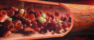
Movement of blood through vessels
The difference between arteries and veins is clearly demonstrated by the mechanism of blood flow. When the heart muscle contracts, blood is forced into the arteries. In the largest of them, the aorta, pressure can reach 150 mm Hg. Art. In the capillaries it decreases significantly to 20. In the vena cava the pressure is minimal and amounts to 3-8 mm Hg. Art.
Types of arteries
There are three types of arteries:
Elastic arteries
Elastic arteries are large vessels leaving the heart. For example, they include the pulmonary artery and aorta. The aorta is the main artery that carries blood away from the heart.
The heart forcibly pumps out blood to move it throughout the body. Elastic arteries must be flexible to cope with the rush of blood. They expand when the heart pumps out blood.
Elastin is a protein found in many tissues that provides flexibility to organs, including elastic arteries.
Muscular arteries
Elastic arteries carry blood to muscular arteries such as the femoral or coronary arteries.
Smooth muscle fibers make up the walls of muscle arteries. Muscles allow these arteries to expand and contract. These changes in size control how much blood moves through the arteries.
Arterioles
Arterioles are the smallest type of arteries. They distribute blood from larger arteries through networks of capillaries.
The outer layer of arterioles also contains smooth muscle, which regulates expansion and contraction.
Main Difference – Veins vs Arteries
Veins and arteries are two types of blood vessels in a closed circulatory system. The main function of blood vessels is to carry blood throughout the body. But arteries and veins differ in their structure and function. Veins are made up of a thin, elastic layer of muscle in their wall, while arteries are made up of a thick, elastic layer of muscle. A thick artery wall is important when dealing with high pressure blood released by the heart. Veins carry oxygen-depleted blood to the heart, while arteries carry oxygenated blood away from the heart. The main difference between veins and arteries is that veins are involved in removing cellular waste from the extracellular environment, whereas arteries are involved in providing nutrients and oxygen to the body's cells .
Key areas covered
1. What are veins - definition, features, functions 2. What are arteries - definition, features, functions 3. What are the similarities between veins and arteries - Brief description of the common features 4. What is the difference between veins and arteries - Comparison of the main differences
- The veins behind the knee hurt
Key words: aorta, arteries, arterioles, blood capillaries, blood pressure, closed circulatory system, veins, venules
Arterial and venous blood
Differences between arterial and venous blood: dark blood flows from the veins, it is thicker, it clots faster, the bleeding is less intense, the stream is smooth and not strained. Venous blood is more suitable for laboratory tests than capillary blood from a finger. It flows through the pulmonary arteries, and in the capillaries of the alveoli it turns into arterial artery.
Which one is darker
Venous blood is darker. Its color depends on the form of hemoglobin. In the arterial blood, it is combined with oxygen (oxyhemoglobin), which gives it a bright scarlet color. The venous contains both oxyhemoglobin and 2 other forms:
- reduced (gave oxygen to cells, but has not yet added carbon dioxide);
- carboxyhemoglobin (a compound with carbon dioxide).
Most of the latter pigment, so the color becomes dark cherry.
Capillary and venous blood: differences
The main differences between capillary blood (from a finger) and venous blood are the contents:
- cells, especially platelets (higher in the venous), leukocytes (higher in the capillary);
- glucose (higher in the veins).
Analysis from a vein is considered more accurate, since it does not contain impurities of tissue fluid or surface epithelium of the skin. Also, circulatory disorders, vascular spasm, and fever can affect the indicators of capillary blood tests. For many types of laboratory diagnostics, a sufficient volume of material is needed, and up to 0.5 ml can be taken from a finger.
Therefore, capillary blood can be used only at the first stage of the examination. It will give reliable results only when determining hemoglobin, red blood cells and ESR. For biochemical and immunological analysis, studies of hormonal levels, as well as, if necessary, an in-depth study of cellular composition, blood from a vein is needed.
Why does venous blood clot faster?
Venous blood clots faster due to high platelet levels. These blood platelets form the basis of the clot, they are connected to each other, and fibrin threads give strength to the formed blood clots.
How to determine the type of bleeding
To determine the type of bleeding, you need to pay attention to the signs that distinguish them:
- Arterial and venous blood: how do they differ in humans?
Through which veins does arterial blood flow?
Arterial blood flows through the veins that leave the lungs to the left atrium. Also, the entire internal venous network of the lungs is filled with hemoglobin, saturated with oxygen. Therefore, the contents in them are exactly the same in characteristics as in the arteries of all other organs (except the lungs).
Where venous blood turns into arterial blood
Venous blood is converted into arterial blood in the capillary network of the alveoli (vesicles) of the lungs. The pulmonary arteries branch into thin capillaries; they entwine the alveolar walls. Carbon dioxide enters the lumen of the pulmonary vesicle, and oxygen from it passes into the blood.
Oxygen molecules join the reduced hemoglobin, which has given up carbon dioxide, and it becomes oxyhemoglobin. Therefore, the blood changes its color and properties - from dark venous to light arterial. Through four pulmonary veins (two left and two right) it goes to the heart.
How arteries differ from veins: features of functioning
Arteries are vessels that provide blood flow from the heart to tissues and organs. The largest artery in the body is called the aorta. It comes directly from the heart. In the arteries, blood moves under high pressure. To withstand it, an appropriate structure of the walls is necessary. They consist of three layers. The inner and outer ones are formed by connective tissue, and the middle one is made of muscle fibers. Thanks to this structure, these vessels are capable of stretching, which means they can withstand high blood pressure.
How does the structure of veins differ from the structure of arteries? First of all, vessels of another type carry blood from organs and tissues to the heart. Having passed through all cells and organs, it is saturated with carbon dioxide, which it carries to the lungs.
Another important question is how the structure of the wall of an artery and vein differs. The latter have a thinner muscle layer and are therefore less elastic. Since blood enters the veins under low pressure, their ability to stretch is not as important.
The amount of blood pressure in different types of vessels is demonstrated by different types of bleeding. With arterial blood, blood is released with force in a pulsating fountain. It is scarlet because it is saturated with oxygen. But with venous it flows out in a slow stream and has a dark color. It is determined by a large amount of carbon dioxide.
The lumen of most veins has specialized pocket valves that prevent blood from flowing in the opposite direction.
- Blood flow in the umbilical cord artery is normal
Main symptoms
What are the symptoms of vascular malformation? Vascular malformations can cause a variety of symptoms depending on their location in the body: A common symptom of all malformations is pain. Venous and lymphatic defects can cause subcutaneous swelling, over which the mole may be located. Skin lesions may ooze lymph or cause bleeding. Lymphatic malformations are usually complicated by an infectious process, requiring treatment with antibacterial drugs. Venolymphatic malformations may be associated with a phenomenon called Klippel-Trenaunay syndrome.
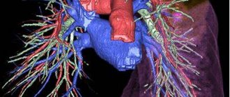
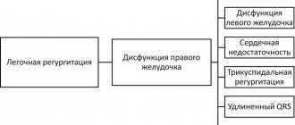
Arteriovenous malformations (AVMs) can also cause pain. They are the most dangerous due to the rapid discharge of blood from the arteries into the veins. Depending on their location, they can also lead to bleeding (eg from the uterus, bladder). Pulmonary arteriovenous malformations are somewhat different in that they act as a shunt that drains blood from the right side of the heart to the left side of the heart without increasing the level of oxygen in the lungs. This leads to symptoms of low oxygen, shortness of breath, and fatigue. Such defects can bleed, resulting in hemoptysis or hemothorax (the appearance of blood in the chest). Additionally, this abnormality can allow blood clots to pass through the lungs and into other arteries in the person's body, thereby causing a stroke or brain abscess. This is an essential reason for immediate treatment of pulmonary arteriovenous malformations.
Different functions - different structure.
The highest blood pressure will be at the blood outlet from the heart (in the left ventricle), slightly lower pressure will be in the arteries, even lower in the capillaries, and the lowest in the veins and at the entrance of the heart (in the right atrium).
The arteries carrying the oxygenated blood pushed out by the heart must resist the high pressure in the circulatory system. That's why they have an elastic membrane. In addition, they must also change their lumen in order to vary the level of blood flow in different organs in response to the actions of the autonomic nervous system - for this they have a well-developed layer of smooth muscle tissue. Therefore, the walls of the arteries are much thicker than the venous ones, they are much more elastic and contain a large number of muscle elements.
The walls of the veins, in turn, are thin and pliable, contain virtually no muscle elements, and ensure the return of blood to the heart. The veins in the lower body have valves that prevent blood from flowing back. Thus, the vascular bed adapts to changing load levels mainly due to changes in the lumen of the arteries.
Circulation
Venous blood (with a low oxygen content) enters the right atrium through the superior and inferior vena cava. The blood then enters the right ventricle, which contracts and pumps it into the pulmonary trunk. The trunk soon divides into the right and left pulmonary arteries, which carry blood to both lungs. The arteries, in turn, break up into lobar and segmental branches, which are further divided into arterioles and capillaries. In the lungs, venous blood is cleared of carbon dioxide and, enriched with oxygen, becomes arterial. It enters the left atrium through the pulmonary veins and then into the left ventricle. From there, under high pressure, the blood is pushed into the aorta, then goes through the arteries to all organs. The arteries branch into smaller and smaller ones and eventually become capillaries. The speed of blood flow and its pressure by this time are significantly reduced. Oxygen and nutrients enter the tissues through the walls of the capillaries from the blood, and carbon dioxide, water and other metabolic products enter the blood. After passing through the network of capillaries, the blood becomes venous. The capillaries merge into venules, then into increasingly larger veins, and eventually the two largest veins - the superior and inferior vena cava - flow into the right atrium. As long as we are alive, this cycle repeats itself over and over again.