Are you experiencing back pain and numbness in your limbs? These are the first signs of osteochondrosis. Insufficient mobility in lifestyle, sedentary work, stress on the neck and spine lead to cartilage wearing out, losing moisture, resulting in micro-tears.
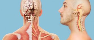
With cervical osteochondrosis, degenerative changes occur in the intervertebral discs. Not only the discs are damaged, but also the vertebrae and joints in the cervical region. If the disease is not treated for a long time, the patient’s general well-being worsens: constant headaches, the appearance of a vertebral hernia, deterioration of cerebral circulation, as a result of which cognitive functions decrease.
Reasons for the development of osteochondrosis
An incorrect sitting position, in which the neck is pulled forward, leads to the development of cervical disease. In this case, excessive pressure occurs on the intervertebral discs, which leads to changes in the nuclei pulposus and compression of blood vessels. This is the position a person takes at a workplace in front of a computer. Therefore, office workers are most often exposed to the development of osteochondrosis of the cervical spine.
In addition, the reasons for the development of pathology may be:
- improper load distribution when carrying bags;
- excessively soft sleeping place (the spine bends in an unnatural shape);
- genetic predisposition;
- lack of vitamins and microelements in the diet;
- endocrine system disorders;
- curvature of the spine and poor posture during active growth of the body;
- cervical vertebrae injuries;
- presence of bad habits.
Prevention
Having gotten rid of osteochondrosis and accompanying hypertension, you should not relax. It is necessary not to forget about preventive measures that have a beneficial effect on blood pressure and the general condition of the spine. The recommendations are simple and accessible:
- It is necessary to give up bad habits - alcohol and smoking, which negatively affect blood circulation.
- A sedentary lifestyle should be changed to a healthy one. The degree of physical activity must be adequate to the health status, capabilities and age of the person. For one, the optimal solution would be morning exercises, for another, jogging in the park and swimming in the pool.
- The diet should be as healthy as possible. To do this, it is worth increasing the amount of healthy food in the daily menu and reducing the amount of harmful food. The basis of nutrition should be vegetables, fruits, lean meat, dairy products, and fish. It is better to avoid fast food, excess salt, carbonated drinks and sweets.
- Do not forget to regularly visit your doctor to identify any pathology at an early stage.
Stages of the disease
Determining whether your pain symptoms are signs of the development of cervical osteochondrosis and what stage of development of the disease can only be determined by an experienced doctor after examination and palpation. In total, cervical osteochondrosis goes through four stages of development:
- The nucleus pulposus, the central part of the intervertebral disc, undergoes moderate dehydration during the first stage. As a result, the supporting and shock-absorbing functions of the intervertebral discs are gradually lost. During the first stage, you will experience pain with sudden movements, hypothermia, and staying in one position for a long time.
- The second stage of development is characterized by the appearance of congestion and spasms, which compress capillaries and blood vessels, preventing normal blood circulation. As a result, the frame of the intervertebral disc becomes thinner, forming a protrusion (bulging forward or backward). Under the influence of excessive load, osteophytes are formed on the cervical vertebrae - bone growths. Pain sensations are localized in one place; with sudden turns and tilts of the head, dislocations of the cervical vertebrae can occur.
- Extrusions are formed as a result of thinning of the intervertebral discs. The edge of the nucleus pulposus breaks the fibrous ring and extends beyond the edges of the vertebral body. Muscles and nerve endings are compressed. Pain is felt in the neck, back and limbs.
- At the fourth stage of the disease, the intervertebral discs are displaced and central and lateral hernias form. In addition, scars form on the discs, which leads to immobility of the affected joint. The patient experiences persistent back pain that radiates to other parts of the body, a constant feeling of fatigue, and asymmetry of parts of the body occurs.
VBSN
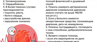
Periodic disturbances in blood supply can also lead to vertebrobasilar vascular insufficiency. VBSD is a reversible process of brain dysfunction. It develops both against the background of osteochondrosis and hypertension. Patients with VBSD experience the following symptoms:
- Nausea and vomiting,
- A sharp deterioration in vision,
- Disorders of consciousness of various types,
- Sudden memory loss,
- Difficulty breathing and swallowing,
- Impaired movement coordination
- Severe dizziness.
The disease can result in a stroke or dyscirculatory encephalopathy.
Symptoms of cervical osteochondrosis
Symptoms of osteochondrosis of the cervical spine manifest themselves differently depending on the stage of development of the pathology. In the early stages it can occur with virtually no symptoms. Pain in the neck and back can only appear if you stay in one position for a long time, sharply bend or turn.
At later stages of development, a crunching sound is heard in the spine, back pain radiates to other limbs, and numbness occurs in parts of the body. When osteophytes and extrusions form, the following occurs:
- headaches in the back of the head and parietal part;
- speech disorders and numbness of the tongue;
- decreased sensitivity of the skin of the neck;
- breathing disorders;
- changes in blood pressure;
- heartbeat disturbances;
- noise and congestion in the ears;
- fainting.
Symptoms of osteochondrosis of the cervical spine in women are much more pronounced than in men. This is due to the fact that women have a predisposition to vascular diseases and a more fragile structure of the bone segments of the spine. Signs of the disease begin to appear when intervertebral discs change. This leads to disruption of normal blood circulation and causes severe headaches, dizziness, and neuroses. An exacerbation of the disease in women often occurs during menopause, when the body is subject to hormonal changes.
The symptoms of cervical osteochondrosis in men are similar to those in women; erectile dysfunction can be observed separately.
Headache with cervical osteochondrosis is caused by poor circulation in the brain and spinal cord. When the vertebrae become misaligned, they compress the arteries and the oxygen content in the blood decreases. Unfortunately, such pain may not go away even after taking strong painkillers. Therefore, it is important to approach solving the problem comprehensively. Dizziness with cervical osteochondrosis may be accompanied by darkening of the eyes and the appearance of tinnitus. This happens because spasming muscles cause a reduction in oxygen supply to the brain.
A lump in the throat with osteochondrosis of the cervical spine, as well as burning, difficulty breathing and muscle spasms are a common occurrence. The disease provokes compression of the nerve fibers of the cervical spine to the head and neck. Disturbances in nerve impulses cause sore throat.
Due to spasms of blood vessels and irritation of nerve endings, jumps in blood pressure occur. Increased lower pressure in osteochondrosis indicates that the blood supply to certain areas of the brain is impaired, since the vertebral artery is compressed by the intervertebral discs. As a result, oxygen starvation occurs and blood pressure rises.
What is intracranial pressure
The brain is surrounded by a fluid called cerebrospinal fluid, which nourishes and protects nerve cells.
Cerebrospinal fluid is continuously produced and flows away from the skull, thereby maintaining a constant pressure. This is intracranial pressure - a certain force that puts pressure on the brain and the walls of the skull. This pressure is changed in mmHg. Art., and normally it is from 10 to 15 mm. If it is higher, this is a reason to be wary, and if the pressure exceeds 25 mm Hg. Art., this can be dangerous for brain function. If the value is more than 35 mmHg. Art. Severe and irreversible changes in the brain are possible - such situations are considered critical.
How is osteochondrosis of the cervical spine diagnosed?
Diagnosis of pathology begins with consultation with a specialist. At the first manifestations of osteochondrosis, consult a rheumatologist, neurologist, surgeon or orthopedic traumatologist. The doctor will ask about the symptoms and the frequency of their occurrence; you need to provide the specialist with a complete medical history and the results of earlier studies (if any). The specialist will conduct a visual examination and palpation, and refer you for tests. During the examination, the doctor pays special attention to neck mobility, muscle tone, skin sensitivity and identifies the most painful areas.
To identify the condition of muscles, ligaments, blood vessels, and to detect inflammatory processes or tumors, an informative and safe diagnostic method is prescribed - MRI of the cervical spine. During an MRI of osteochondrosis, the patient lies on a special sliding table with his back. Rollers are placed on the patient's head to relieve muscle tension, and the limbs are secured with belts. Any slight movement during the procedure can affect the quality of the result. Next, the table moves into the tomograph area. The procedure does not cause pain. The tomograph makes a lot of noise during scanning, so you can use headphones to avoid discomfort.
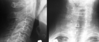
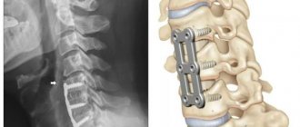
If MRI is contraindicated, there are other diagnostic methods such as computed tomography and radiography. X-ray is suitable only for primary diagnosis and does not provide a layer-by-layer image of the affected tissue. However, this study is the simplest and most economical, allowing you to examine the patient’s body in several projections. Due to the strong radiation exposure to the body, radiography cannot be performed frequently.
During a computed tomography scan, a scan is performed using one or more beams of ionizing rays. They pass through the human body and are recorded by detectors. The detectors move along the patient's body in opposite directions and record up to 6 million signals. The image displays tissues of different densities with precise definition of the boundaries of organs and the affected areas in the form of a section. The procedure allows you to obtain a layer-by-layer image.
Types of exercises
For hypertensive patients, two types of exercises have been developed aimed at normalizing blood pressure:
- Isotonic,
- Isometric.
Isotonic exercises also have another name - cardio exercises. They consist of tensing the main muscle groups of the arms and legs. This leads to burning calories, strengthening muscle tissue and improving posture. Exercising requires a lot of oxygen, which puts more strain on the heart. The result is a decrease in blood pressure.
Isometric or strength exercises aim to build muscle mass in the body. Most often, such exercises are performed in the gym under the supervision of a personal trainer. Doctors approach their prescription with caution: an increase in body weight often provokes an increase in blood pressure. Another disadvantage is that hypertensive patients are not recommended to lift weights, and the isometric set of exercises is designed taking into account the use of heavy sports equipment.
Breathing exercises are gentle and effective. It has no contraindications, but with its help you can increase the flow of oxygen to the lungs, improve the flow of lymph and establish normal metabolism. It does not help cope with osteochondrosis, but it saturates the brain with oxygen, which reduces the symptoms of the underlying disease to a minimum, improves overall well-being and elevates mood.
And for this we have all the most progressive and effective methods:
- shock wave therapy
- dosed traction and massage using the Ormed device
- manual therapy
- kinesio taping
- carboxytherapy
- ozone therapy (intravenous administration of ozonized saline solution and subcutaneous local administration of ozone)
- mesoinjection therapy with vascular drugs
- chronomagnetic complex "Multimag"
- physiotherapeutic techniques (high-top, ultrasound, electrophoresis)
- medical therapeutic blockades