Classification
Heart failure is a very dangerous condition in which the heart is unable to sufficiently perform its functions.
As a result, the organs and all tissues of the body do not receive the required amount of blood, which supplies oxygen and nutrients. This disease has several stages of development, the most dangerous, the third, is decompensation. Heart failure in the decompensation stage often leads to death. In this case, the heart fails to cope with its task not only during physical or emotional stress, but even if the person is in a calm state. This form is irreversible, and therefore so dangerous.
Decompensated heart failure is characterized by:
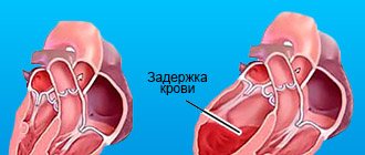
- Enlargement of the myocardium, or rather, its stretching and thinning.
- Fluid retention in the body.
- Rapid decrease in cardiac output.
- Swelling of the myocardium.
The chronic form of decompensation is a long-term process, that is, this pathology develops over the course of years, and at the same time progresses. Its peculiarity lies in the fact that due to any damage to the organ (necrosis, inflammation, dysplasia), the myocardial cells change. This affects its functioning. But the cells that are not yet affected do the work of compensating for the dysfunction of the affected myocytes. Then decompensation occurs, and the heart is not able to pump blood in the required quantity.
Decompensated heart failure is divided by location:
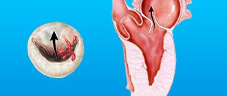
- Pathology of the left ventricle. Diastolic type - in this case the cavity is not able to receive the required amount of blood. This is fraught with overload of the left atrium and congestion in the lungs. But cardiac output into the aorta is still preserved. Systolic type - the left ventricle is dilated, cardiac output is reduced. The contractile function of the ventricle is impaired.
- Pathology of the right ventricle. It is characterized by a stagnant process in the systemic circulation, while the small circulation is insufficiently supplied with blood.
- Mixed form. It is very rare. It is characterized by dysfunction of both the left and right ventricles.
Acute heart failure
DEFINITION.
Acute heart failure, which occurs as a consequence of impaired myocardial contractility, a decrease in systolic and cardiac output, is manifested by several extremely severe clinical syndromes: cardiogenic shock, pulmonary edema, acute decompensated cor pulmonale, etc.
ETIOLOGY AND PATHOGENESIS.
The contractility of the myocardium decreases either as a result of its overload with an increase in the hemodynamic load on the left or right heart, or due to a decrease in the functioning mass of the myocardium or a decrease in the compliance of the chamber walls. Acute heart failure develops when:
- impairment of diastolic and/or systolic myocardial function due to the development of infarction (the most common cause), inflammatory or dystrophic diseases of the myocardium, as well as tachycardia, tachy- and bradyarrhythmias;
- sudden occurrence of myocardial overload in the corresponding part of the heart due to a rapid significant increase in resistance on the outflow tract (in the aorta - hypertensive crisis; in the pulmonary artery - massive thromboembolism of the branches of the pulmonary artery, a prolonged attack of bronchial asthma with the development of acute pulmonary emphysema, etc.) or volume load (increased mass of circulating blood, for example, with massive fluid infusions - a variant of the hyperkinetic type of hemodynamics);
- acute disturbances of intracardiac hemodynamics due to rupture of the interventricular septum or the development of aortic, mitral or tricuspid insufficiency (septal infarction, infarction or avulsion of the papillary muscle, bacterial endocarditis with perforation of the valve leaflets, rupture of the chordae, trauma);
- increased load (physical or psycho-emotional stress, increased inflow in a horizontal position, etc.) on the decompensated myocardium in patients with more or less severe chronic congestive heart failure due to congenital or acquired heart defects, post-infarction cardiosclerosis, hypertrophic or dilated cardiomyopathy.
A decrease in myocardial contractile function leads to a number of compensatory changes in hemodynamics:
- to maintain cardiac output with a decrease in stroke volume, heart rate increases, which is accompanied by a shortening of diastole, a decrease in diastolic filling and leads to an even greater drop in stroke volume;
With a decrease in ventricular contractility, the pressure in the atria and veins increases, resulting in the formation of stagnation in that part of the bloodstream that precedes the chamber of the decompensated myocardium. Increased venous pressure contributes to an increase in diastolic filling of the corresponding chamber and, according to the Frank-Starling law, shock ejection, but an increase in preload leads to an increase in myocardial energy consumption and the progression of decompensation. Acute congestive left ventricular failure is manifested by an increase in pressure in the pulmonary artery system (which is aggravated by the Kitaev reflex - narrowing of the pulmonary arterioles in response to increased pressure in the left atrium), deterioration of external respiration and blood oxygenation, and, when the hydrostatic pressure in the pulmonary capillaries exceeds the oncotic and osmotic pressure , leads first to interstitial and then to alveolar pulmonary edema;
When cardiac output decreases, maintaining a sufficient level of blood pressure is achieved by increasing peripheral resistance. However, this leads to an increase in afterload and a deterioration in tissue perfusion (including the perfusion of vital organs - heart, kidneys, brain), which is especially pronounced when compensatory mechanisms are insufficient and blood pressure decreases.
An increase in peripheral resistance, shunting and sequestration of blood and a slowdown in tissue blood flow, which are primarily characteristic of shock, contribute to the exudation of the liquid part of the blood into the tissue, and therefore hypovolemia, hemoconcentration develop, deterioration of the rheological properties of the blood and conditions are created for the development of thrombotic complications.
With various clinical variants, certain variants of hemodynamic disturbances may come to the fore.
CLINICAL PICTURE AND CLASSIFICATION.
Depending on the type of hemodynamics, the affected chamber of the heart and some features of pathogenesis, the following clinical variants of acute heart failure are distinguished:
| A) with a stagnant type of hemodynamics: - right ventricular (venous congestion in the systemic circulation), - left ventricular (cardiac asthma, pulmonary edema); | B) with hypokinetic 1 type of hemodynamics (small output syndrome - cardiogenic shock): - arrhythmic shock, - reflex shock, - true shock. 1The clinical picture of cardiogenic shock can develop with hypovolemia preceding a heart attack (against the background of active diuretic therapy, profuse diarrhea, etc.) |
Since myocardial infarction is one of the most common causes of acute heart failure, its classification in this disease is of interest (Table 8).
Table 8 Classification of acute heart failure during myocardial infarction (based on Killip T. & Kimball J., 1967)
| Class | Clinical signs of deficiency | Frequency % | Mortality % | Principles of pharmacological treatment |
| I | There are no wheezing or third sounds in the lungs | 33 | 8 | Not required |
| II | Wheezing in the lungs no more than 50% of the surface or the third tone | 38 | 30 | Reduce preload using primarily diuretics |
| III | Crackles in the lungs over more than 50% of the surface (often a picture of pulmonary edema) | 10 | 44 | Reducing preload with diuretics and nitrates, and if ineffective, increasing cardiac output with non-glycoside inotropic agents |
| IV | Cardiogenic shock | 19 | 80-100 | Depending on the clinical variant, severity and type of hemodynamics, different combinations of infusion and inotropic therapy |
Acute congestive right ventricular failure
manifested by venous stagnation in the systemic circulation with increased systemic venous pressure, swelling of the veins (best noticeable in the neck) and liver, and tachycardia; swelling may appear in the lower parts of the body (in a horizontal position - on the back or side). Clinically, it differs from chronic right ventricular failure by intense pain in the liver area, aggravated by palpation. Signs of dilatation and overload of the right heart are determined (expansion of the borders of the heart to the right, systolic murmur over the xiphoid process and protodiastolic gallop rhythm, emphasis of the second tone on the pulmonary artery and corresponding ECG changes). A decrease in left ventricular filling pressure due to right ventricular failure can lead to a drop in the minute volume of the left ventricle and the development of arterial hypotension up to the picture of cardiogenic shock (see).
With pericardial tamponade, constrictive pericarditis, the pattern of congestion in a large circle is not associated with contractile insufficiency of myocardial contractile function, and treatment is aimed at restoring diastolic filling of the heart.
Biventricular failure, when congestive right ventricular failure accompanies left ventricular failure, is not discussed in this section, since its treatment differs little from the treatment of severe acute left ventricular failure.
Acute congestive left ventricular failure
clinically manifests: paroxysmal shortness of breath, painful suffocation and orthopnea, occurring more often at night, sometimes - Cheyne-Stokes breathing, cough (first dry, and then with sputum, which does not bring relief), later - foamy sputum, often colored pink, pallor, acrocyanosis, hyperhidrosis and is accompanied by agitation and fear of death. In case of acute congestion, moist rales may not be heard at first, or a meager amount of fine bubbling rales is detected over the lower parts of the lungs; swelling of the mucous membrane of small bronchi can manifest itself as a moderate picture of bronchial obstruction with prolongation of exhalation, dry wheezing and signs of pulmonary emphysema. For differential diagnostics with bronchial asthma, dissociation between the severity of the condition and (in the absence of a pronounced expiratory nature of shortness of breath and “silent zones”) the paucity of the auscultatory picture can serve. Loud, varied moist rales over all the lungs, which can be heard at a distance - bubbling breathing, are characteristic of a detailed picture of alveolar edema. Possible acute expansion of the heart to the left, the appearance of a systolic murmur at the apex of the heart, a proto-diastolic gallop rhythm, as well as an emphasis on the second tone on the pulmonary artery and other signs of load on the right heart up to the picture of right ventricular failure; Tachycardia up to 120-150 per minute is possible. Blood pressure, depending on the initial level, can be normal, high or low.
The picture of acute congestion in the pulmonary circulation, which develops with stenosis of the left atrioventricular orifice, is essentially left atrial failure, but is traditionally considered together with left ventricular failure.
Cardiogenic shock
– clinical syndrome characterized by arterial hypotension (SBP less than 90-80 mm Hg, or 30 mm Hg below the “working” level in people with arterial hypertension, decreased pulse pressure and signs of a sharp deterioration in microcirculation and tissue perfusion, including blood supply to the brain and kidneys (lethargy or agitation, a drop in diuresis of less than 20 ml per hour, cold skin covered with sticky sweat, pallor, gray cyanosis, marbled skin pattern); sinus tachycardia, which has a compensatory nature.
A decrease in cardiac output with a clinical picture of cardiogenic shock can be observed in a number of pathological conditions not associated with insufficiency of myocardial contractile function - with acute obstruction of the atrioventricular orifice by an atrial myxoma or thrombus of a prosthetic valve, with pericardial tamponade, with massive pulmonary embolism. These conditions are often combined with the clinical picture of acute right ventricular failure. Pericardial tamponade and atrioventricular orifice obstruction require immediate surgical intervention; drug therapy in these cases can only worsen the situation. In addition, the picture of shock during myocardial infarction is sometimes imitated by dissecting aortic aneurysm (see), which requires differential diagnosis, since it requires a fundamentally different therapeutic approach.
There are three main clinical variants of cardiogenic shock:
- arrhythmic shock develops as a result of a drop in cardiac output due to tachycardia/tachyarrhythmia or bradycardia/bradyarrhythmia; after stopping the rhythm disturbance, adequate hemodynamics are quickly restored;
- reflex shock (pain collapse) develops as a reaction to pain and is characterized by a rapid response to pain therapy; absence of signs of congestive heart failure, deterioration of tissue perfusion (in particular, gray cyanosis); pulse pressure usually exceeds a critical level;
- true cardiogenic shock develops when the volume of damage exceeds 40-50% of the mass of the left ventricular myocardium (more often with anterolateral and repeated infarctions, in people over 60 years of age, against the background of arterial hypertension and diabetes mellitus), characterized by a detailed picture of shock that is resistant to therapy , often combined with congestive left ventricular failure; Depending on the chosen diagnostic criteria for this condition, the reported mortality rates (in the absence of surgical treatment) range from 80-100%.
In some cases, especially with the development of myocardial infarction in patients receiving diuretics, the developing shock is hypovolemic in nature, and adequate hemodynamics are relatively simply restored by replenishing the circulating volume.
DIAGNOSTIC CRITERIA.
One of the most consistent signs of acute heart failure is sinus tachycardia (in the absence of sick sinus syndrome, complete AV block or reflex sinus bradycardia); characterized by expansion of the borders of the heart to the left or right and the appearance of the third tone at the apex or above the xiphoid process.
In acute congestive right ventricular failure, the following have diagnostic value:
- swelling of the neck veins and liver,
- Kussmaul's sign (swelling of the jugular veins on inspiration),
- intense pain in the right hypochondrium,
- ECG signs of acute overload of the right ventricle (type S1-Q3, increasing R wave in leads VI,II and formation of a deep S wave in leads V4-6, depression STI,II,aVL and elevation STIII,aVF, as well as in leads V1,2 ; possible formation of blockade of the right bundle branch, negative T waves in leads III, aVF, V1-4) and the right atrium (high pointed waves PII, III).
In acute congestive left ventricular failure, the following are of diagnostic importance:
- shortness of breath of varying severity up to suffocation,
- paroxysmal cough, dry or with foamy sputum, foaming from the mouth and nose,
- orthopnea position,
the presence of moist rales heard over the area from the posterior-lower sections to the entire surface of the chest; local small-bubble rales are characteristic of cardiac asthma; with advanced pulmonary edema, large-bubble rales are heard over the entire surface of the lungs and at a distance (bubbling breathing)
Cardiogenic shock at the prehospital stage is diagnosed based on:
- SBP drops less than 90-80 mmHg. (or 30 mm Hg below the “working” level in people with arterial hypertension),
- decrease in pulse pressure - less than 25-20 mm Hg,
- signs of impaired microcirculation and tissue perfusion - a drop in diuresis of less than 20 ml per hour, cold skin covered with sticky sweat, pallor, gray cyanosis, marbled skin pattern, in some cases - collapsed peripheral veins.
ALGORITHM FOR TREATMENT OF ACUTE HEART FAILURE AT THE PREHOSPITAL STAGE.
For any clinical variant of acute heart failure, prompt correction of the condition that led to the development of such a formidable complication is indicated:
If the cause is cardiac arrhythmia, the basis for normalizing hemodynamics and stabilizing the patient’s condition is restoring normal heart rate.
a) For paroxysms of tachycardia and tachyarrhythmia, electrical pulse therapy is indicated, and if it is impossible to carry out it in the shortest possible time, specific antiarrhythmic therapy is indicated, depending on the nature of the rhythm disturbance (see chapter “ ARRHYTHMIAS ”)
b) In case of tachysystolic form of constant form of atrial fibrillation, atrial fibrillation of unknown duration or paroxysm of atrial fibrillation more than a day old, it is necessary to carry out rapid digitalization by administering digoxin intravenously in an initial dose of 1 ml of 0.025% solution,
c) with sinus bradycardia and sinoatrial block, it is sufficient to increase heart rate by intravenous administration of 0.3-1 ml of 0.1% atropine solution. If it is ineffective and with other bradyarrhythmias - slow rhythm from the AV connection (replacement), AV block of the II-III degree, electrical pacing is indicated. The impossibility of its implementation serves as an indication for drug treatment (for more details, see the chapter “ ARRHYTHMIAS ”);
If the cause is myocardial infarction, then one of the most effective methods of combating decompensation is the rapid restoration of coronary blood flow through the affected artery, which in prehospital care can be achieved using systemic thrombolysis (see chapter “MYOCARDIAL INFARCTION ” );
If acute heart failure is the result of acutely developed disturbances of intracardiac hemodynamics due to trauma, myocardial rupture, or damage to the valve apparatus, emergency hospitalization by a special team to a specialized surgical hospital to provide surgical care is indicated.
However, in practice, it is more often necessary to limit oneself (at least at the first stage of care) to pathogenetic and symptomatic therapy. The main task is to maintain adequate pumping function of the heart, for which different approaches are used depending on the clinical variant of acute heart failure. In any case, oxygen therapy plays a certain role in the fight against hypoxemia - inhalation of humidified oxygen through a nasal catheter at a rate of 6-8 l/min.
Treatment of acute congestive right ventricular failure consists of correcting the conditions that are its cause - pulmonary embolism (see), status asthmaticus (see), etc. This condition does not require independent therapy.
The combination of acute congestive right ventricular failure with congestive left ventricular failure is an indication for therapy according to the principles of treatment of the latter.
The combination of acute congestive right ventricular failure with small output syndrome (cardiogenic shock), caused by a decrease in blood flow into the pulmonary circulation and left ventricle, may require fluid infusion, sometimes coupled with inotropic therapy.
In the treatment of acute congestive left ventricular failure, the following areas are distinguished:
- Reducing preload on the myocardium and pressure in the pulmonary artery, for which they use appropriate body position and medications that have a venous vasodilating effect - Lasix, morphine, nitrates.
- Dehydration.
- Suppression of the respiratory center, reducing the work of the respiratory muscles and thereby providing physical rest to the patient. Suppression of the respiratory center helps to relieve the so-called “respiratory panic” (inappropriately deep and frequent breathing), leading to a further increase in shifts in the acid-base balance.
- Anti-foaming.
- Inotropic therapy (according to strict indications).
- Combating increased membrane permeability (if standard therapy is ineffective).
- Correction of microcirculatory disorders (as an auxiliary measure).
- Treatment of acute congestive heart failure begins with the administration of sublingual nitroglycerin in a dose of 0.5-1 mg (1-2 tablets) and giving the patient an elevated position (in case of an unexpressed picture of congestion - an elevated head end, in case of advanced pulmonary edema - a sitting position with legs down) ; these measures are not performed in cases of severe arterial hypotension.
- A universal pharmacological agent for acute congestive heart failure is furosemide (Lasix, Urix), which reduces the hemodynamic load on the myocardium due to venous vasodilation within 5-15 minutes after IV administration. Thanks to the diuretic effect that develops later, the preload decreases even more. Furosemide is administered intravenously as a bolus without dilution in a dose of 20 mg for minimal signs of congestion to 200 mg for extremely severe pulmonary edema.
- The more pronounced the tachypnea and psychomotor agitation, the more indicated is the addition of a narcotic analgesic to therapy - morphine, which, in addition to venous vasodilation and reducing preload on the myocardium, already 5-10 minutes after administration reduces the work of the respiratory muscles, suppresses the respiratory center, which provides an additional reduction in load on the heart. Its ability to reduce psychomotor agitation and sympathoadrenal activity also plays a certain role; the drug is administered intravenously in fractional doses of 2-5 mg (for which 1 ml of a 1% solution is adjusted to 10 ml with isotonic sodium chloride solution and 2-5 ml are administered) with repeated administration if necessary after 15 minutes. Contraindications are respiratory rhythm disturbances (Cheyne-Stokes breathing), severe depression of the respiratory center, acute obstruction of the respiratory tract, chronic decompensated cor pulmonale, and cerebral edema.
- Severe congestion in the pulmonary circulation in the absence of arterial hypotension, resistant to therapy, or any degree of acute congestive left ventricular failure during myocardial infarction, as well as pulmonary edema against the background of a hypertensive crisis without cerebral symptoms, are indications for intravenous drip administration of nitroglycerin or isosorbide dinitrate. The infusion is carried out under constant monitoring of blood pressure and heart rate at an initial dose of 10-15 mcg/min, followed by an increase every 3-5 minutes by 10 mcg/min until the desired effect is achieved or side effects appear, in particular, a decrease in blood pressure to 90 mmHg . Art. (every 10 mg of the drug is diluted in 100 ml of 0.9% sodium chloride solution; 1 drop of the resulting solution contains 5 mcg of the drug). Contraindications to the use of nitrates are uncompensated arterial hypotension, hypovolemia, pericardial constriction and cardiac tamponade, pulmonary artery obstruction, and inadequate cerebral perfusion.
- With the development of acute left ventricular failure against the background of a hypertensive crisis with cerebral symptoms, intravenous drip administration of a mixed vasodilator of myotropic action sodium nitroprusside (50 mg in 250 ml of 5% glucose solution) is indicated under the control of blood pressure and heart rate at an initial dose of 0.5 mcg/kg min, or 20 mcg/min with its increase every 5 minutes by 5 mcg/min until the expected effect is achieved (average dose - 1-3 mcg/kg min), the maximum rate of administration (5 mcg/kg min) or the development of side effects. Unlike nitrates, sodium nitroprusside not only reduces preload, but, by increasing arterial flow to tissues, in particular, increasing cerebral and renal blood flow, it also reduces afterload, which leads to a reflex increase in cardiac output. Sodium nitroprusside is more likely than nitrates to cause steal syndrome; Contraindications to its use are coarctation of the aorta and arteriovenous shunts; it requires special caution in old age.
- Modern vasodilating therapy, including powerful diuretics, has reduced to a minimum the importance of bloodletting and the application of venous tourniquets to the extremities, however, if it is impossible to carry out adequate therapy due to the lack of drugs, these methods not only can, but should be used, especially with rapidly progressing pulmonary edema ( bloodletting in a volume of 300-500 ml).
- In case of acute congestive left ventricular failure, combined with cardiogenic shock or with a decrease in blood pressure during therapy that has not given a positive effect, the addition of non-glycoside inotropic agents to treatment is indicated - intravenous administration of dobutamine at a dose of 2.5-10 mcg/kg min, dopamine 5 -20 mcg/kg/min. Persistent hypotension with SBP below 60 mm Hg. requires the addition of norepinephrine infusion (see “Treatment of cardiogenic shock” ).
- The means of directly combating foaming during pulmonary edema are “defoamers” - substances that ensure the destruction of foam, reducing surface tension. The simplest of these means is alcohol vapor. Alcohol is poured into a humidifier, passing oxygen through it, supplied to the patient through a nasal catheter or breathing mask at an initial rate of 2-3 liters, and after a few minutes - at a rate of 6-8 liters of oxygen per minute (less effective is the use of cotton wool moistened with alcohol, inserted into a mask); the disappearance of bubbling breathing is observed within a period of 10-15 minutes to 2-3 hours; the simplest method - spraying alcohol in front of the patient's mouth using any pocket inhaler or a regular spray bottle - is the least effective; in extreme cases, intravenous administration of 5 ml of 96% ethyl alcohol in the form of a 33% solution is possible.
- Persistent signs of pulmonary edema during stabilization of hemodynamics may indicate an increase in membrane permeability, which requires the administration of glucocorticoids for membrane stabilizing purposes (4-12 mg of dexamethasone).
- In the absence of contraindications, in order to correct microcirculatory disorders, especially with long-term intractable pulmonary edema, heparin is indicated - 5000 IU intravenously as a bolus, then drip at a rate of 1 thousand IU/hour with continued therapy in the hospital (see chapter “ MYOCARDIAL INFARCTION ” ).
Treatment of cardiogenic shock involves increasing cardiac output and improving peripheral blood flow in the following ways.
- Relief of adverse reflex effects on hemodynamics.
- Combating heart rhythm disturbances.
- Ensuring adequate venous return and diastolic filling of the left ventricle, combating hypovolemia and disturbances in the rheological properties of blood.
- Restoration of adequate tissue perfusion of vital organs.
- Stimulation of myocardial contractility with non-glycoside inotropic agents.
In the absence of signs of congestive heart failure (shortness of breath, moist rales in the posterior lower parts of the lungs), the patient must be given a horizontal position or even a Trendelenburg position (with the head end lowered), which contributes to an increase in venous return, an increase in cardiac output, and also an improvement in cerebral blood flow during centralized circulation .
Regardless of the characteristics of the clinical picture, it is necessary to provide complete analgesia (see chapter “ MYOCARDIAL INFARCTION ”).
Stopping rhythm disturbances (see above) is the most important measure to normalize cardiac output, even if, after restoration of normosystole, there is no restoration of adequate hemodynamics. Bradycardia may indicate increased vagal tone and requires immediate intravenous administration of 0.5-1 ml of 0.1% atropine solution.
To combat hypovolemia that occurs with right ventricular infarction or with previous dehydration (long-term use of diuretics, profuse sweating, diarrhea), intravenous isotonic sodium chloride solution is used in an amount of up to 200 ml over 10-20 minutes with repeated administration of a similar dose if there is no effect or complications.
The lack of effect from the entire complex of therapeutic measures, including the active fight against hypovolemia, or the combination of cardiogenic shock with congestive heart failure is an indication for the use of inotropic agents from the group of pressor amines.
a) dopamine (dopmin) at a dose of 1-5 mcg/kg min has a vasodilating effect, at a dose of 5-15 mcg/kg min - vasodilating and positive inotropic (and chronotropic) effects, and at a dose of 15-25 mcg/kg min - positive inotropic (and chronotropic) and peripheral vasoconstrictive effects. 200 mg of the drug is dissolved in 400 ml of 5% glucose solution (1 ml of the mixture contains 0.5 mg, and 1 drop - 25 mcg of dopamine); the initial dose is 3-5 mcg/kg min with a gradual increase in the rate of administration until the effect is achieved, the maximum dose (25 mcg/kg min) or the development of complications (most often sinus tachycardia exceeding 140 per minute, or ventricular arrhythmias). Contraindications to its use are thyrotoxicosis, pheochromocytoma, cardiac arrhythmias, hypersensitivity to disulfide, previous use of MAO inhibitors; If you have previously taken tricyclic antidepressants, the dose should be reduced.
b) dobutamine (Dobutrex), unlike dopamine, does not affect dopaminergic receptors, has a more powerful positive inotropic effect and a less pronounced ability to increase heart rate and cause arrhythmias. 250 mg of the drug is diluted in 500 ml of 5% glucose solution (1 ml of the mixture contains 0.5 mg, and 1 drop - 25 mcg of dobutamine); in monotherapy it is prescribed at a dose of 2.5 mcg/kg/min. with an increase every 15-30 minutes. by 2.5 mcg/kg/min. until an effect, side effect is obtained or a dose of 15 mcg/kg/min is achieved, and when dobutamine is combined with dopamine - in the maximum tolerated doses. Contraindications to its use are idiopathic hypertrophic subaortic stenosis, stenosis of the aortic mouth;
c) if it is not possible to use other pressor amines or if dopamine and dobutamine are ineffective, norepinephrine can be used in an increasing dose not exceeding 16 mcg/min (if combined with an infusion of dobutamine or dopamine, the dose should be halved). Contraindications to its use are thyrotoxicosis, pheochromocytoma, previous use of MAO inhibitors; if you have previously taken tricyclic antidepressants, the dose should be reduced;
If there are signs of congestive heart failure and in the case of using inotropic drugs from the group of pressor amines, the administration of peripheral vasodilators - nitrates (nitroglycerin or isosorbide dinitrate at a rate of 5-200 mcg/min) or sodium nitroprusside (at a dose of 0.5-5 mcg/kg) is indicated /min).
In the absence of contraindications, in order to correct microcirculatory disorders, especially with long-term intractable shock, heparin is indicated - 5000 IU intravenously as a bolus, then drip at a rate of 1 thousand IU/hour with continued therapy in the hospital (see chapter “ MYOCARDIAL INFARCTION ”).
COMMON THERAPY ERRORS.
Acute heart failure is a life-threatening condition, and therefore incorrect therapy can be fatal. All identified treatment errors are due to outdated recommendations , which are partially preserved in some modern standards of medical care at the prehospital stage.
The most common mistake in all clinical types of acute heart failure is the prescription of cardiac glycosides. In conditions of hypoxemia, metabolic acidosis and electrolyte disorders, which are invariably present in acute heart failure and cause increased sensitivity of the myocardium to digitalis, glycosides increase the risk of developing serious rhythm disturbances. The inotropic effect is achieved late and affects both the left and right ventricles, which can lead to an increase in pulmonary hypertension.
In acute heart failure , attempts to stop paroxysmal arrhythmias with drugs rather than by electrical cardioversion are dangerous, since most of the antiarrhythmic drugs used have a pronounced negative inotropic effect (exceptions include lidocaine for ventricular paroxysmal tachycardia and magnesium sulfate for ventricular paroxysmal tachycardia of the “pirouette” type). Equally dangerous are drug attempts to combat bradyarrhythmias, taken instead of cardiac pacing, which is not always effective and can be fraught with the development of fatal arrhythmias or an increase in myocardial oxygen demand.
In acute left ventricular failure ( both in small output syndrome and in congestive hemodynamics), glucocorticoid hormones are still widely used.
In case of cardiogenic shock, they are inferior to modern drugs in their effect on hemodynamics, but their use in large doses worsens potassium deficiency and increases the risk of arrhythmias, even fatal ones, and in case of myocardial infarction, myocardial ruptures become more frequent and scarring processes worsen (their use may be justified only in acute myocarditis).
In case of congestive left ventricular failure, aminophylline is traditionally used, to reduce pressure in the pulmonary artery system, stimulate diuresis and unloading is not justified, since modern drugs with vasodilating and diuretic activity (see above) are much more effective in this regard and, unlike aminophylline, are not increase myocardial oxygen demand and do not have an arrhythmogenic effect. Recommendations for the use of aminophylline to combat broncho-obstruction accompanying edema also seem unconvincing, since the obstruction is caused not so much by bronchospasm as by edema of the mucous membrane. In addition, obstruction, by increasing expiratory resistance, increases pressure in the alveoli, which partially prevents further exudation of fluid.
In case of cardiogenic shock , mesaton is relatively often used, which has very narrow indications only for the reflex nature of arterial hypotension caused by peripheral vasodilation. Mezaton does not increase cardiac output, but causes only peripheral vasoconstriction, which in most cases leads to worsening disturbances in the blood supply to vital organs, increasing the load on the myocardium and worsening the prognosis.
Relatively often one has to deal with the administration of pressor amines during cardiogenic shock without a preliminary attempt to compensate for hypovolemia, which, with inadequate blood volume, is fraught with the development of a critical state of microcirculation with a fatal deterioration in prognosis. However, excessive use of plasma expanders can provoke the development of congestive heart failure.
Unlike chronic, acute right ventricular failure should not resort to bloodletting.
INDICATIONS FOR HOSPITALIZATION.
Acute circulatory failure is a direct indication for hospitalization in the intensive care unit (unit) or cardiac intensive care unit.
If possible, patients with cardiogenic shock should be hospitalized in a hospital with a cardiac surgery department, because modern ideas about the treatment of this condition are clearly associated with aortic balloon counterpulsation and early surgical intervention.
Transportation is carried out on a stretcher in a horizontal position (for cardiogenic shock and right ventricular failure) and in a sitting position for congestive left ventricular failure.
previous article
Point of view about Beta blockers
next article
The Ministry of Health of the Republic of Belarus is 100 years old!
Top
Symptoms
Pathology exhibits quite a lot of symptoms. There are no specific signs by which the disease can be accurately diagnosed.
Decompensated heart failure is a consequence of other heart diseases, so the clinical picture is quite varied, as some symptoms overlap with others.
The main symptoms of decompensated heart failure:
- Dyspnea. It manifests itself under light loads, but if there is no appropriate treatment, the pathology progresses and is disturbing, even if the person is in a calm state. This occurs due to congestion in the lungs.
- Swelling of the legs and arms. They are explained by impaired outflow of fluid from the body. In this case, the heart muscle is also affected.
- Dry cough.
- Pain or discomfort in the right hypochondrium.
- Ascites is dropsy in the abdominal cavity. At the same time, the stomach and weight of a person increase significantly.
- General weakness, feeling of overwork.
- Cold feet and hands.
- Small amount of urine, rare urge to urinate.
During examination, doctors detect wheezing in the lungs, arrhythmia, and congestive processes. There is an increase in pressure in the jugular vein.
Detection methods
Since symptoms of cardiopulmonary failure may indicate a number of other diseases, cardiologists, to determine an accurate diagnosis, carry out a number of necessary measures, which include:
- Study of patient complaints.
- X-ray examination of the lungs, which allows you to determine changes in the pulmonary system and the size of the heart.
- If the previous study did not lead to an accurate diagnosis, an additional CT scan of the chest is performed.
- An ultrasound is required to determine the extent of heart damage.
- To determine the pressure in the lungs and heart, invasive manometry and catheterization are performed.
- In advanced cases, it is necessary to conduct an ECG, which will show the degree of changes in the organs.
Causes
The causes of chronic heart failure in the decompensation stage are various diseases of the cardiovascular system that were not treated in time.
Decompensated heart failure occurs for the following reasons:
- Hypertension, or rather, a hypertensive crisis can trigger an irreversible process.
- Congenital heart defects. These are abnormalities of the heart valves, as a result of which cardiac function fails.
- Myocarditis.
- Stable tachyarrhythmia.
- Hypertrophic type cardiomyopathy.
The above reasons relate to cardiac pathologies. Other factors can also cause heart failure:
- Chronic alcoholism.
- Poisoning of the body of a bacterial-toxic type.
- Bronchial asthma, which was not treated and became advanced.
- Incorrect treatment or its absence at the first stage of the pathology.
- Obesity.
- Diabetes.
Clinical course of the disease
HF can have an acute onset and a chronic course. The signs are:
- Dyspnea (shortness of breath) during exercise or at rest, suffocation
- Weakness, lethargy
- Swelling of the feet, ankles and legs
- Swelling of the neck veins
- Pain in the right hypochondrium
- Cardiopalmus
- Inability to perform usual physical activities
- Persistent cough with light-colored or pinkish sputum
- Need to urinate at night
- Ascites
- Loss of appetite or nausea
- Inability to concentrate, absent-mindedness
- Chest pain relieved by taking aminophylline
- Sudden attacks of suffocation with coughing and discharge of foamy pink sputum
Emergency medical attention is necessary if the patient experiences:
- Chest pain
- Loss of consciousness or severe weakness
- Fast or irregular heartbeat associated with shortness of breath, dizziness, or chest pain
- Sudden cough with pink sputum or coughing up blood
- Bluishness around the mouth, tip of the nose and fingers
If you have already been diagnosed with heart failure or another cardiac disease and experience the symptoms described above, seek medical help.
Treatment
Before prescribing a treatment method and specific medications, the doctor conducts a full examination. This includes studying the medical history, since if the last stage of the disease has developed, then the person already has a medical history. A physical examination is also performed.
The patient must undergo blood and urine tests. Instrumental studies include x-ray and echocardiography. Modern diagnostic methods include MRI and CT. They are very informative and can be prescribed instead of x-rays and ultrasound. After the necessary studies, medications are prescribed.
Treatment should be aimed at eliminating decompensation of the body and restoring its basic functions, neutralizing stagnant processes. Therapy must be carried out under the supervision of doctors, that is, only in a hospital setting. The patient needs bed rest; any stress (physical and emotional) must be excluded. The patient should periodically sit down or take a few steps. This will help prevent stagnation and the formation of blood clots.
Drug therapy
Decompensated heart failure is treated with a complex of various drugs. It is important to take:
- ACE inhibitors.
- Beta inhibitors. These drugs help reduce myocardial demand.
- Cardiac glycosides. These drugs help increase cardiac output.
- Aldosterone antagonists. These drugs increase blood pressure and remove excess fluid from the body.
- Antiarrhythmic drugs.
It is important to take antihypertensive drugs, drugs that affect the rheological quality of blood and lipid metabolism. Additionally, diuretics are required.
For advanced heart failure, other medications are also prescribed. Since the whole body suffers from cardiac dysfunction, it is necessary to take hepatoprotectors, immunomodulators and vitamin complexes.
It is important that a complex of drugs is prescribed by a qualified specialist, because all heart medications have a fairly large list of side effects. Only a doctor can prescribe therapy based on all the individual characteristics of the patient’s body in order to reduce the likelihood of such actions affecting the body.
Surgical operations
Most often, surgical treatment is prescribed for cardiac decompensation. These methods include:
- vascular bypass;
- coronary angioplasty;
- installation of a defibrillator or pacemaker.
Radical surgical treatment is prescribed to patients who have irreversible changes in the structure of organs. This may require a lung, heart or large artery transplant.
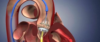
In case of heart failure of the left ventricle, patients are implanted with a special device that functions for it. Some time ago, doctors installed this device temporarily, before a heart transplant, but now it has been proven that it significantly prolongs the life of patients.
Heart failure in the stage of decompensation is a serious condition in which in most cases death occurs. Therefore, self-medication in this case is simply contraindicated. According to statistics, about 75% of men and 62% of women do not live more than 5 years with this pathology. But such figures are due to the fact that people do not consult doctors in a timely manner.
First aid for pulmonary edema
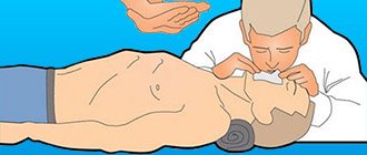
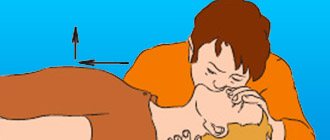
Unlike the chronic form, the acute form of the pathology requires immediate assistance during an attack. A person is not able to help himself, so he needs help from the outside. First of all, you should call an emergency team. While waiting for her, the person should be seated on a chair and given a Nitroglycerin tablet. If the attack occurs indoors, be sure to open all windows to provide fresh air flow.
If a person gets worse, cardiopulmonary resuscitation methods are required. To do this, rhythmically press on the chest area or do 30 presses and mouth-to-mouth breathing (alternating these actions). The main goals of these measures are to normalize blood circulation and restore respiratory function. If you lack the skills, you can simply massage the sternum.
Connection between the vascular and cardiac systems
To know what cardiovascular failure is, you need to understand the mechanism of interaction between the heart and blood vessels.
The heart normally functions as a pump that pumps blood using the capabilities of the heart muscle - the myocardium.
Arterioles, the smallest arteries, transport oxygen to tissues and can change the direction of blood through a spasmodic contraction: the tissues will not receive enough oxygen, and the blood will be directed into the vessels.
If the vascular lumen increases due to the tone of the arteriole walls, blood circulation accelerates.
There are mechanisms that regulate tone:
- The impact of an impulse sent by the nervous system;
- Hormones produced by the adrenal glands (norepinephrine, adrenaline), and others (serotonin, angiotensin II);
- Substances arising from oxidative reactions.
Connection between the vascular and cardiac systems
These mechanisms begin to work in stressful situations, when most of the blood passes into the peripheral vessels. Some toxic substances and medications also change the tone.
Capillaries, which are even smaller vessels, transport blood cells and remove waste products. About 12% of blood is contained in them. If the tone in the vessels is weakened, the amount of blood in the capillaries will increase.
The bulk of the blood is concentrated in the veins, which adapt to changes in blood levels.
Venous pressure controls the flow of blood into the right atrium and increases when:
- Heart failure;
- The presence of an obstruction in the pulmonary artery (blockage by a blood clot);
- Excessive fluid content.
Decreases when:
- Intense blood loss;
- Reducing the tone of the walls of peripheral vessels;
- A sharp decrease in the amount of fluid with diarrhea and vomiting.
Excessive vascular tone in the periphery greatly increases the load on the heart muscle, which requires more effort to pump blood.
If the capillary tone decreases, the bulk of the blood is retained and a sufficient amount does not flow to the heart, which leads to oxygen starvation of the myocardium.
The essence of the concept
To put it simply, heart failure is a syndrome of ventricular dysfunction or failure of the pumping function of the heart, in which the myocardial tissue is slowly replaced by fibrous tissue. Either one ventricle or both can work poorly: problems with the left one cause shortness of breath, cardiac asthma and fatigue, and problems with the right one cause swelling of the limbs and abdominal cavity.
In fact, cardiovascular failure (CVF) is a complication of cardiac diseases: hyper- and hypotension, arrhythmias, heart attack, valvular lesions, myocarditis and cardiomyopathies.
At the same time, the situation with its prevalence in the world is such that in the latest revision of the International Classification of Diseases (ICD 10), heart failure is already considered as an independent disease, and it is assigned a separate code - I50.
In Russia, SSN is a “women’s disease”, and in developed countries it is more common in men
Heart failure has a cascade of negative long-term effects:
- processes that retain H2O and Na in tissues are enhanced;
- renal blood flow decreases, including from taking medications that normalize blood pressure;
- the synthesis of aldosterone-II increases, leading to the deposition of interstitial collagen in blood vessels and myocardium, as well as the development of fibrosis in their tissues;
- the production of norepinephrine and vasopressin increases - hormones that accelerate the death of myocardial cells programmed by nature;
- hypertrophic remodeling of vascular and coronary walls is accelerated;
- the synthesis of nitric oxide, which is responsible for maintaining normal afterload indicators, decreases;
- the use of free fatty acids unreasonably increases, and glucose utilization decreases.
SHF enhances the progress of renal dysfunction, thereby aggravating its own course. A separate term has been coined for this condition – cardiorenal syndrome.
For your information. As long as a woman's body synthesizes large amounts of estrogen, her heart is relatively protected from cardiovascular disease. After menopause, the weaker sex catches up and is ahead of men in these statistics. But hormone replacement therapy does not help prevent heart failure, and if present, it can cause reactive pulmonary embolism and death.
The domestic reality is that among 1,500 people, 175 have SSN. Half of them need to be treated in a hospital 2-3 times a year, otherwise they will die quickly. Such regular hospital treatment is especially important in old age.
Diagnostics
To make a diagnosis, the doctor must perform the following actions and analyze the results of the following studies:
- Collecting anamnesis - what complaints are bothering you, when they appeared, how they change under the influence of stress or medications; presence of concomitant diseases
- Examination – presence of edema, dilated veins on the anterior abdominal wall and neck
- Physical examination - auscultation and percussion of the lungs determines the level of fluid in the pleural cavity without the use of x-rays. Tapping the cardiac borders allows you to assess the size of the heart, especially the right half. Listening to murmurs is necessary to diagnose valve defects and changes in the density and structure of lung tissue.
- General clinical tests - blood for cellular composition, lipids, protein, glucose, bilirubin, coagulogram and others.
- Assessment of blood gas composition - percentage of oxygen and carbon dioxide.
- Study of external respiration function (spirography)
- X-ray of the chest - provides information about the condition of the chest organs
- Electrocardiogram
- ECHO-KG
- Catheterization of the right ventricle to measure pressure in the branches of the pulmonary artery
- Lung biopsy
- CT, MRI of the chest - visualize the size, structure of the heart and its relationship with neighboring organs, as well as the structure of the lungs and the condition of the pleural cavity.
- Angiography of the pulmonary vessels - allows you to evaluate the patency of the arteries by introducing an iodine-containing substance through a catheter under the control of an X-ray machine.
Prevention
In order to prevent the development, as well as to prevent the progression of acute heart failure that has already occurred, it is recommended to adhere to a number of measures:
- timely consultation with a cardiologist if cardiac pathology is suspected;
- sufficient physical activity (regular, but not exhausting);
- balanced diet;
- body weight control;
- timely treatment and prevention of diseases that can lead to acute heart failure;
- rejection of bad habits.
Video from YouTube on the topic of the article: