Heparin
Heparin sodium is administered intravenously (as a continuous infusion or repeated boluses) or subcutaneously. Heparin sodium should not be administered intramuscularly due to the risk of developing intramuscular hematomas.
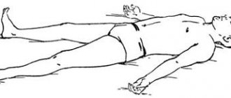
Subcutaneous injections are preferably performed in the anterior abdominal wall. As an exception, other injection sites can be used (outer thigh, shoulder) if subcutaneous adipose tissue is sufficiently developed. It is not recommended to re-inject sodium heparin into previous injection sites.
Recommended therapeutic (full) doses of heparin sodium for adult
patients:
| Continuous intravenous infusion | Initial dose | 5000-10000 IU IV bolus |
| Continuous infusion | 20000-40000 IU/day (administration rate is about 1000 IU/hour) | |
| Bolus intravenous introduction | Initial dose: | 10000 ME |
| Maintenance doses | 5000-10000 IU every 4-6 hours | |
| Subcutaneous introduction | Initial dose: | 333 IU/kg (with a body weight of less than 75 kg - 20,000 IU, with a body weight of 75-90 kg - 25,000 IU, with a body weight of 90-105 kg - 30,000 IU, with a body weight of more than 105 kg - 35,000 IU) |
| Maintenance doses | 250 IU/kg (15,000-25,000 IU) every 12 hours. |
Laboratory monitoring of the effectiveness and safety of sodium heparin therapy
The dose of heparin sodium should be adjusted based on laboratory blood clotting parameters. When using heparin sodium, it is necessary to monitor the activated partial thromboplastin time (aPTT) or blood clotting time (BCT). The administered dose of heparin sodium is considered adequate if the aPTT is 1.5-2.0 times higher than normal values or if the patient's ICT is 2.5-3.0 times higher than control values.
With continuous intravenous infusion
sodium heparin, it is recommended to determine the initial aPTT, then determine the aPTT every 4 hours, followed by increasing or decreasing the rate of sodium heparin infusion until the target aPTT level is reached (1.5-2 times higher than normal), then determine the aPTT every 6 hours.
With bolus intravenous administration
heparin sodium, it is recommended to determine the initial aPTT, then determine the aPTT before each bolus injection, followed by an increase or decrease in the administered dose of heparin sodium.
When administered subcutaneously
sodium heparin, it is recommended to monitor the aPTT 4-6 hours after injection with a subsequent increase or decrease in the administered dose of sodium heparin.
When using sodium heparin in low doses to prevent thromboembolic complications, it is not necessary to monitor the aPTT.
Use of heparin sodium in special clinical situations
Primary percutaneous coronary angioplasty for acute non-ST segment elevation coronary syndrome and ST-segment elevation myocardial infarction
: Heparin sodium is administered intravenously as a bolus at a dose of 70-100 U/kg (if the use of glycoprotein IIb/IIIa receptor inhibitors is not planned) or at a dose of 50-60 U/kg (when used together with glycoprotein IIb/IIIa receptor inhibitors).
Thrombolytic therapy for ST-segment elevation myocardial infarction:
Heparin sodium is administered intravenously as a bolus at a dose of 60 U/kg (maximum dose 4000 U), followed by intravenous infusion at a dose of 12 U/kg (not more than 1000 U/hour) for 24-48 hours. The target APTT level is 50-70 sec or 1.5-2.0 times higher than normal; APTT monitoring 3.6, 12 and 24 hours after the start of therapy.
Prevention of thromboembolic complications after surgery using low doses of sodium heparin:
s/c, deep into the fold of the skin of the abdomen. The initial dose is 5000 IU 2 hours before surgery. In the postoperative period: 5000 IU every 8-12 hours for 7 days or until the patient’s mobility is completely restored (whichever comes first). When using sodium heparin in low doses to prevent thromboembolic complications, it is not necessary to monitor the aPTT.
Application in cardiovascular surgery during operations using extracorporeal circulation systems:
the initial dose of sodium heparin is not less than 150 IU/kg body weight. Next, sodium heparin is administered by continuous intravenous infusion at a rate of 15-25 drops/min, 30,000 IU per 1 liter of infusion solution. The total dose of heparin sodium is usually 300 IU/kg body weight (if the expected duration of the operation is less than 60 minutes) or 400 IU/kg body weight (if the expected duration of the operation is 60 minutes or more).
Use in hemodialysis:
Initial dose of heparin sodium: 25-30 IU/kg (or 10,000 IU) intravenous bolus, then continuous infusion of heparin sodium 20,000 IU/100 mg sodium chloride solution at a rate of 1500-2000 IU/hour (unless otherwise indicated in the instructions for use of the systems for hemodialysis).
Use of heparin sodium in pediatrics:
Adequate controlled studies of the use of heparin sodium in children have not been conducted. The recommendations presented are based on clinical experience.
Initial dose: 75-100 units/kg IV bolus over 10 minutes
Maintenance dose: children aged 1-3 months - 25-30 Units/kg/hour (800 Units/kg/day), children aged 4-12 months - 25-30 Units/kg/hour (700 Units/kg/day day), children over 1 year old - 18-20 units/kg/hour (500 units/kg/day) intravenously.
The dose of heparin sodium should be adjusted based on blood coagulation parameters (target aPTT 60-85 seconds).
Content
- 1 Heparin 1.1 Chemical properties and mechanism of action 1.1.1 Related glycosaminoglycans
- 1.1.2 Sources
- 1.3.1 Other properties of heparin
- 2.1 Pharmacokinetics
- 2.3.1 Bleeding
Read also[edit | edit code]
- Treatment of thrombosis
- Hirudin
- Fibrinolytics
- Thrombosis
- Antiplatelet agents
- Plasma substitutes
- Hemostasis
- Drugs that affect blood clotting
- Hemostatics (coagulants, fibrinolysis inhibitors)
- Agents that reduce vascular permeability
- Anticoagulant blood system
- Direct anticoagulants
- Indirect anticoagulants
- Agents affecting fibrinolysis
- Agents affecting platelet aggregation
Heparin[edit | edit code]
Historical reference
. In 1916, medical student McLane, who was studying the nature of ether-soluble procoagulants, was lucky enough to discover a phospholipid anticoagulant. Shortly thereafter, Govell, in whose laboratory McClain worked, discovered a water-soluble glycosaminoglycan, named heparin because of its high content in the liver (Jaques, 1978). Heparin's success in preventing coagulation in vitro led to its later use in the treatment of venous thrombosis.
Chemical properties and mechanism of action[edit | edit code]
Heparin
- glycosaminoglycan found in mast cell granules. During its synthesis, a polymer consisting of alternating D-glucuronic acid and N-acetyl-glucosamine residues is formed from various UDP sugars (Bourin and Lindahl, 1993). Approximately 10-15 such glycosaminoglycan chains (200-300 monosaccharides each) are attached to the protein part of the molecule, forming a proteoglycan with a molecular weight of 750,000-1,000,000. Then modification of the glycosaminoglycan chains occurs: N-deacetylation and N-sulfation of glucosamine residues, epimerization D-glucuronic acid into L-iduronic acid, O-sulfation of residues of these acids in position 2, O-sulfation of glucosamine residues in positions 3 and 6 (Fig. 55.2). Since these reactions do not affect all monosaccharides, the structure of the resulting molecules is quite diverse. The glycosaminoglycan chains of heparin transferred to mast cell granules are cleaved by p-glucuronidase into fragments with a molecular weight of 5000-30,000 (on average about 12,000, that is, 40 monosaccharides) within a few hours.
Related glycosaminoglycans[edit | edit code]
Heparan sulfate is present on the cell membrane of most eukaryotic cells and in the extracellular matrix. It is synthesized from the same repeating disaccharide sequences as heparin (D-glucuronic acid and N-acetylglucosamine), but undergoes less modification and therefore contains more D-glucuronic acid and N-acetylglucosamine and fewer sulfate groups. Heparan sulfate also has anticoagulant properties in vitro, but at much higher concentrations.
Dermatan sulfate is a polymer of L-iduronic acid and N-acetylgalactosamine with varying degrees of O-sulfation of L-iduronic acid at position 2 and galactosamine at positions 4 and 6. Like heparan sulfate, dermatan sulfate is present on the cell membrane and in the extracellular matrix and has anticoagulant properties in vitro.
Sources[edit | edit code]
Heparin is usually obtained from bovine lung or porcine intestinal lining. Such drugs may contain a small admixture of other glycosaminoglycans. Although the composition of heparins from different manufacturers is somewhat different, their biological activity is approximately the same (about 150 units/mg). 1 unit is taken to be the amount of heparin that prevents the clotting of 1 ml of citrated sheep plasma within an hour after adding 0.2 ml of 1% CaCl2.
Low molecular weight heparins with a molecular weight of 1000–10,000 (average 4500, i.e. 15 monosaccharides) are obtained from a conventional preparation by gel filtration, ethanol precipitation or partial depolymerization using nitrous acid and other reagents. Low molecular weight heparins differ from regular heparins and from each other in pharmacokinetic properties and mechanism of action (see below). Their activity is usually determined by inhibition of factor Xa.
Physiological role[edit | edit code]
Heparin is found in tissues inside mast cells. It appears to be required for the storage of histamine and some proteases within the granules of these cells (Humphries et al., 1999; Forsberg et al., 1999). Once released from mast cells, heparin is quickly taken up and destroyed by macrophages. It cannot be detected in the plasma of healthy people. However, in patients with systemic mastocytosis with massive degranulation of mast cells, a slight prolongation of the aPTT is sometimes observed, presumably associated with the release of heparin into the bloodstream.
Heparan sulfate molecules on the surface of endothelial cells and in the extracellular matrix of the subendothelial layer interact with antithrombin III, preventing thrombus formation. With malignant neoplasms, bleeding is sometimes observed, caused by heparan sulfate or dermatan sulfate entering the bloodstream (probably due to tumor disintegration).
Mechanism of action[edit | edit code]
In 1939, Brinkhaus et al. discovered that the anticoagulant effect of heparin is mediated by one of the plasma components and called it the heparin cofactor. Thirty years later, it was discovered to be antithrombin III, a plasma protein that rapidly inactivates thrombin in the presence of heparin (Olson and Bjork, 1992). Antithrombin III is a glycosylated single-chain polypeptide with a molecular weight of approximately 58,000, homologous to the serpin family (spin yrothease inhibitors), in particular agantitrypsin. Antithrombin III is synthesized in the liver, its serum concentration is 2.6 µmol/l. It is active against factors of the intrinsic and general coagulation mechanisms (in particular, 1Xa, Xa and thrombin), but has little effect on factor Vila. The mechanism of the inhibitory effect of antithrombin III is as follows. The listed coagulation factors, as already mentioned, are proteases. Antithrombin III acts as their substrate: active coagulation factors attack a specific peptide bond between arginine and serine in the reactive center of its molecule. However, cleavage of this bond does not occur, and a stable complex is formed from the coagulation factor and antithrombin III in an equimolar ratio. As a result, the coagulation factor loses its proteolytic activity.
Heparin accelerates the interaction of antithrombin III with thrombin by more than 1000 times due to the fact that it serves as a matrix linking both proteins. Binding to heparin also changes the conformation of antithrombin III, making its reactive site more accessible to thrombin (Jin et al., 1997). After the formation of the thrombin-antithrombin III complex, the heparin molecule is released. The region of the heparin molecule responsible for binding to antithrombin III is a pentasaccharide sequence containing a glucosamine residue O-sulfated at position 3 (Fig. 55.2). This structure is found in approximately 30% of heparin molecules and, less commonly, in heparan sulfate. Other glycosaminoglycans (dermatan sulfate, chondroitin sulfates) lack this structure and are not able to activate antithrombin III. Heparins with a molecular weight of less than 5400 (containing less than 18 monosaccharides) cannot bind both antithrombin III and thrombin and therefore do not accelerate the inactivation of the latter. At the same time, shown in Fig. 55.2 pentasaccharide catalyzes the inhibition of factor Xa by antithrombin III (apparently, only conformational changes in antithrombin III are sufficient for this). This explains the anticoagulant effect of low molecular weight heparins, most of whose molecules are too short to bind thrombin.
Antithrombin III quickly (T1/2 <0.1 s) inhibits factors 1Xa, Xa and thrombin at plasma heparin concentrations of 0.1 - 10 units/ml. At the same time, the aPTT and thrombin time (plasma clotting time when thrombin is added) are prolonged; PV changes less. Factor Xa on the surface of platelets (as part of the prothrombinase complex) and thrombin bound to fibrin are not inhibited by heparin and antithrombin III.
Figure 55.2. Antithrombin-binding region of the heparin molecule.
Thus, heparin accelerates the inactivation of factor Xa and jumbin only after they are released from their binding sites. Platelet factor 4, released from α granules during platelet aggregation, interferes with the binding of antithrombin III to heparin and heparan sulfate, promoting thrombus formation at the site of clotting.
When the concentration of heparin or dermatan sulfate is above 5 units/ml, their inhibitory effect on thrombin is mediated primarily by the heparin cofactor P. Heparin also stimulates the suppression of thrombin activity by plasminogen antiactivator 1, protein C inhibitor and nexin-1 protease and factor Xa activity by an inhibitor of the extrinsic coagulation mechanism. The concentrations of the last four inhibitors in plasma are more than 100 times less than the concentration of antithrombin III. IV administration of heparin increases the concentration of the inhibitor of the extrinsic coagulation mechanism several times (possibly causing its release from binding sites on the endothelium).
Other properties of heparin[edit | edit code]
High doses of heparin may prolong bleeding time by interfering with platelet aggregation. It is not clear whether the antiplatelet effect of heparin contributes significantly to the bleeding it causes. Heparin clarifies chylous plasma, causing the release of lipoprotein lipase into the bloodstream, which breaks down triglycerides into fatty acids and glycerol. This phenomenon is observed even at low concentrations of heparin, insufficient to exhibit an anticoagulant effect. After discontinuation of the drug, rebound hyperlipoproteinemia is possible.
Heparin inhibits the growth of many cells in culture, including endothelial and vascular smooth muscle cells, as well as renal mesangial cells. In animal experiments, it prevented the proliferation of vascular smooth muscle cells after damage to the endothelium of the carotid arteries. This effect of heparin is in no way related to its anticoagulant activity (Wright et al., 1989).
Acid and basic fibroblast growth factors have a high affinity for heparin. These factors stimulate the growth of smooth muscle, endothelial and other mesenchymal cells, as well as angiogenesis. Heparin itself suppresses the growth of capillary endothelial cells, but at the same time potentiates the effect of acidic fibroblast growth factor on these cells (Sudhal-tere et al., 1989). This effect does not depend on its anticoagulant activity, but on the size and degree of sulfation of heparin molecules. Heparan sulfate on the surface of mesenchymal cells serves as a low-affinity binding site for basic fibroblast growth factor, and in the extracellular matrix it stabilizes this factor and acts as a depot from which basic fibroblast growth factor is released under the action of heparin sulfate lyase or excess heparin. In addition, like heparin, it is necessary for the biological activity of basic fibroblast growth factor, facilitating its binding to a high-affinity receptor with its own tyrosine kinase activity (Yayon et al., 1991).