Intestinal infarction is a disease in which blockage of the bloodstream of the mesentery occurs, and then, due to acute disruption of the blood supply, necrosis of the intestinal wall. The disease is also called thrombosis of visceral vessels, mesenteric infarction, intestinal ischemia. With a heart attack of the small intestine, pain occurs in the central region or right half of the abdomen, large intestine - in the left half, rectum - in the lower left.
general description
Intestinal infarction is an acute surgical disease, which is based on the cessation of the flow (or outflow) of blood to the intestine.
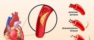
Intestinal infarction is a serious disease in which death is not uncommon. The main cause of intestinal infarction is blockage of the lumen of a blood vessel by a thrombus or atherosclerotic plaque.
Intestinal infarction, as a rule, occurs in older patients with severe heart pathology (atrial fibrillation, infective endocarditis, atherosclerosis). It is extremely rare that intestinal infarction may occur in young women taking combined oral contraceptives.
Classification in connection with the state of the circulatory system
Depending on the state of the circulatory system, intestinal infarction can have three stages of development of changes.
Table. Classification of intestinal infarction according to the state of the circulatory system.
| Stage name | Short description |
| The mildest stage of a heart attack, it is possible to restore physiological functions without any negative consequences. Patients may recover spontaneously and not be aware of the danger of their situation. Sometimes light conservative therapy is sufficient. Due to such mild consequences of this stage, medicine does not have accurate statistics; most cases are simply not recorded. | |
| As a result of a decrease in the volume of blood supplied to the intestinal tissue, a number of diseases can appear: ulcers on the walls, abdominal toad, colitis, enteritis. If treatment has not begun or does not correspond to the actual cause, then the patient’s condition will inevitably worsen, there may be bleeding due to micro-breaks of the small intestine, wall phlegmon, stenosis, etc. | |
| The last and most difficult stage of the disease. Gangrene and complete rupture of the intestines begin, purulent peritonitis and severe surgical sepsis spread. It is at this stage that patients most often end up on the operating table, but the likelihood of recovery is minimal. |
Diagnostics
Important factors for establishing the correct diagnosis are advanced age, the presence of severe atherosclerosis, especially atherosclerosis of the abdominal aorta and lower extremities, and thromboembolic processes.
Instrumental diagnostic methods:
- X-ray examination - survey radiography of the abdominal organs.
- Laparoscopy.
- Aortography.
- Selective mesentericography, which allows you to diagnose any type of circulatory disorders at the earliest possible time.
Signs of intestinal infarction
The main symptom of intestinal infarction is pain syndromes. They have different intensities depending on the stage of development of the pathology.
- Ischemic stage
. The most severe pain, in intensity, can only be compared with a volvulus. Even today, medicine does not have effective means to eliminate such pain; even drugs do not help. The mute condition is alleviated by antispasmodics, but their effect is short-term and does not completely relieve the syndrome. The patient cannot restrain his screams, is very restless, pulls his legs to his stomach, panics and is afraid of death. The duration of this stage is from six to twelve hours. Upon external examination, severe pallor of the skin is noticeable; if patients have heart problems, then its cyanosis may increase. Due to the fact that the mesenteric artery is completely blocked, the pressure increases sharply, the jump reaches 80 mm Hg. Art. And this is against the backdrop of the fact that the pulse noticeably slows down. The leukocyte count increases to 12×109/l. The tongue becomes white, but the abdomen remains soft and there is no swelling. - Heart attack stage
. It begins immediately after the end of the acute pain stage and lasts 12–24 hours. Necrosis begins in the intestines, due to which the vital activity of pain receptors gradually ceases. This results in less pain. The patient rejoices, is in a stage of euphoria, it seems to him that all the troubles are behind him. Unfortunately, even inexperienced doctors often think that all problems have been solved and no special help is required anymore. Another feature is that intoxication of the body leads to inappropriate behavior of patients, they can laugh for no reason, increase activity, etc. The pressure returns to normal, the pulse returns to normal levels. The catastrophic development of the disease can be seen only on the basis of a general blood test. The number of leukocytes at this stage can reach 40×109/l. - Stage of peritonitis
. It occurs 18–36 hours after arterial occlusion, the pain gradually increases, the patient finds it difficult to move, and the condition worsens upon palpation. At this stage, the chances of survival are minimal, even urgent surgical measures have a 50% fatal outcome. If surgery is done later, the mortality rate is 100%. At this stage, endotoxemia and dehydration develop, and metabolic acidosis begins in the body. Patients become more mobile and enter a delirious state.
In the initial stages there is reflex vomiting; the presence of blood in the stool is important for making a diagnosis. As already mentioned, at the first stage of the disease the patient feels very severe pain, comparable to the symptoms of volvulus. But there is one difference. During a heart attack, intestinal patency is maintained; during volvulus, the evacuation function is absent. Moreover, the removal of feces can occur on an instinctive level; stool retention indicates that the disease has already reached the stage of peritonitis.
Some diseases that affect the human body require a special procedure to be performed as part of the overall treatment plan - colonic lavage with an enema. Read more in the article: “enema solution at home.”
Incidence (per 100,000 people)
| Men | Women | |||||||||||||
| Age, years | 0-1 | 1-3 | 3-14 | 14-25 | 25-40 | 40-60 | 60 + | 0-1 | 1-3 | 3-14 | 14-25 | 25-40 | 40-60 | 60 + |
| Number of sick people | 0 | 0 | 0 | 4 | 6 | 7.8 | 8.7 | 0 | 0 | 0 | 4 | 6 | 7.8 | 8.7 |
How is the disease treated?
Advertising:
Treatment of mesenteric infarction must begin as quickly as possible; its timeliness determines the patient’s chances of survival and how serious the consequences will be. The goal of treatment is to eliminate the blockage of the vessel and remove the affected area of the intestine. In the first hours after the onset of a heart attack, it is necessary to begin thrombolytic therapy, which helps dissolve blood clots that have blocked the vessel. Medicines are used that activate fibrinolysis, i.e., the resorption of blood clots - streptokinase, streptodecase, urokinase and other anticoagulants.
At the same time, infusion therapy is started - intravenous infusion of drugs that stabilize blood circulation, replace the volume of circulating blood, and promote detoxification. In case of a heart attack caused by non-occlusive causes, the administration of antispasmodics is indicated to improve visceral blood flow. Attention! Photo of shocking content. To view, click on the link. The above methods relate to conservative therapy, and in this case, although they play an important, but auxiliary role.
In case of infarction of a section of the intestine, surgical intervention is required, and the less time passes from the start of drug therapy to surgery, the higher the chances of a favorable outcome. As the disease progresses, the patient's condition worsens, but at a certain point a period of imaginary well-being begins - the pain gradually weakens or disappears, but this is a poor prognostic sign. Surgical treatment consists of removing the affected area of the intestine, as well as restoring the blood supply to the affected area of the intestine. For peritonitis, the abdominal cavity is also rinsed with saline and antiseptics.
Classification
To determine the most effective treatment plan, it is important to know the full diagnosis, including the shape and stage of the heart attack. The disease is classified according to its course, location and degree of circulatory disturbance, and predominant symptoms.
According to the course, acute and chronic forms of the disease are distinguished.
Typically, intestinal vascular ischemia occurs against the background of progression of cardiovascular pathology in people over 70 years of age.
Depending on the vessels in which the circulatory disturbance occurred, three types of heart attack are distinguished:
- arterial - blood flow is disrupted in the mesenteric arteries; in most cases this leads to a heart attack within 6–8 hours;
- venous – damage occurs in the mesenteric veins, such a violation does not lead to a heart attack immediately, but after 1–4 weeks;
- mixed - characterized by impaired blood flow first in the arteries and then in the veins.
According to the degree of blood flow disturbance:
- compensated;
- subcompensated;
- decompensated heart attack.
Compensation is a process in which blood supply is maintained even when a vessel is damaged by additional vessels. In a compensated disorder, unaffected vessels completely take over the blood supply; in a subcompensated disorder, the blood supply is not fully restored; in a decompensated disorder, the blood flow stops completely.
Mesenteric infarction is manifested by acute abdominal pain
Acute disturbance of mesenteric circulation
Treatment of acute disorders of mesenteric circulation in the vast majority of cases involves emergency surgery, which should be undertaken as soon as the diagnosis is made or there is a reasonable suspicion of this disease.
The futility of using only conservative treatment methods has been proven by many years of practical experience. Only active surgical tactics provide a real chance of saving the lives of patients.
Surgical intervention should solve the following problems: 1) restoration of mesenteric blood flow; 2) removal of areas of the intestine that have undergone destruction; 3) fight against peritonitis. The nature and extent of surgical intervention in each specific case are determined by a number of factors: the mechanism of disturbance of mesenteric circulation, the stage of the disease, the localization and extent of intestinal lesions, the general condition of the patient, surgical equipment and the experience of the surgeon. The range of surgical procedures can vary from purely vascular surgery to purely resection interventions. In most cases, you have to combine both of these approaches. Restoring blood flow through the mesenteric arteries within 4-6 hours from the moment of occlusion (which is quite rare) usually leads to the prevention of intestinal gangrene and restoration of its functions. In case of irreversible changes in a more or less extended section of the intestine, in addition to its removal, surgery on the mesenteric vessels may still be necessary to restore the blood supply to its still viable sections.
The main stages of surgical intervention in this pathological condition should be considered: 1) surgical access, 2) inspection of the intestine and assessment of its viability, 3) inspection of the main mesenteric vessels, 4) restoration of mesenteric blood flow, 5) intestinal resection according to indications, 6) drainage and sanitation abdominal cavity. Let's take a closer look at each of them.
Surgical access should provide the possibility of revision of the entire intestine, exposure of the great vessels of the mesentery and retroperitoneal space, and sanitation of all parts of the abdominal cavity. A wide median laparotomy seems optimal.
Intestinal inspection necessarily precedes active surgical actions. The subsequent actions of the surgeon depend on the correct determination of the localization, extent and severity of ischemic intestinal damage. Detection of total gangrene of the small intestine forces us to limit ourselves to a trial laparotomy. The detection of ischemic disorders of the small and right half of the large intestine dictates the need for revision of the trunk of the superior mesenteric artery. The presence of necrotic changes in the sigmoid colon suggests exposure of the inferior mesenteric artery.
Assessment of intestinal viability is based on known clinical criteria: coloring of the intestinal wall, determination of peristalsis and pulsation of mesenteric vessels. This assessment in cases of obvious necrosis is quite simple. Determining the viability of an ischemic bowel is much more difficult. Mesenteric circulation disorders are characterized by a “mosaic pattern” of ischemic disorders: neighboring areas of the intestine may be in different circulatory conditions. That is why a repeated thorough examination of the intestine is often necessary after the vascular stage of surgery, and sometimes during relaparotomy one day after the first operation.
Revision of the main mesenteric vessels is the main distinguishing feature of surgical intervention in cases of suspected mesenteric circulation disorder. It begins with examination and palpation of the vessels near the intestine. Normally, pulsation is clearly visible to the eye. If mesenteric blood flow is impaired, pulsation along the edge of the intestine usually disappears. It is also difficult to detect it due to the developing swelling of the mesentery and intestinal wall. It is convenient to determine pulsation along the mesenteric edge by clasping the intestine with the thumb, index and middle fingers of both hands. To identify the pulsation of the mesenteric vessels located closer to its root, including the superior mesenteric artery, you should place one hand under the mesentery, and lightly squeeze it from above with the fingers of the other.
The pulsation of the trunk of the superior mesenteric artery is determined using the thumb and index fingers of the right hand. To do this, the thumb, feeling the pulsation of the aorta, is moved as high as possible to the site of the artery immediately to the right of the duodenum-jejunal flexure. By moving the small intestine to the right, you can determine the pulsation of the aorta and inferior mesenteric artery.
Sometimes (with swelling of the mesentery, systemic hypotension, severe obesity) it may be necessary to isolate the trunks of the mesenteric vessels. This is also necessary for intervention on them, aimed at restoring blood circulation in the intestines.
Exposure of the superior mesenteric artery can be done from two approaches: anterior and posterior. For embolism of the superior mesenteric artery, an anterior approach is usually used (Fig. 8.4). To do this, the transverse colon is brought into the wound and its mesentery is stretched. The mesentery of the small intestine is straightened, moving the intestinal loops to the left and down. The posterior layer of the parietal peritoneum is dissected longitudinally from the ligament of Treitz along the line connecting it to the ileocecal angle. The middle colic artery can be used as a guide, exposing it towards the mouth, gradually reaching the main arterial trunk. Depending on the location of the embolus, the artery is isolated either only in the area of origin of the middle colic artery (embolism of the first segment), or up to the bifurcation in the area of origin of the ileocolic artery (embolism of segments II and III).
With a posterior approach, the superior mesenteric artery is exposed to the left of the root of the mesentery of the small intestine. To do this, the intestinal loops are moved to the right and down. The ligament of Treitz is stretched and cut. The duodenojejunal flexure is mobilized. Next, the parietal peritoneum is dissected above the aorta. The left renal vein is located, above which the mouth of the superior mesenteric artery is located.
To isolate the inferior mesenteric artery, the parietal peritoneum is dissected longitudinally above the aorta. The trunk of the artery is found along the left lateral contour of the aorta.
Methods for restoring mesenteric blood flow depend on the nature of the vascular occlusion. Embolectomy from the superior mesenteric artery is usually performed from an anterior approach. After covering its trunk and mobilized branches using tourniquets, vascular scissors or a scalpel, a transverse arteriotomy is performed. It must be performed in such a way that, if necessary, catheter revision of the middle colon, ileocolic and at least one of the intestinal branches can be carried out through it. Embolectomy is performed using a Fogarty balloon catheter. The arteriotomy is sutured with separate sutures using a synthetic thread on an atraumatic needle. After bowel resection, embolectomy can also be performed through the stump of one of the transected branches of the superior mesenteric artery.
Vascular operations for arterial thrombosis are technically more difficult (thrombin-thymectomy, bypass surgery, and reimplantation of the artery into the aorta have to be performed) and give worse results. Due to the predominant localization of thrombosis in the first segment of the trunk of the superior mesenteric artery, a posterior approach to the vessel is indicated. The aorta is pressed parietally in the area of the artery mouth. It is opened with a longitudinal incision. Using instruments (vascular forceps, scissors, dissector), the thrombus is removed along with the intima. The distal edge of the intima is sutured with U-shaped sutures. The operation is completed by sewing a patch of autovein or synthetic material into the arteriotomy hole. Bypass operations are safer from the point of view of surgical risk. Rethrombosis of the superior mesenteric artery occurs less frequently after these interventions.
Prosthesis of the superior mesenteric artery is indicated when it has thrombosis over a significant extent. The graft can be sewn between the ends of the artery after resection of the area in the first segment or between the aorta and the distal end of the artery, and also connected to the right internal iliac artery. If thrombosis, which has developed against the background of a stenotic plaque, is localized at the mouth of the artery, replantation of the superior mesenteric artery into the aorta is justified.
Thrombectomy is indicated for thrombotic occlusion of the portal vein with transition to the superior mesenteric vein (descending thrombosis) or for occlusion of its trunk with an ascending nature of thrombosis. In this case, the operation is aimed mainly at preventing thrombosis of the portal vein. Thrombectomy can be performed through a longitudinal phlebotomy on the venous trunk or through a large intestinal branch after bowel resection.
Intestinal resection for disorders of mesenteric circulation can be used as an independent intervention or together with vascular operations. As an independent operation, resection is indicated for: thrombosis and embolism of the branches of the superior or inferior mesenteric arteries; venous thrombosis limited in extent; non-occlusive disorders of mesenteric blood flow. With the listed disorders, the extent of intestinal damage is, as a rule, insignificant, therefore, after resection, digestive disorders usually do not occur.
The predominance of high occlusions and late timing of surgical interventions in acute disorders of the mesenteric circulation quite often determine the use of subtotal resections of the small intestine. When performing resection for infarction, it is necessary to follow some technical rules. Along with the intestine affected by the infarction, it is necessary to remove the altered mesentery with thrombosed vessels, so it is not crossed along the edge of the intestine, but significantly away from it. In case of thrombosis of the branches of the superior mesenteric artery or vein, after incising the peritoneum 5-6 cm from the edge of the intestine, the vessels are isolated, crossed between clamps and ligated. For extensive resections with intersection of the trunk of the superior mesenteric artery or vein, a wedge-shaped resection of the mesentery is performed.
When resection is performed within viable tissue, an end-to-end anastomosis is performed according to the generally accepted technique. If there is a significant discrepancy in the diameters of the ends of the resected small intestine, a side-to-side anastomosis is formed. Recently, we have increasingly begun to resort to the practice of delayed anastomosis. The basis for such tactics are doubts about the exact determination of intestinal viability and the extremely serious condition of the patient during surgery. In such a situation, the operation is completed by suturing the stumps of the resected intestine and active nasointestinal drainage of the afferent part of the small intestine. After the patient’s condition has been stabilized against the background of intensive therapy (usually every other day), during relaparotomy the viability of the intestine in the resection area is finally assessed; if necessary, reresection is performed, and only after that an interintestinal anastomosis is applied.
When the cecum and ascending colon are affected, it is necessary to perform a right hemicolectomy along with resection of the small intestine. In this case, the operation is completed with ileotransversostomy.
Necrosis localized in the left half of the colon requires resection of the sigmoid colon (for thrombosis of the branches of the inferior mesenteric artery or non-occlusive disorder of the mesenteric circulation), or left-sided hemicolectomy (for occlusion of the trunk of the inferior mesenteric artery). Due to the serious condition of the patients and the high risk of failure of the primary colonic anastomosis, the operation, as a rule, should be completed with a colostomy.
If intestinal gangrene is detected, it is advisable to use the following procedure for surgical intervention. First, resection of clearly necrotic intestinal loops is performed with wedge-shaped excision of the mesentery, leaving areas of questionable viability. In this case, the operation on the vessels is delayed by 15-20 minutes, but the delay is compensated by the greater convenience of the operation, since swollen, non-viable intestinal loops make intervention on the mesenteric vessels difficult. In addition, this procedure of the operation prevents a sharp increase in endotoxemia after restoration of blood flow through the vessels of the mesentery and, to some extent, prevents the development of purulent peritonitis and possible phlegmon of the mesentery. The stumps of the resected intestine are sutured with UKL-type devices and placed in the abdominal cavity. Then intervention is performed on the vessels. After eliminating the vascular occlusion, it is possible to finally assess the viability of the remaining intestinal loops, and also decide on the need for additional intestinal resection and the possibility of anastomosis.
The rules for performing intestinal resection in cases where mesenteric blood flow is restored surgically are somewhat different (than without its restoration). Only obvious areas of intestinal gangrene are subject to removal; the resection boundary may be closer to necrotic tissue. In such a situation, the tactic of delayed anastomosis during relaparotomy is especially justified.
If, for one reason or another, surgery on the mesenteric vessels is not performed, intestinal resection must be performed within the area supplied by the vessel in which the occlusion occurred.
It is advisable to complete the intervention on the intestines with nasointestinal intubation, which is necessary to combat postoperative paresis and to carry out selective decontamination of the remaining part of the intestinal tract. This measure is a necessary component of relieving endotoxicosis.
Sanitation and drainage of the abdominal cavity should be carried out at the final stage of the operation. They are necessary as a measure to prevent purulent peritonitis or treat it if it has already developed. This procedure is performed in the same way as for other forms of secondary peritonitis. It is described in detail in Chapter XIII.
Intensive therapy. Improving the results of treatment of patients with acute disorders of mesenteric circulation largely depends on the effective provision of resuscitation measures in the pre- and postoperative periods, as well as adequate anesthesia during surgery.
Preoperative preparation includes a wide range of activities aimed at: (1) restoring the effective volume of circulating blood and improving tissue perfusion; (2) normalization of cardiac activity; (H) correction of metabolic disorders; (4)reduction of endotoxemia. The scope of preoperative preparation depends on the stage of the process, the presence and severity of concomitant diseases.
At the stage of intestinal ischemia and infarction, to prepare patients, measures aimed at reducing total peripheral resistance, replenishing the volume of circulating blood and combating metabolic disorders are used. Resuscitation measures for deep destructive changes in the intestine involving the peritoneum in the process do not differ from those for peritonitis of other etiologies (see Chapter XIII).
The rapidity of development of irreversible changes in the intestine is a factor dictating the need for emergency surgery and limiting the time of intensive therapy in the preoperative period (no more than 1-2 hours). Therapy must be continued during surgery. During surgery, close attention should be paid to adequate replacement of blood loss, especially after restoration of blood circulation in the intestine, which leads to massive hemorrhage into the wall and lumen of the intestine; to neutralize acidic metabolites washed out of the ischemic intestine.
In the postoperative period, intensive care includes measures aimed at improving systemic and tissue circulation, maintaining adequate gas exchange and oxygenation, correcting metabolic disorders, combating toxemia and bacteremia. It must be taken into account that resection of non-viable intestine does not eliminate severe systemic disorders, which may even worsen in the immediate postoperative period, including due to the loss of a significant part of the intestine, the role of which in the regulation of homeostasis is indisputable.
The main directions of therapy in the postoperative period can be formulated as follows.
1. Correction of hemodynamic disorders, maintaining an adequate volume of circulating blood, stabilizing cardiac output and improving the energy of myocardial contractions, normalizing tissue blood flow. Rheologically active agents and anticoagulants (rheopolyglucin, pentoxifylline, unfractionated or low molecular weight heparins) are of great importance in restoring effective blood circulation.
2. Normalization of gas exchange and ensuring adequate oxygenation, which in many cases can be achieved by using artificial ventilation.
3. Elimination of shifts in the water-electrolyte and acid-base state, which requires parenteral administration of an adequate amount of fluid, the use of buffer preparations, electrolytes and necessary microelements.
4. Replenishing the energy and metabolic needs of the body with the help of high-calorie infusions, amino acid and protein solutions. The patient should receive at least 4000 calories per day.
5. Prevention and treatment of acute renal and liver failure, which threaten many patients in the immediate postoperative period.
6. Treatment of intestinal paresis (long-term nasointestinal drainage, ubretide).
7. Rational use of antibacterial drugs.
With a favorable course, severe intoxication practically disappears by the 2-3rd day of the postoperative period. Hemodynamics stabilize, congestion in the stomach and thirst disappear, a feeling of hunger appears, peristalsis is restored, and gases begin to pass away. The well-being of patients noticeably improves after restoration of intestinal function. Leukocytosis gradually decreases and ESR decreases, the band shift disappears, the content of urea and bile pigments in the blood differs little from normal values. Diuresis is restored, proteinuria and cylindruria are reduced. In an uncomplicated postoperative period, these indicators are normalized by 7-10 days after surgery.
Low resistance of patients predisposes to the development of all kinds of complications (abdominal surgical sepsis, pneumonia, pulmonary embolism). These complications can be prevented by intensive therapy. At the same time, any conservative measures in case of relapse or progression of vascular occlusion are useless. That is why the main diagnostic efforts in the postoperative period should be aimed at identifying ongoing intestinal gangrene and peritonitis.
In patients with ongoing gangrene of the intestine, persistent leukocytosis and a pronounced band shift with a tendency to increase are observed, and the ESR increases. The development of jaundice and the progressive accumulation of nitrogenous wastes in the blood are perhaps the most characteristic signs of ongoing intestinal gangrene and indicate deep toxic damage to the liver and kidney parenchyma. Urine output progressively decreases to the point of anuria, despite large amounts of fluid administered and significant doses of diuretics. Urine examination reveals the development of toxic nephrosis, manifested in persistent and increasing proteinuria, cylindruria and microhematuria. Reasonable suspicions of ongoing intestinal gangrene serve as an indication for an immediate transition to active surgical actions - relaparotomy.
Early targeted (programmed) relaparotomy is performed to monitor the condition of the abdominal cavity when, after reconstructive vascular surgery, areas of intestine of questionable viability are left, or to perform a delayed anastomosis. The time for relaparotomy is from 24 to 48 hours.
Improving the results of treatment of acute disorders of mesenteric circulation primarily depends on the timely hospitalization of patients in surgical hospitals. Hospitalization should be carried out at the slightest suspicion of this disease. In the surgical department, the diagnosis must be confirmed or completely excluded within a few hours. Monitoring patients until signs of peritonitis appears is unacceptable. For diagnosis, instrumental methods (angiography, laparoscopy) should be used. As soon as the diagnosis is established, it is necessary to undertake emergency surgical intervention aimed at eliminating vascular occlusion and removing necrotic sections of the intestine. Based on our experience, we can state that only active diagnostic and treatment tactics can prevent an unfavorable outcome of this disease.