What species are found?
So, all heart tumors can be divided into 3 types:
- Primary benign – these include myxomas, rhabdomyomas, lipomas, teratomas, angiomas, papillary fibroelastomas.
- Primary malignant - represented mainly by sarcomas (angiosarcomas, fibrosarcomas); endothelioma and mesothelioma are also found.
- Secondary malignant ones are metastases of cancer of other organs (breast, lungs, stomach). Their appearance in the heart indicates a deeply advanced oncological process.
Let's take a closer look at the most common types - myxoma, rhabdomyoma, angiosarcoma.
Myxoma
Myxoma is the most common primary tumor of the heart (about 50%). Despite their similar names, cardiac myxoma and mitral valve myxomatosis are completely different diseases.
Myxoma is more often observed in women between 30 and 50 years old. It can be located in any cavity, but is more common in the left atrium. In approximately 3% of cases, the pathology develops as part of a hereditary syndrome called Carney complex. Such patients have a large number of pigment spots on the skin, fibroadenomatosis of the mammary glands and Itsenko-Cushing syndrome, caused by abnormal overproduction of hormones of the adrenal cortex (obesity, purple stretch marks on the skin of the abdomen and thighs, high levels of glucose in the blood).
Typically, myxoma is quite large in size - from 2 to 15 cm, which is why it can almost completely block the heart chamber.
Approximately 20% of patients with a small tumor are asymptomatic. One of the main signs of the disease is “myxoma intoxication.” A person’s body temperature rises, joints begin to ache, he loses weight, the level of hemoglobin in the blood decreases, and “inflammatory markers” appear - leukocytosis, high ESR and C-reactive protein. At first glance, it may seem that the patient has a chronic infection (for example, tuberculosis) or an autoimmune disease from the field of rheumatology.
The development of such symptoms is explained by the body’s immune reaction to proteins produced by the tumor. After removal of the myxoma, these signs quickly disappear.
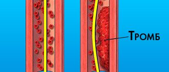
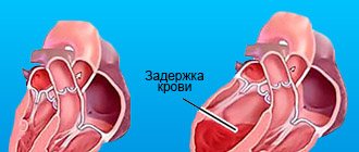
The two main problems that myxoma brings are obstruction of blood flow inside the heart and thrombotic complications.
Due to the fact that less blood enters the left ventricle, people with myxoma often experience dizziness and loss of consciousness. The rapid growth of the tumor leads to the rapid development of symptoms of chronic heart failure (CHF) - shortness of breath, constant fatigue, swelling of the legs, heaviness in the right side, etc. Characteristic features of CHF with myxoma are the speed of onset (from 3 to 6 months) and resistance to drug therapy.
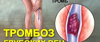
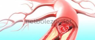
A common complication is cardioembolism. Blood clots form on the surface of the tumor, which clog the arteries, stopping blood flow in different parts of the body. Due to the loose structure of the neoplasm, its fragments can separate and float freely.
If I suspect myxoma, the first step I will perform is cardiac auscultation. Almost always I am able to listen to a pathological diastolic murmur, which becomes more intense when standing.
To exclude Carney complex, I prescribe tests to determine the level of hormones of the adrenal cortex - small and large dexamethasone tests, salivary cortisol, 24-hour urine, etc.
Routine diagnostic methods (ECG and radiography) are useless for detecting not only myxoma, but also all other heart tumors. Imaging research methods are considered more informative in this regard, i.e., during which we can see both the organ itself and its cavities. Such diagnostic methods include:
- Echocardiography (ultrasound of the heart, Echo-CG) is the main method for diagnosing myxomas. On the monitor, the tumor is defined as a rounded formation with clear edges, usually located in the area of the interatrial or interventricular septum in the vicinity of the valve leaflets. The myxoma is located on a stalk, due to which it can move from the atrium to the ventricle and back. Transesophageal echocardiography is sometimes required .
- Computer or magnetic resonance imaging (CT, MRI) are additional studies. I prescribe them when the ultrasound picture is unclear, or to better assess the extent of tumor growth.
The only treatment for myxoma is surgical removal. Abdominal surgery is performed under general anesthesia - the chest is opened, the patient is connected to a heart-lung machine.
To avoid relapses, the tumor is completely removed along with the underlying tissues. If the resulting defect in the heart wall is large, it is covered with a “patch” of pericardium or synthetic material. Then the cavities are actively washed and the liquid is sucked out. This is done to prevent the remnants of tumor cells from clogging small vessels. After the myxoma is removed, its sample is necessarily sent to a histology laboratory.
It often happens that due to the large size of the formation, the valves are damaged, so the patient may need prosthetics or plastic surgery followed by taking anticoagulants ( Warfarin ). If myxoma occurs as part of the Carney complex, then removal of both adrenal glands and lifelong use of hormonal drugs ( Corteff , Cortineff ) are additionally required.
Rhabdomyoma
Rhabdomyoma is the most common benign heart tumor in children under 15 years of age. In approximately half of the cases it is combined with such a severe hereditary disease as tuberous sclerosis. The neoplasm consists of several dense nodes growing from the muscular wall of the ventricles or the interventricular septum. They can occupy from 25 to 80% of the chamber volume.
Due to the fact that the tumor grows from the myocardial wall, it compresses the conduction system of the heart, so the symptoms of arrhythmias come to the fore in a person with rhabdomyoma:
- attacks of rapid heartbeat, accompanied by a feeling of fear and lack of air;
- irregular pulse;
- feeling of “fading”, interruptions in heart function;
- dizziness, darkening of the eyes;
- loss of consciousness followed by a convulsive seizure resembling epilepsy (Morgagni-Adams-Stokes attack).
With a large size of the formation that creates a circulatory disorder, signs of CHF appear.
To diagnose rhabdomyoma, I prescribe 2 instrumental studies:
- electrocardiography – on ECG film I can often detect various rhythm and conduction disturbances (supraventricular and ventricular tachycardia, extrasystoles, atrioventricular and intraventricular blockades);
- echocardiography - ultrasound of the heart shows round-shaped formations in the thickness of the myocardium, which can grow into the lumen of the chambers.
In some cases, due to tumor degeneration, rhabdomyomas decrease in size, and in children under 4 years of age they may completely disappear. If there are no symptoms, then no treatment is needed.
Heart cancer
Primary malignant neoplasms of the heart are mainly represented by sarcomas. Men are susceptible to them 3 times more than women. In sarcomas, the tumor is often located in the right parts of the organ.
Due to its rapid growth, symptoms of heart cancer appear fairly quickly. Signs of chronic heart failure and arrhythmias quickly appear (literally within a few weeks). Fluid often accumulates in the pericardial sac, which can lead to cardiac tamponade (compression) and very rapid death.
Since this is a malignant tumor, its appearance will be accompanied by general (constitutional) symptoms:
- weakness;
- lack of appetite;
- weight loss;
- prolonged fever.
As in the diagnosis of other heart tumors, echocardiography plays a decisive role, but to confirm sarcoma it is necessary to take a piece of the formation (biopsy) and conduct a histological examination.
Unfortunately, in 80% of patients, at the time of diagnosis of cardiac sarcoma, the tumor has already spread beyond the organ and metastasized. This greatly reduces the success of treatment.
Sometimes, in the absence of metastases, they resort to such a serious operation as heart and lung transplantation. It allows patients to live longer by 3–4 years.
Symptoms of heart tumors
Clinical signs of heart tumors are nonspecific. Sometimes they can resemble signs of common heart disease: chest pain, heart murmurs, arrhythmias, conduction disturbances, pericardial effusion, or cardiac tamponade.
The nature of symptoms observed with heart tumors is most closely related to the location of the tumor. Thus, if localized in the walls, heart failure may occur (difficulty breathing, shortness of breath during any physical activity, dizziness, tachycardia, etc.)
The tumor can affect the functioning of the heart valves. If a small part of it comes off and enters the lungs and brain through the bloodstream and clogs a blood vessel, a stroke will occur.
In the secondary form, noticeable weight loss, sweating, and increased fatigue are observed.
What is the prognosis for life?
With myxomas, the risk of death is approximately 30%. The most common immediate causes are stroke and heart failure. However, the prognosis for cardiac myxoma can be considered favorable, since the patient has a chance of a full recovery thanks to surgical excision of the tumor. If the operation is performed correctly, the risk of relapse of the disease is minimal (about 4%). Life after removal of cardiac myxoma (I mean its duration) may be practically no different from that of a healthy person.
Rhabdomyomas mostly regress, that is, they decrease in size, and sometimes completely disappear without any treatment. Still, patients are at risk of dying from severe cardiac arrhythmias.
The prognosis for cardiac sarcoma is extremely unfavorable. Most patients die within a year of their first symptoms.
Material and methods
Retrospective analysis of 47 medical records of patients who underwent surgery for a heart tumor from 2000 to 2014 according to demographic indicators, clinical data, laboratory and instrumental examination indicators before surgery and during the hospital period after surgery, assessment of the macroscopic description of the tumor and its histological examination . Mathematical data analysis was performed in SPSS version 17. Quantitative variables were examined for normality of distribution using the Kolmogorov-Smirnov test and the Shapiro-Wilk test. Data are presented as M ± SD
.
To compare quantitative variables with a normal distribution, a paired t
-test was used, and to compare frequencies, a nonparametric χ2 test was used.
Statistical significance was set at p
< 0.05.
Mix diagnostics
The main diagnostic methods are cardiac ultrasound and transesophageal echocardiography , which allow visualization of the size, location and mobility of the tumor
CT and MRI of the heart are more often used to diagnose tumors .
To confirm the diagnosis of atrial myxoma, the following tests may be ordered:
- Troponin level determination;
- determining the level of electrolytes in the blood;
- chest x-ray;
- Pulse oximetry.
In order to exclude pulmonary embolism, a pulmonary ventilation and perfusion study is performed using scanning.
Treatment of cardiac myxoma
Treatment for myxoma is only surgical. When a diagnosis of myxoma is made, surgery should be performed urgently. Surgical treatment is performed on an open heart using a heart-lung machine. The tumor is removed along with the pedicle, its attachment site, and the area of the heart surrounding the pedicle.
Remember! Timely cardiac examination can save your life! Go through a routine medical examination of your body, and also immediately contact a cardiologist if you have any symptoms indicating disturbances in the functioning of the cardiovascular system!