Extrasystole
History and objective examination
The main objective method for diagnosing extrasystole is an ECG study, however, it is possible to suspect the presence of this type of arrhythmia during a physical examination and analysis of the patient’s complaints.
When talking with the patient, the circumstances of the occurrence of arrhythmia are clarified (emotional or physical stress, in a calm state, during sleep, etc.), the frequency of episodes of extrasystole, and the effect of taking medications. Particular attention is paid to the history of past diseases that can lead to organic heart damage or their possible undiagnosed manifestations. During the examination, it is necessary to find out the etiology of extrasystoles, since extrasystoles with organic heart damage require different treatment tactics than functional or toxic ones. When palpating the pulse on the radial artery, an extrasystole is defined as a prematurely occurring pulse wave followed by a pause or as an episode of pulse loss, which indicates insufficient diastolic filling of the ventricles.
When auscultating the heart during extrasystole, premature I and II sounds are heard above the apex of the heart, while the I tone is strengthened due to low filling of the ventricles, and the II sound is weakened as a result of a small ejection of blood into the pulmonary artery and aorta.
Instrumental diagnostics
The diagnosis of extrasystole is confirmed after an ECG in standard leads and daily ECG monitoring. Often, using these methods, extrasystole is diagnosed in the absence of patient complaints. Electrocardiographic manifestations of extrasystole are:
- premature occurrence of the P wave or QRST complex; indicating a shortening of the pre-extrasystolic coupling interval: with atrial extrasystoles, the distance between the P wave of the main rhythm and the P wave of the extrasystoles; with ventricular and atrioventricular extrasystoles - between the QRS complex of the main rhythm and the QRS complex of the extrasystoles;
- significant deformation, expansion and high amplitude of the extrasystolic QRS complex during ventricular extrasystole;
- absence of the P wave before the ventricular extrasystole;
- following a complete compensatory pause after a ventricular extrasystole.
Holter ECG monitoring is a long-term (over 24-48 hours) ECG recording using a portable device attached to the patient’s body. Registration of ECG indicators is accompanied by keeping a diary of the patient’s activity, where he notes all his sensations and actions. Holter ECG monitoring is performed for all patients with cardiac pathology, regardless of the presence of complaints indicating extrasystole and its detection with a standard ECG.
The identification of extrasystole, not recorded on the ECG at rest and during Holter monitoring, can be done by the treadmill test and bicycle ergometry - tests that determine rhythm disturbances that appear only during exercise. Diagnosis of concomitant cardiopathology of an organic nature is carried out using ultrasound of the heart, stress echo-CG, and MRI of the heart.
Supraventricular extrasystole - symptoms and treatment
NVE may be benign. In this case, the risk of sudden death is very low, sometimes the patient does not even feel the rhythm disturbance. Such extrasystole does not always require treatment.
If possible, the etiological factor must be eliminated:
- normalize sleep;
- limit or completely stop taking provoking medications and drinks;
- quit smoking:
- normalize thyroid function in hyperthyroidism;
- adjust the level of potassium in the blood;
- remove the gallbladder in case of cholelithiasis;
- Avoid a horizontal position after eating if you have a hiatal hernia;
- normalize blood pressure;
- increase physical activity according to the body’s capabilities;
- Avoid excessive physical activity (weightlifting, heavy lifting).
The patient is advised to establish a daily routine. The diet should be supplemented with foods rich in potassium and magnesium; they have a beneficial effect on the cardiovascular system.
| Products containing potassium | Products containing magnesium |
| ⠀•⠀dried apricots; ⠀•⠀cocoa powder; ⠀•⠀wheat bran; ⠀•⠀raisins; ⠀•⠀sunflower seeds; ⠀•⠀nuts (pine nuts, almonds, peanuts, walnuts); ⠀•⠀legumes (peas, lentils, beans); ⠀•⠀jacket potatoes; ⠀•⠀avocado; ⠀•⠀ceps; ⠀•⠀bananas; ⠀•⠀citrus fruits; ⠀•⠀Brussels sprouts and kohlrabi; ⠀•⠀milk and fermented milk products; ⠀•⠀cereals (oatmeal, buckwheat, pearl barley, rice); ⠀•⠀fruits (peaches, pears, watermelon, apples, prunes, apricots, melon); ⠀•⠀chicory; ⠀•⠀vegetables (carrots, spinach, green onions, eggplant, cucumbers); ⠀•⠀chicken eggs; ⠀•⠀fish and meat; ⠀•⠀apple juice. | ⠀•⠀oil (sesame, flaxseed, peanut); ⠀•⠀cheese (Dutch, Poshekhonsky, goat, with mold); ⠀•⠀cottage cheese (low-fat and low-fat, curd cheese); ⠀•⠀bitter chocolate; ⠀•⠀almost all types of meat; ⠀•⠀fish (halibut, sturgeon, perch, haddock, cod, saury); ⠀•⠀duck eggs; ⠀•⠀cereals (rolled oats, chickpeas, peas, buckwheat, brown rice, lentils); ⠀•⠀fruits and berries (cherries, kiwi, pineapple, feijoa, raspberries, pears, peach, persimmon); ⠀•⠀many varieties of tea (for example, “Ivan-tea”) and juices; ⠀•⠀ginger; ⠀•⠀mustard; ⠀•⠀vanilla. |
Indications for antiarrhythmic therapy are:
1. Poor tolerance of supraventricular extrasystole. In this case, it is necessary to determine in what situations and at what time of day heart rhythm disturbances most often occur, and then time the drug intake to this time.
2. The occurrence of VVC (not necessarily frequent) in patients with heart defects (primarily mitral stenosis) and other organic heart diseases. In such patients, atrial overload and dilatation progress. Supraventricular extrasystole in this case serves as a harbinger of the occurrence of atrial fibrillation.
3. Supraventricular extrasystole, which arose as a result of a long-term etiological factor in patients without previous organic heart disease and atrial enlargement (with thyrotoxicosis, inflammatory process in the heart muscle, etc.). If antiarrhythmic treatment (along with etiotropic treatment) is not carried out, the risk of persistent EVE increases. Frequent supraventricular extrasystole in such situations is potentially malignant in relation to the development of atrial fibrillation.
4. Frequent (700-1000 extrasystoles per day or more) EVA also requires antiarrhythmic therapy, even if it is regarded as idiopathic, since there is a risk of complications. The approach in these cases should be differentiated. It is also possible to refuse antiarrhythmic therapy if there are reasons for this:
- absence of subjective symptoms and complaints;
- borderline number of extrasystoles;
- intolerance to antiarrhythmic drugs;
- signs of sick sinus syndrome or impaired AB conduction.
Antiarrhythmic drugs used for EVA:
- Beta blockers (Metoprolol, Bisoprolol), calcium antagonists (Verapamil). It is pathogenetically justified to prescribe drugs from this group to patients with hyperthyroidism, a tendency to tachycardia, when EVE occurs against the background of stress and is provoked by sinus tachycardia. Beta blockers are indicated for ischemic heart disease, arterial hypertension, and sympathoadrenal crises. "Verapamil" is prescribed for concomitant bronchial asthma, variant angina, nitrate intolerance, and patients with coronary artery disease.
- “Belloid”, “Teopek” are indicated for patients with vagal-mediated VHE, which develops at night against the background of a decrease in heart rate. These drugs increase the rhythm and are prescribed at night.
- Sotalol (“Sotalex”, “Sotagexal”). It is necessary to select a dose depending on blood pressure and heart rate, the duration of the PQ and QT intervals. Indicated for a combination of ventricular extrasystole and ventricular extrasystole.
- Antiarrhythmics of IA and IC classes (“Dizopyramide”, “Allapinin”, “Propanorm”, “Etatsizin”). Use is not indicated in patients with coronary artery disease who have recently suffered a myocardial infarction due to its arrhythmogenic effect on the ventricles.
- Amiodarone (“Cordarone”). Amiodarone is the most effective antiarrhythmic drug available. Can be prescribed to patients with organic heart damage.
- If monotherapy is insufficiently effective (i.e., using one antiarrhythmic), combinations of drugs can be used.
If the prescribed therapy has a good effect, antiarrhythmics should not be discontinued quickly. Treatment lasts several weeks (months). If there is a threat of developing atrial fibrillation or if there are episodes of it in the anamnesis, NVE therapy is carried out for life. In the case of continuous antiarrhythmic therapy, the minimum effective doses are selected. Patients with an undulating course of EVE should strive to discontinue the antiarrhythmic during periods of improvement (excluding cases of severe organic myocardial damage). The withdrawal of antiarrhythmics is carried out gradually with a decrease in dosage and number of doses per day. After discontinuation, the patient is recommended to have the drug with him (the “pill in his pocket” strategy) for the purpose of rapid administration when the arrhythmia resumes [11].
If there is no effect from antiarrhythmic therapy, with frequent NVE (up to 10,000 per day), the issue of surgical treatment is considered - radiofrequency ablation of arrhythmogenic foci (destruction of foci using electric current) [5].
What does it represent?
According to the International Classification of Diseases (ICD-10), supraventricular extrasystole has code 149.4. It is included in the registry of cardiac arrhythmias in the heart diseases section.
Extrasystole can be of different types, but the main ones are atrial and ventricular. When an additional contraction out of turn is caused by an impulse that comes from the ventricular system, they speak of ventricular extrasystole. These attacks are accompanied by an interruption in the heart rhythm, which is accompanied by weakness and dizziness.
According to ongoing studies, single extrasystoles can also occur in healthy people; 50% of the patients studied had positive results.
Therefore, we can say that this condition can be physiological and occur in healthy people. Stress may be the main cause of functional impairment. In addition, drinking alcohol and energy drinks, smoking, etc. can also provoke extrasystole. In these cases, the pathology is considered harmless and quickly goes away on its own.
As for pathological ventricular arrhythmia, it develops against the background of serious diseases and requires mandatory treatment and monitoring by a specialist.
Causes
Pathology can occur for various reasons.
Cardinal (heart) causes:
- ischemic disease – lack of blood supply and oxygen starvation;
- myocardial infarction – a certain area of the heart muscle dies and is replaced by scar tissue;
- cardiomyopathy – damage to the heart muscle;
- myocarditis - an inflammatory process in the heart muscle;
- congenital and acquired defects - violation of the structure of the organ;
- heart failure - the heart cannot fully pump blood, that is, it cannot cope with its main function.
The disease can be caused by taking medications , especially those that are taken uncontrolled and for a long time, for example:
- anti-arrhythmia drugs;
- cardiac glycosides;
- diuretics.
Hormonal ailments can also provoke extrasystole:
- diabetes;
- adrenal gland diseases;
- thyrotoxicosis.
Other reasons:
- electrolyte imbalance – change in the ratio of sodium, magnesium, potassium salts;
- the toxic effects of alcohol and nicotine on the human body;
- failures in the functioning of vegetation ;
- prolonged oxygen starvation - observed with apnea, anemia, bronchitis;
- idiopathic - the disease occurs without any specific cause or there is no way to find out.
Classification
The disease differs according to the following characteristics.
- According to the location of the source of excitation:
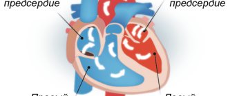
- atrioventricular - the ectopic focus is located in the atrioventricular septum (between the ventricles and atria);
- atrial – the focus is located in the atrium (in the upper part of the heart).
- According to the frequency of rhythmic contractions in 1 minute:
- single – extra contractions are observed in an amount of no more than 5;
- multiple – more than 5 extra contractions;
- group - extrasystoles follow one after another;
- steam room – 2 additional contractions are observed one after another.
- By the number of ectopic foci:
- monotopic – single focus;
- polytopic - several foci.
- By appearance:
- early – observed at the moment of atrial contraction;
- medium - occurs between the contraction of the atria and ventricles;
- late - appears after contraction of the ventricle or at the moment when the heart muscle completely relaxes.
- By order:
- ordered - normal contractions alternate with extrasystoles;
- disordered - no pattern can be traced.
Regarding the classification according to the type of bigeminy, supraventricular extrasystole can be:
- Ventricular - most often observed in older people. Extrasystoles occur after each contraction of the heart under the influence of excitation sent by the ventricles.
- Supraventricular - additional contraction occurs under the influence of an impulse from the atria or atrioventricular node. It can be observed in healthy people during sports, poisoning, or stress.
Symptoms
Symptoms of the disease are:
- shortness of breath, feeling of suffocation;
- dizziness – blood flow is reduced and oxygen starvation occurs;
- increased sweating and weakness;
- hot flashes that have no basis;
- disruptions in heart rhythm - beats outside the heart, and so on.
Important! The main symptom of supraventricular extrasystole is a feeling of temporary cardiac arrest, which in most cases causes panic attacks, anxiety, and paleness of the skin in patients.
Quite often, supraventricular extrasystole accompanies most cardiac, autonomic and psycho-emotional disorders.
On a note! The insidiousness of supraventricular extrasystole is that for a long time the pathology does not manifest itself with clear clinical signs, the disease proceeds unnoticed until it causes complications.
Treatment and recommendations
If the patient is diagnosed with single systoles and there are no other cardiac pathologies, treatment for supraventricular extrasystole is not carried out. Recommended lifestyle adjustments:
- quitting smoking and alcohol;
- moderate physical activity;
- proper nutrition;
- minimizing stress;
- a full night's rest;
- positive emotions;
- walks in the open air.
When diagnosing heart pathology, treatment should be aimed at eliminating the root cause. It is possible to prescribe symptomatic therapy - drugs with a sedative effect, antiarrhythmic drugs.
Consequences and complications
With timely detection of pathology, adequate therapy and lifestyle changes, the prognosis for supraventricular extrasystole is favorable. If the patient does not have concomitant illnesses, death from extrasystole cannot occur. But if the clinical picture is ignored and treatment is refused, the following complications may develop - coronary heart disease, atrial fibrillation and other life-threatening diseases.
Important! Despite the fact that supraventricular extrasystole, as an independent pathology, does not pose a threat, consultation with a doctor should be mandatory.
LiveJournal
Features in children
Supraventricular extrasystole in childhood can manifest itself in the following types:
- group;
- steam room
It can be observed in young children, schoolchildren and puberty.
Causes:
- thyroid pathology;
- inflammation in the myocardium;
- hormonal changes;
- autonomic disorders;
- heart pathologies;
- stress;
- physical and emotional fatigue;
- VSD;
- helminthic infestations;
- lack of microelements;
- diabetes;
- the influence of certain medications;
- smoking and drug addiction.
As for the signs of extrasystole, in a child they are more pronounced than in adults, and are often accompanied by headaches and dizziness. There is also a decrease in appetite and worsening sleep, the child complains of aching pain in the heart area. Much rarer symptoms are shortness of breath and difficulty breathing.
Diagnostics
The first thing you should pay attention to when diagnosing supraventricular extrasystole is the clinical signs. This pathology is characterized by unpleasant sensations in the chest, which cause significant discomfort, but cannot be called pain.
To confirm the diagnosis, the patient must undergo an ECG and 24-hour Holter monitoring.
On the ECG, supraventricular extrasystole looks like this:
- expansion and deformation of the atrial P wave ahead of time;
- the P wave is followed by the normal ventricular complex;
- the compensatory pause is incomplete;
- if the origin of the extrasystole is atrioventricular, the P wave may not be detected or may be observed after the ventricular complex.
Reference! Supraventricular extrasystole on the ECG manifests itself clearly and leaves no doubt for making a diagnosis.