Tests and diagnostics
To identify the true cause of changes in the myocardium, it is recommended to undergo a full examination, which includes:
- General analysis and biochemical blood test. Based on the results, it will be possible to talk about the presence of an inflammatory process in the body, the functioning of the renal system and kidneys, and the level of cholesterol , which forms plaques in the coronary arteries.
- ECG. Characteristic changes during the examination make it possible to determine further examination tactics. In some cases, it is recommended to perform an ECG with stress, or organize daily ECG monitoring.
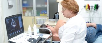
- EchoCG. Ultrasound examination of the heart allows you to assess the condition of the valve apparatus of the heart, identify areas of damage, and evaluate the pumping function of the heart.
- Myocardial scintigraphy. The radioisotope research method shows areas of accumulation of a special substance to identify areas of damage and determine their nature.
Changes in the myocardium on the ECG make it possible to determine further tactics for examining the patient to establish an accurate diagnosis and select the correct therapy.
Left ventricular aneurysm on ECG*
Another important nuance, without which it is impossible to reveal the topic of scar changes, is “post-infarction aneurysm of the left ventricle.”
Its main symptom is the presence of ST segment elevation in leads where there are cicatricial changes (pathological Q waves). Unlike elevation during a heart attack (infarction with ST elevation), such elevation is not accompanied by reciprocal ST depression and does not disappear after the resolution of myocardial infarction, that is, it exists constantly.
It must be said that it is impossible to distinguish an aneurysm from an akinesia zone (an area of the heart that does not contract) on an ECG . But in an era when cardiac ultrasound had not yet appeared or was unavailable, the changes described above were associated exclusively with an aneurysm. We will not change anything so as not to cause confusion, we accept this definition, but with the above-mentioned amendment , and we will use the term “aneurysm” in further interpretation of the ECG.
Below is a typical ECG showing a “left ventricular aneurysm at the apex.”
Causes
Dystrophic changes in the myocardium
Such changes in the ECG are formed due to insufficient nutrition of cardiomyocytes, which inevitably leads to a decrease in the contractility of the left ventricle. Diffuse-dystrophic changes in the myocardium are observed with:
- pathologies of the endocrine system: diabetes mellitus , adrenal dysfunction, disorders of the thyroid gland;
- pathologies of the renal system and liver: excessive amounts of toxic metabolic products negatively affect the functioning of the heart;
- chronic diseases of infectious origin: changes can be observed with tuberculosis , influenza , malaria , etc.;
- chronic iron deficiency anemia : constant oxygen starvation affects the functioning of cardiomyocytes;
- with an unbalanced diet, with vitamin deficiency in the diet;
- with excessive nervous and physical overload;
- with fever and concomitant dehydration;
- in case of poisoning with alcohol, medications or chemical components.
Metabolic changes in the myocardium
What it is? Characteristic nonspecific changes on the ECG are formed as a result of disturbances in intracellular metabolic processes associated with potassium and sodium ions.
Metabolic changes are associated with dystrophy of the heart muscle and appear when:
- ischemia , which is reflected on the cardiogram in the form of deviations of the T wave. Its polarity and shape changes in the leads corresponding to the damaged areas;
- myocardial infarction : the location of the ST segment changes on the ECG, which is located either above or below the isoline;
- death, necrosis of the myocardium, which is characterized by the appearance of an abnormal Q wave.
Scar changes
Areas of scar tissue form at the site of a former inflammatory process, necrosis, as a result of which normal, healthy cardiomyocytes lost their contractility and were replaced by connective tissue that does not have elasticity. Focal cicatricial changes on the ECG indicate a previous myocardial infarction .
- The lower wall of the left ventricle is characterized by changes in leads: II, III and a VF (indicates damage to the right, less often the left circumflex coronary artery).
- The anterior septal region is characterized by changes in leads: V1 and V2 (the left descending septal branch is damaged), or V2-V4 (the left descending coronary artery or its branches is involved).
- The anterior-lateral region is characterized by changes in leads: I, aVL, V4-V6 (the circumflex artery or the left descending artery is damaged).
- Anterior widespread infarction is characterized by changes in leads: I, aVL, V1-V6 (the left descending coronary branch is damaged).
Moderate inflammatory changes in the myocardium
Characteristic changes are observed in myocarditis, in which the voltage of the waves in all leads decreases and rhythm disturbances are recorded. Moderate left ventricular changes may occur after:
- typhus , diphtheria ;
- rheumatism , after streptococcal infection ( tonsillitis , tonsillitis , scarlet fever );
- infections caused by the Coxsackie virus, rubella , influenza , measles ;
- exacerbation of an autoimmune disease ( systemic lupus erythematosus , scleroderma ).
Brown myocardial atrophy
This is what a macropreparation is called during histological examination. Characteristic pathological changes in the myocardium are formed as a result of a long-term lack of blood supply, which is observed in debilitating diseases, cachexia , drug abuse, increased physical activity, and also in old age. lipofuscin, is deposited . Its granules are a product of impaired metabolism in cardiac muscle cells, weakened by insufficient nutrition and blood supply.
How to determine diffuse myocardial damage?
30.09.2021
In inflammatory diseases of the myocardium, diffuse lesions of the heart muscle are very common. This pathology may be accompanied by tachycardia , shortness of breath or other signs of heart failure.
Development mechanism
Promotes the development of diffuse changes in the myocardium (myocardial dystrophy or myocarditis). Also, the disease can be caused by the development of connective tissue of the heart muscle instead of fibers, which manifests itself as a consequence of metabolic and inflammatory disorders.
In some cases, these changes are caused by excessive physical exertion, the use of certain medications, and disturbances in water-salt metabolism. In this case, uniform damage to the heart muscle occurs in the ventricles, atria and interventricular septa.
Signs of myocardial damage
With diffuse myocardial lesions, the following syndromes occur:
- Abramov-Fiedler syndrome;
- cardiomegaly;
- Berardinelli syndrome;
- Bernheim syndrome;
- Bouveret syndrome;
- Buyot's syndrome;
- Wenckebach syndrome.
Abramov-Fiedler syndrome is characterized by an acute onset, anxiety, shortness of breath, and tachycardia . There may be diarrhea or respiratory symptoms even before the onset of illness. Cardiomegaly is caused by glycogen storage. Hypergenitalism appears, but secondary sexual characteristics are absent. Berardinelli syndrome occurs with acromegaloid gigantism, atrophy of adipose tissue, muscle hypertrophy, hypertension and liver .
With Bernheim syndrome, a special type of heart failure occurs, occurring without signs of pulmonary stasis, with ventricular hypertrophy and venous stagnation. Bouveret syndrome is accompanied by paroxysmal tachycardia , which leads to circulatory failure. For Buyot syndrome, the main symptom is the presence of an inflammatory process of connective tissue localized in the cardiovascular system. Wenckebach syndrome includes a symptom complex of atrial manifestations, ventricular manifestations, as well as signs of proximal tachycardia .
Diagnostics
The diagnosis is made after instrumental examination methods, laboratory tests , ultrasound and ECG . The ECG reveals a change in the T wave, a decrease in voltage, a disturbance in rhythmic activity and atrioventricular conduction. Ultrasound examination makes it possible to identify the degree of damage to the heart muscle, determine the pathological focus and prescribe appropriate treatment.
Treatment
Treatment of diffuse changes in the myocardium is carried out in a hospital . The main goal is to eliminate the main cause that caused myocarditis : it could be rheumatism , sepsis, etc. Among medications, corticosteroid hormones to provide an antiallergic effect. If there are signs of heart failure, cardiac glycosides are prescribed. In the presence of edema, diuretics are used. In addition, vitamins, substances that improve metabolism, cocarboxylase and ATP are prescribed.
Nutrition for myocarditis should be special. It is recommended to eat only easily digestible foods rich in vitamins that do not cause bloating : vegetables, boiled fish and meat, and dairy products. The amount of salt and liquid is limited to the minimum norm.
If the patient’s condition is normal, exercise therapy from the first days of his treatment.
To prevent myocarditis, it is necessary to treat the diseases that cause this pathology and try to lead a healthy lifestyle.
Published in Cardiology Premium Clinic
Dystrophic disorders
These changes in the myocardium of the left ventricle are provoked by a lack of nutrients, without which the heart muscle will not function normally. In medicine, this condition is referred to as cardiac dystrophy, and it appears due to the following factors:
- disruptions in the functioning of the kidneys and liver, because these organs are responsible for metabolic processes;
- diabetes;
- diseases of the endocrine system;
- shocks affecting the central nervous system;
- serious loads;
- low hemoglobin level;
- infectious pathologies of a chronic nature;
- intoxication;
- unhealthy diet, causing vitamin deficiency;
- frequent consumption of alcoholic beverages.
- long-term use of drugs.
- Features of the ECG during myocardial infarction - the procedure and signs of the disease
Often this condition is detected in adolescents during exams, when they are mentally overstrained. But in young children, changes in the myocardium are considered normal, and all because metabolic processes are not yet perfect. Also, older people may suffer from such changes, because metabolic processes in their bodies slow down.
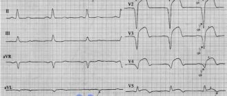
EXAMPLE 3
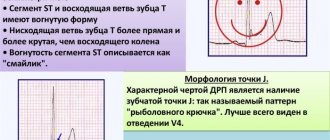
The first complexes were recorded with little interference, so we pay attention to the second and third complexes in sections V1-V3. The red frame shows an example of what the complexes in these leads should look like normally.
The morphology of V1 here is not very different from the norm, but V2 and V3 are distinguished by the absence of R waves. After a short isoline following the P wave, a deep negative wave begins (which, as you know, is called Q). The Q wave is jagged, which is characteristic of cicatricial changes. More attentive people have already noticed that “pathological Q” is also present in lead V4. If it is carefully measured, its parameters will be outside the normal range. In addition, there should not be such an obvious Q in this lead.
Thus, here we are talking about cicatricial changes in the anteroseptal region.
Pathogenesis
Changes in the ECG are not a disease, but only a manifestation of some pathological processes occurring in the myocardium. With shifts in the biochemical activity in the heart cells, their contractility changes, which is reflected in the cardiogram when recording the conduction of impulses. Cardiomyocyte function can be impaired during inflammatory processes, for example, with myocarditis . Taking certain medications also affects the functioning of the heart muscle.
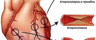
Long-term diabetes mellitus can gradually lead to atherosclerosis . Not only large vessels are affected, but also the coronary arteries that supply the myocardium. With inflammatory pathology in the gastrointestinal tract, the absorption of nutrients is impaired, which also negatively affects metabolic processes in cardiomyocytes.
Is there a danger with changes in the myocardium?
Moderate changes in the myocardium are a fairly common pathology that gradually affects healthy cells of the heart muscle. Doctors say that this leads to the development and progression of heart failure. Let's consider whether it is worth showing concern when diagnosing such a pathology, what it is: moderate disturbances in the myocardium.
Such changes may not always be dangerous. Especially if it does not manifest itself with pronounced symptoms. Often pathology is discovered during a regular medical examination. If a person does not feel any problems with the functioning of the heart, then most likely there is no need to show concern. The reason for a visit to a cardiologist should be:
- pain in the heart area;
- heart rhythm disturbances;
- dyspnea;
- high or low blood pressure;
- weakness, drowsiness.
EXAMPLE 4
Here we see a similar picture, only the Q waves are so pronounced that it can be confusing.
After the P wave there is immediately a negative wave, which cannot be anything other than a Q wave. Here the Q wave is pathological (given the duration and amplitude). That is, here we are talking about cicatricial changes in the anteroseptal-apical region. The fact that there are no scar changes in lead V6 does not allow us to talk about lateral localization, although in this patient it is necessary to make sure that the electrodes are applied correctly; perhaps V6 is not in place and then the scar zone will expand to the lateral wall.
In such doubtful cases, I recommend that you describe what you see - pathological Q waves in leads V1-V5 and therefore “tie” them to the walls of the heart.