If a local area of the heart muscle fails to work (necrosis, hibernation, sclerosis, stunning), this is manifested on the ECG by the appearance of a pathological Q wave.
A Q wave is considered pathological if its duration exceeds 0.03 s and its amplitude is greater than 1/4 of the R wave. An exception is lead aVR.
Changes in “pathological waves” must be present in two adjacent leads (for example V1+V2, V5+V6, AVL+I, etc.)
If a pathological Q is suspected in lead III, then it should be duplicated in lead aVF and not disappear with a deep breath!
Scar changes cannot be judged in the presence of left bundle branch block
In fact, these are the main criteria and now we will analyze them in more detail.
General information
Electrocardiography is the simplest diagnostic method for studying the functioning of the heart.
The essence of the examination is to record electrical impulses that accompany the contractile and restorative functions of the myocardium, called “depolarization” and “repolarization.” What do myocardial changes on an ECG mean?
Specific abnormalities in the electrocardiogram can be recorded during a routine medical examination and characterize the condition of the myocardium at the time of the examination. The function of the myocardium is to synthesize by cardiomyocytes , due to which the cavity contracts and ensures normal blood supply to the entire body. This process is carried out due to the cellular exchange of sodium and potassium ions in the cell. The functioning of the cardiac conduction system is recorded on an electrocardiogram using electrodes fixed on the limbs and chest.
Pathogenesis
Changes in the ECG are not a disease, but only a manifestation of some pathological processes occurring in the myocardium. With shifts in the biochemical activity in the heart cells, their contractility changes, which is reflected in the cardiogram when recording the conduction of impulses. Cardiomyocyte function can be impaired during inflammatory processes, for example, with myocarditis . Taking certain medications also affects the functioning of the heart muscle.
Long-term diabetes mellitus can gradually lead to atherosclerosis . Not only large vessels are affected, but also the coronary arteries that supply the myocardium. With inflammatory pathology in the gastrointestinal tract, the absorption of nutrients is impaired, which also negatively affects metabolic processes in cardiomyocytes.
Dystrophic disorders
These changes in the myocardium of the left ventricle are provoked by a lack of nutrients, without which the heart muscle will not function normally. In medicine, this condition is referred to as cardiac dystrophy, and it appears due to the following factors:
- disruptions in the functioning of the kidneys and liver, because these organs are responsible for metabolic processes;
- diabetes;
- diseases of the endocrine system;
- shocks affecting the central nervous system;
- serious loads;
- low hemoglobin level;
- infectious pathologies of a chronic nature;
- intoxication;
- unhealthy diet, causing vitamin deficiency;
- frequent consumption of alcoholic beverages.
- long-term use of drugs.
- Features of the ECG during myocardial infarction - the procedure and signs of the disease
Often this condition is detected in adolescents during exams, when they are mentally overstrained. But in young children, changes in the myocardium are considered normal, and all because metabolic processes are not yet perfect. Also, older people may suffer from such changes, because metabolic processes in their bodies slow down.
Classification
Depending on the size and location of the changed cardiomyocytes, the following are distinguished:
- Diffuse changes. Such a widespread lesion indicates the presence of multiple foci of altered cardiomyocytes. Characteristic signs are present in all leads on the ECG.
- Focal changes. Lesions are recorded only in certain leads that correspond to specific areas of the myocardium. Focal changes are a kind of clearly limited areas of connective, scar tissue, which are inert to conduct electricity.
When diagnosing changes on the ECG, a functional diagnostics doctor and cardiologist can state:
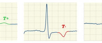
- early ventricular repolarization, which is manifested by a negative “T” wave;
- a decrease in the voltage of the r wave, which characterizes the contractility of the myocardium;
- rhythm disturbances;
- conduction disturbances.
Depending on these indicators, the causes of changes in the ECG are determined by nature:
- inflammatory;
- cicatricial;
- dystrophic;
- metabolic.
EXAMPLE 1
When assessing the ventricular complexes, we notice a more or less pronounced QIII wave. We measure its width and height. As you can see, its width approaches 0.03 s., but does not exceed the norm, we also see that its amplitude (height) is about 1-1.1 mm, which is less than 1/4 of the R wave, which is equal to 4. 5 mm. We also see that in lead aVF the Q wave also falls within the normal range.
Thus, we conclude that these are not scar changes.
Causes
Dystrophic changes in the myocardium
Such changes in the ECG are formed due to insufficient nutrition of cardiomyocytes, which inevitably leads to a decrease in the contractility of the left ventricle. Diffuse-dystrophic changes in the myocardium are observed with:
- pathologies of the endocrine system: diabetes mellitus , adrenal dysfunction, disorders of the thyroid gland;
- pathologies of the renal system and liver: excessive amounts of toxic metabolic products negatively affect the functioning of the heart;
- chronic diseases of infectious origin: changes can be observed with tuberculosis , influenza , malaria , etc.;
- chronic iron deficiency anemia : constant oxygen starvation affects the functioning of cardiomyocytes;
- with an unbalanced diet, with vitamin deficiency in the diet;
- with excessive nervous and physical overload;
- with fever and concomitant dehydration;
- in case of poisoning with alcohol, medications or chemical components.
Metabolic changes in the myocardium
What it is? Characteristic nonspecific changes on the ECG are formed as a result of disturbances in intracellular metabolic processes associated with potassium and sodium ions.
Metabolic changes are associated with dystrophy of the heart muscle and appear when:
- ischemia , which is reflected on the cardiogram in the form of deviations of the T wave. Its polarity and shape changes in the leads corresponding to the damaged areas;
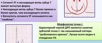
- myocardial infarction : the location of the ST segment changes on the ECG, which is located either above or below the isoline;
- death, necrosis of the myocardium, which is characterized by the appearance of an abnormal Q wave.
Scar changes
Areas of scar tissue form at the site of a former inflammatory process, necrosis, as a result of which normal, healthy cardiomyocytes lost their contractility and were replaced by connective tissue that does not have elasticity. Focal cicatricial changes on the ECG indicate a previous myocardial infarction .
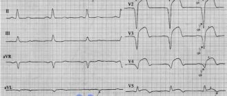
- The lower wall of the left ventricle is characterized by changes in leads: II, III and a VF (indicates damage to the right, less often the left circumflex coronary artery).
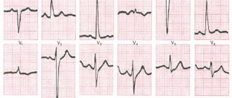
- The anterior septal region is characterized by changes in leads: V1 and V2 (the left descending septal branch is damaged), or V2-V4 (the left descending coronary artery or its branches is involved).
- The anterior-lateral region is characterized by changes in leads: I, aVL, V4-V6 (the circumflex artery or the left descending artery is damaged).
- Anterior widespread infarction is characterized by changes in leads: I, aVL, V1-V6 (the left descending coronary branch is damaged).
Moderate inflammatory changes in the myocardium
Characteristic changes are observed in myocarditis, in which the voltage of the waves in all leads decreases and rhythm disturbances are recorded. Moderate left ventricular changes may occur after:
- typhus , diphtheria ;
- rheumatism , after streptococcal infection ( tonsillitis , tonsillitis , scarlet fever );
- infections caused by the Coxsackie virus, rubella , influenza , measles ;
- exacerbation of an autoimmune disease ( systemic lupus erythematosus , scleroderma ).
Brown myocardial atrophy
This is what a macropreparation is called during histological examination. Characteristic pathological changes in the myocardium are formed as a result of a long-term lack of blood supply, which is observed in debilitating diseases, cachexia , drug abuse, increased physical activity, and also in old age. lipofuscin, is deposited . Its granules are a product of impaired metabolism in cardiac muscle cells, weakened by insufficient nutrition and blood supply.
EXAMPLE 2
Here is another Q in the same lead III. But here, after measurements, we see that its amplitude is not just 1/3 or 1/2 R, but almost equal to it - this is not the norm. In addition, the duration (width) of 0.04 s also goes beyond what is permitted. In the adjacent lead aVF, the Q wave is also pathological - it has a height of 1/3 R and a width of about 0.04 s. This gives us every right to talk about scar changes in the posterior wall (this is the area controlled by these leads)
Symptoms
Moderate and minor changes in the ECG may not manifest themselves clinically and may be found on the cardiogram during routine medical examinations and medical examinations. With pronounced changes, special symptoms appear:
- chest pain of a pressing, burning nature, indicating an attack of angina pectoris ;
- swelling of the lower extremities, shortness of breath with minimal physical activity indicate the progression of heart failure ;
- a feeling of interruptions in the work of the heart, arrhythmias ;
- fatigue, pale skin;
- sticky sweat;
- tremors , weight loss in a short time.
Is there a danger with changes in the myocardium?
Moderate changes in the myocardium are a fairly common pathology that gradually affects healthy cells of the heart muscle. Doctors say that this leads to the development and progression of heart failure. Let's consider whether it is worth showing concern when diagnosing such a pathology, what it is: moderate disturbances in the myocardium.
Such changes may not always be dangerous. Especially if it does not manifest itself with pronounced symptoms. Often pathology is discovered during a regular medical examination. If a person does not feel any problems with the functioning of the heart, then most likely there is no need to show concern. The reason for a visit to a cardiologist should be:
- pain in the heart area;
- heart rhythm disturbances;
- dyspnea;
- high or low blood pressure;
- weakness, drowsiness.
Tests and diagnostics
To identify the true cause of changes in the myocardium, it is recommended to undergo a full examination, which includes:
- General analysis and biochemical blood test. Based on the results, it will be possible to talk about the presence of an inflammatory process in the body, the functioning of the renal system and kidneys, and the level of cholesterol , which forms plaques in the coronary arteries.
- ECG. Characteristic changes during the examination make it possible to determine further examination tactics. In some cases, it is recommended to perform an ECG with stress, or organize daily ECG monitoring.
- EchoCG. Ultrasound examination of the heart allows you to assess the condition of the valve apparatus of the heart, identify areas of damage, and evaluate the pumping function of the heart.
- Myocardial scintigraphy. The radioisotope research method shows areas of accumulation of a special substance to identify areas of damage and determine their nature.
Changes in the myocardium on the ECG make it possible to determine further tactics for examining the patient to establish an accurate diagnosis and select the correct therapy.