Why is the physiological maturity of a newborn more important than a healthy weight or being born at term?
The most common criterion by which young mothers assess the condition of their baby is height and weight.
Born 3500 grams and 54 cm - a “hero”, and if 2800 grams - then sympathy and words of support immediately appear in the eyes of those around him: “It’s okay, he’ll gain more, but the birth went well.” But is it really important how much a baby weighs at birth? How will his height and weight affect his further development, resistance to diseases and psycho-emotional state?
Neonatologists and pediatricians, who have been observing the growth and development of children for years, have come to the conclusion that external signs in the form of weight and height do not provide much information about the child. Much more important is physiological maturity.
Heart formation
So, a new life was born. Whether you wanted it or not, whether the fruit of your love is desired or not - it doesn’t matter anymore. The egg formed in the ovary passed through the tubes, settled in the uterine mucosa, received and merged with the sperm. This is already a fertilized egg that will grow and eventually become your child.
This life, while still only one cell, carries all the information contained in your genes, i.e. the smallest protein molecules, and in your partner's genes. We will return to this later. But now, the cells have merged, and in the first two weeks after conception, the processes of formation of cellular systems begin, which will then turn into tissues and organs.
As the amazing poet Dmitry Kedrin once wrote:
“There is still no trace of nausea or spots. And your belt is just as narrow, just look in the mirror. But you, by elusive, secret feminine signs, frightenedly guessed what was inside you ... "
At first, new life has the shape of a disk. Sometimes such a small protein disc can be seen in the yolk of a broken chicken egg. It is called an embryo and in the first days it is just a collection of wise cells that know exactly what they need to do. With each subsequent hour there are more and more cells. They connect and fold into certain shapes, first forming two tubes, then merging into one. This tube folds and descends from the primary disk to form a loop called the “primary cardiac loop.” The loop quickly lengthens, significantly outstripping the growth and increase in the number of cells surrounding it, and lies to the right, in the form of such a ring as the ring of a mooring rope, which is thrown onto the bollard when mooring a boat or vessel. This loop normally lies only on the right, otherwise the future heart will lie not on the left, but on the right of the sternum. And on the 22nd day after conception, the first contraction occurs in the thickened lower part of the loop. The heart began to beat. You can try to remember what happened to the future mother then. What condition was she in? What was happening to her? And, if you, like the vast majority of married and non-family couples, did not pay attention to this, I can guarantee that you will not remember. You will say: “So what?” - and you will be right. As a rule, nothing. But still, think about it. The first days may not solve anything. But the next ones will decide a lot.
The cardiovascular system of the fetus is formed first of all its systems, because the fetus needs its own blood circulation for the full development of its other organs. The development and formation of the cardiovascular system begins in the third week and mainly ends by the eighth week of the embryo’s life, i.e. occurs within five weeks.
We will briefly describe these stages, but now let’s ask ourselves the question: “What is 4–5 weeks of pregnancy today?” The woman is not yet sure whether she is pregnant, especially if she is not looking forward to this event too much. She does not change her lifestyle, habits, sometimes harmful ones. She can work in hard and hazardous work or do hard physical work at home. She can carry a viral infection in the form of influenza on her feet. Usually a couple doesn’t think yet, tries not to think about the future, but it – this future – not only lives, but also beats, contracts, grows. But wait to punish yourself - there may be other reasons. More about them later. In the meantime, let us remember: today in the world they believe that a child’s life begins not from the moment of his birth, but from the moment of conception.
So, on the 22nd day, the future heart begins to pulsate, and on the 26th day, independent blood circulation begins in the body of the fetus, whose length is 3 millimeters. Thus, by the end of the fourth week the fetus has a contracting heart and circulation. So far it is one stream, one curved tube, in the bend of which lies the “motor” - the heart. But every minute processes take place in it that lead to final formation. It is very important to understand that these processes flow simultaneously in three-dimensional space and in order for “everything to come together correctly and accurately,” they need to be completely synchronized. Moreover, if this did not happen, i.e. at some point something did not connect where it was needed, the growth and development of the heart does not stop. Everything is going as usual. After all, when some musician in the orchestra suddenly plays a false note, the orchestra will still finish the symphony. But the false sound will fly away and be forgotten, and few will pay attention to it, and the developing heart will remember it. And now the growing septum has nowhere to attach, or the valve has nothing to hold onto. This is how birth defects are formed. In order for the heart to become four-chambered and not two-chambered (as in the third week), it is necessary for its partitions to grow (interatrial and interventricular), so that the common arterial trunk is divided into the aorta and pulmonary artery, so that inside the common ventricle it is divided into right and left so that the aorta connects to the left ventricle so that the heart valves are fully formed. All this happens between the 4th and 8th weeks of pregnancy (at this time the length of the fetus reaches only 3.5–4 cm). By the end of the second month of pregnancy, the “inch” (3.5 cm) embryo is already fully formed. Obviously, the earlier in this process a disruption of normal development occurred, the more the heart is deformed, i.e. the more severe his congenital defect. The later this happened, the smaller the structural change will be and the easier it will be to correct the defect in the future.
Quoted from the book by G. E. Falkovsky, S. M. Krupyanko. The heart of a child. A book for parents about congenital heart defects
How to get treatment at the Scientific Center named after. A.N. Bakuleva?
Online consultations
What is physiological maturity?
Physiological maturity implies the baby’s readiness for independent life outside the mother’s body. The concept extends to assessing the functioning of the main systems - nervous, cardiovascular, musculoskeletal, respiratory.
Even to a person far from science, it is obvious: in order for a baby to grow and develop harmoniously, his body must itself maintain a constant temperature, the lungs must exchange oxygen and carbon dioxide in the blood, and the heart must fully saturate all organs with blood.
Why is it so important to correctly assess physiological maturity? To preserve it in mature babies and help “mature” those who have deviations.
The more the baby corresponds to the concept of maturity, the less often he will get sick. Its correct development from the first days is insurance against endless viral infections, allergic reactions to drugs and diathesis.
Musculoskeletal reflexes
These are motor reflexes that are characteristic of physiologically mature children. They represent the baby’s reaction to irritation of various parts of his skin.
- Robinson's reflex or "grasp" reflex
If you put an adult’s finger into the baby’s palm, he grabs it so tightly that the doctor can calmly lift the child on this finger. Because simultaneously with the contraction of the palm muscles, the overall tone of the body and all skeletal muscles are strengthened.
- Plantar reflex
The doctor touches the skin of the baby’s foot with line movements, that is, irritates it along the inner edge of the sole. In response to the impact, the baby extends the big toe and bends all the other toes. At the same time, it can be noted that the child bends his legs at the knee and hip joints, and the contractile activity of the remaining muscles of his body increases.
- Difficulty straightening the leg at the knee
If a child’s leg is bent at the hip joint, it is difficult to straighten at the knee joint. This is explained by the fact that in a physiologically mature infant, the tone of the flexor muscles always prevails over the tone of the extensor muscles.
- "Crawl"
If you put a child on his tummy and put your palm on his soles, he reflexively pushes away from it. A crawling effect is created, although the baby remains in place.
- Negative reaction to support
The baby, who is lifted vertically above the table, held under the arms, does not lean on it. Even if his legs are lowered to the table. The child bends his legs and pulls them towards his tummy.
- Heel reflex, or Arshavsky reflex
With moderate pressure on the baby's heel bone, a generalized reaction of the body occurs in the form of extensor motor activity, screaming and crying grimace.
- General motor activity of the baby
During sleep, physiologically mature infants exhibit spontaneous motor activity. It occurs due to periodic changes in the composition of the baby’s blood. Its manifestations are a separate trembling of the arms or trembling of the legs, more with a tendency to extension than to flexion.
What may indicate childhood heart disease?
First of all, it is painful sensations in the sternum, as well as in the back in the area between the shoulder blades. Sometimes the pain is so severe that it may be symptoms of coronary artery disease. As a rule, pain appears after active activities or sports, and can sometimes become more active at night when the body is in an uncomfortable position. Among the additional symptoms that indicate that you urgently need to visit a pediatric cardiologist in Saratov are:
- persistent cough;
- frequent fever;
- dyspnea;
- the child complains of headaches;
- severe motion sickness in transport;
- increased fatigue.
During your appointment, an experienced cardiologist will carefully listen to the heart’s function to determine whether the child has a heart rhythm disorder. Heredity is taken into account, as well as:
- how the pregnancy progressed;
- were there any complications during childbirth;
- whether there have been previous infections.
Note!
It would seem that it is easier to check a baby’s reflexes during breastfeeding: the baby is busy, not distracted, and the reflexes arise independently. In reality, it turns out that when feeding, the severity of all the reflexes described above decreases. Therefore, such untimely testing may mistakenly classify a physiologically mature infant as immature.
- When sucking, you can check only one reflex - sucking: stroking the baby's cheeks leads to increased sucking.
While eating, shuddering, flapping of arms and other physical activity of the baby are the absolute norm. Babies who actively move their limbs during feedings gain weight and grow in length faster. If the baby is swaddled tightly so that he does not “scare himself” while eating, the processes of metabolism in his body and growth slow down sharply.
Muscle activity, which is triggered and regulated by the child’s nerve centers, is the main factor on which the further development of his brain, the increase in brain mass and the intellectual development of the baby in later life depend.
Respiration rate
The respiratory rate in infants who are physiologically mature is 35-42 breaths per minute.
Heart rate
A baby's heart beats 135-140 times per minute at rest. This surprising heart rate is the absolute norm for infancy.
Arterial pressure
Measuring blood pressure in mature newborns in the first days after birth shows values of 80-85 mm Hg. for “upper” pressure, and 45 mm Hg. for "bottom".
Pediatrician tactics for critical congenital heart defects in newborns
In the structure of infant mortality, developmental anomalies occupy third place, and half of the mortality cases are determined by congenital heart defects (CHD). Among children who died from congenital heart disease and malformations of large vessels, 91% of patients were infants of the first year of life, of which 35% of deaths occurred in the early neonatal period (up to 6 days). About 70% of children die during the first month of life [3, 4].
The scale of the problem is emphasized by the high frequency of congenital heart disease: in different countries this figure varies from 0.6% to 2.4% per year in children born alive; taking into account intrauterine fetal death and early miscarriages, the overall frequency of congenital heart disease is 7.3% [1, 3].
Prenatal diagnosis. In order to reduce infant mortality, prenatal ultrasound screening is used, which makes it possible to detect most congenital heart diseases before the 24th week of gestation. If a defect is suspected, a targeted ultrasound of the fetus is performed using an expert-class device. The main goal is to prevent the birth of children with inoperable defects - hypoplastic left heart syndrome (HLHS), hypertrophic cardiomyopathy with signs of organic myocardial damage, multiple fetal malformations. The prenatal consultation should offer termination of pregnancy only if an incurable defect is accurately diagnosed [2, 4].
Classification. In the neonatal period (sometimes in the first days, hours or minutes after birth), defects called critical manifest themselves, since in 95–100% of cases they are accompanied by life-threatening conditions and determine early neonatal mortality. The group of critical defects includes transposition of the great vessels (TMS), TGV, atresia of the tricuspid valve or pulmonary artery with an intact interventricular septum (IVS), preductal coarctation of the aorta, common truncus arteriosus, single ventricle, double origin of the great vessels from the right ventricle and others [1 , 5].
Considering the high mortality rate of newborns and infants from congenital heart disease, a classification has been created for this age group of patients based on the definition of the leading clinical syndrome, the effectiveness of therapeutic tactics and determining the timing of surgical intervention [3, 4].
Syndromic classification of congenital heart disease in newborns and children of the first year of life (Sharykin A. S., 2005)
- CHD manifested by arterial hypoxemia (chronic hypoxemia, hypoxemic status) are “ductus-dependent” defects.
- CHD, predominantly manifested by heart failure (acute heart failure, congestive heart failure, cardiogenic shock).
- CHD manifested by heart rhythm disturbances (complete atrioventricular block, paroxysmal tachycardia).
These conditions can be combined, aggravating the severity of the condition of children; 50% of these children require surgical or therapeutic intervention in the first year of life.
Hemodynamics. Critical defects are characterized by ductus-dependent pulmonary or systemic circulation; they are united by a sudden sharp deterioration of an apparently healthy child at birth, associated with a decrease in blood flow through the ductus arteriosus. Ductus-dependent pulmonary circulation in TMS, atresia (or critical pulmonary artery stenosis) with an intact IVS provides blood flow through the duct into the pulmonary circulation, and when it is limited or stopped, severe arterial hypoxemia and acute hypoxia of organs and tissues develop.
Clinic of congenital heart disease with pulmonary ductus-dependent circulation
The anatomy of one of the most common critical defects - transposition of the great vessels - consists in the incorrect origin of the aorta - from the right and pulmonary arteries - from the left ventricle, which contributes to the separation of the circulatory circles: arterial blood circulates in the pulmonary circulation system, and venous blood circulates in the systemic circulation system.
The supply of oxygen to life-supporting organs is possible only if there are functioning fetal communications - ductus arteriosus, interatrial defect. This communication between the circulation does not provide compensation for hypoxemia. In order to compensate for the deficiency of peripheral circulation, the minute volume of blood flow increases, overload of the small circle occurs (this happens faster in the presence of a defect in the IVS), and pulmonary hypertension quickly develops. That is why, during the management of the patient, constant monitoring of symptoms of arterial hypoxemia and monitoring of clinical signs of heart failure (HF) is necessary -
The natural course of the vice is very severe. The child is born at term with normal body weight, but in the first hours after birth diffuse cyanosis of the skin appears, especially pronounced in the periphery - cyanosis of the face, hands, and feet. The condition of extreme severity is caused by severe arterial hypoxemia. Dyspnea and tachycardia appear 1–2 hours after clamping the umbilical cord. There is a progressive deterioration of the condition. The child is lethargic, lethargic, and easily cools down.
When fetal communications are closed, acute hypoxia leads to the development of multiple organ failure and death of the newborn within a few hours. As the child survives for several weeks, heart failure progresses. Severe malnutrition quickly develops. It should be noted that in the case of adequate observation and treatment tactics, as well as timely - up to a month - surgical correction of the defect in a child (since only during this period is radical correction possible by the method of arterial switching of the great vessels), physiological hemodynamics, growth and development rates are completely restored, physical and subsequently social adaptation. If the defect is corrected later, the outcomes are less favorable.
Diagnostic criteria for TMS include:
- Electrocardiographic signs of hypertrophy of the right atrium and right ventricle are a high P wave in the “right” leads - III, V1-3, deep S waves in the “left” - I, V5-6 and high R waves in leads III, V1-3.
- X-ray reveals cardiomegaly and an “ovoid” shape of the heart with a narrow vascular bundle as a result of the alignment of the contours of large vessels (photo).
- According to echocardiography, there is a parallel course of the outflow sections of the ventricles - the pulmonary artery and the aorta.
- The hyperoxide test is negative - when trying to supply 100% oxygen through a mask in patients with “blue” defects, after 10–15 minutes pO2 increases by no more than 10–15 mmHg. Art. (whereas in lung diseases the increase in pO2 is up to 100–150 mm Hg).
Scheme for examining a newborn child with suspected congenital heart disease:
- examination of the patient (with assessment of symptoms of hypoxemia and/or heart failure);
- assessment of pulsation in all extremities;
- auscultation of the heart and lungs (dynamic control of heart rate, breathing);
- measurement of blood pressure (BP) in all extremities (further dynamic control).
In addition, monitoring a child involves monitoring blood gases (pO2, pCO2), oxygen saturation (SatO2) using pulse oximetry and metabolic indicators - pH, BE. Gas exchange in the lungs is not impaired if PaO2 is in the range of 60–80 mmHg. Art., SaO2 - 96–98%. Arterial hypoxemia develops when PaO2 is less than 60 mm Hg. Art. and a hemoglobin saturation level of 85–75%.
Tasks of a pediatrician (neonatologist):
- ensure a reduction in the body's oxygen needs by creating thermal and physical comfort - incubator conditions, with an elevated position of the upper body;
- swaddling with the chest and arms free;
- limiting energy costs for physiological load (tube feeding);
- support of blood flow through the ductus arteriosus (infusion of fluids, prostaglandin E);
- correction of metabolic changes, if necessary, artificial ventilation of the lungs (ALV) without adding oxygen to the inhaled mixture, in a mode that excludes hyperventilation and with simultaneous infusion of the drug prostaglandin E (calculation of the drug dose is described below). When deciding whether to prescribe mechanical ventilation, it is necessary to take into account that oxygen has a vasoconstrictor effect on the ductus arteriosus, which makes oxygen therapy dangerous in this group of patients;
- if there is a threat of closure of ductus-dependent defects, the volume of infusions and feeding is increased to 110–120% of normal needs against the background of constant assessment of diuresis. It has been established that a 5% increase in body weight in a newborn in 1–2 days stabilizes the function of the ductus arteriosus.
Transportation to a cardiac surgery center is optimal during the first weeks and first month of life. It is first necessary to inform the cardiac surgery hospital about a patient with congenital heart disease with ductus-dependent circulation. The observation period until transfer and transportation to the center is carried out against the background of infusion of the drug prostaglandin E (Alprostan, Vazaprostan).
Clinical picture of congenital heart disease with systemic ductus-dependent circulation (the group of defects includes HFRS, severe coarctation of the aorta, interruption of the aortic arch). The most positive example of defects in this group is pronounced preductal coarctation, which occupies from 1% to 10% of critical congenital defects. With this defect, blood flow from the proximal part (below the origin of the ductus arteriosus) to the distal part is sharply limited or completely absent. The hemodynamic disorder, accordingly, lies in the fact that a small volume of blood enters the descending aorta (into the greater circle) only from the pulmonary artery through the ductus arteriosus. When the ductus arteriosus closes, hypoperfusion of organs and tissues and multiple organ failure acutely develop. Clinic: a full-term newborn with a sharp deterioration in the first few days of life - adynamia, cold extremities, a symptom of hypoperfusion of peripheral tissues (“white spot”), low filling pulse, high blood pressure in the arms and low or not determined in the legs, shortness of breath, tachycardia, oliguria with increasing azotemia, hepatomegaly with increased transaminases, necrotizing enterocolitis.
Let us consider the diagnosis and optimal therapeutic tactics for a patient with severe coarctation of the aorta using a specific clinical example.
Full-term newborn A. was delivered to the intensive care unit in serious condition: lethargic, does not suckle, pale skin, tachypnea 120 breaths per minute, breathing is symmetrical in all fields, no wheezing. Heart sounds are sonorous, 167 per minute, gentle systolic murmur in the third intercostal space to the left of the sternum, hepatomegaly (liver +5 cm from under the edge of the rib, dense). Diuresis is reduced, there is no peripheral edema. Blood pressure in the arms - 127/75 mm Hg. Art., pulsation in the femoral artery is not detected. SatO2 - 98%.
From the anamnesis: the condition suddenly worsened on the 14th day of life, when the child became lethargic, severe shortness of breath appeared, and was hospitalized by ambulance. A boy from a second, normal pregnancy, term birth with a weight of 3220 g, an Apgar score of 5 (9) points. He was discharged from the maternity hospital in satisfactory condition and was breastfed. Periodically there were episodes of anxiety and flatulence.
Upon admission, the child is intubated and undergoes mechanical ventilation with a low oxygen content in the inhaled mixture. An examination in the hospital revealed cardiomegaly (cardiothoracic index - 80%), depleted pulmonary pattern, and, according to electrocardiography, combined overload of both ventricles. Echocardiography revealed hypoplasia of the aorta below the origin of the left subclavian artery (and above the localization of the ductus arteriosus), in a place typical for the ductus arteriosus - punctate blood flow (closing ductus arteriosus). After 6 hours, the child’s condition worsened: oliguria developed, creatinine increased to 213 mmol/l, and transaminases increased by 4–5 times the laboratory norm. Humoral activity has not been established.
Rationale for diagnosis and tactics: given the clinical picture of severe respiratory and subsequently multiple organ failure in combination with cardiomegaly, systemic arterial hypertension, renal and liver failure, according to clinical data, coarctation of the aorta should have been suspected. A sudden deterioration in the child’s condition in the absence of signs of infection allows one to think about ductus-dependent systemic circulation; taking into account the imaging data of the heart and blood vessels, the diagnosis is made: “CHD, preductal coarctation of the aorta, arterial hypertension stage 2, secondary, multiple organ failure.”
From the moment the diagnosis is confirmed by echocardiography, it is necessary to begin therapy with Vazaprostan 0.02 (with an increase in dose to 0.05 mcg/kg/min) in order to restore blood flow through the ductus arteriosus. With this defect, the shunt is directed from the pulmonary artery to the descending aorta, and only this small portion of blood provides the entire systemic circulation.
Dose calculation and administration method. The child’s weight upon admission is 3220 g. 1 ampoule contains 20 mcg of Vazaprostan. In this case, drug administration began with a dose of 0.02 mcg/kg/min, that is, 0.02 ´ 3.2 = 0.064 mcg/kg/min was required. Over an hour, the dose of the drug was 0.064 ´ 60 = 3.8 mcg/hour. To administer the drug, 1 ampoule (20 mcg of Vazaprostan) was diluted in 20 ml of physiological solution (1 mcg in 1 ml). If there was no effect within two hours, the dose was increased to 0.04–0.05 mcg/kg/min: accordingly, the rate of drug administration increased to 7.6–9.5 ml/hour. In this case, therapy control was very indicative - after 6 hours of infusion, an improvement in the condition was noted, an increase in the blowing systolic murmur in the second intercostal space on the left, and an increase in the size of the flow through the ductus arteriosus according to echocardiography. Transferred to a maintenance dose of Vazaprostan - 0.01 and then 0.005 mcg/kg/min, which was maintained throughout the entire period of observation in the pediatric hospital and during transportation during transfer to the cardiac surgery hospital. In this clinical case, it was impossible to completely withdraw from mechanical ventilation (given the severity of respiratory failure); on the 5th day of hospitalization (19th day of life), the child was transferred to the clinic named after him during therapy. Meshalkin, where the defect was successfully corrected - the formation of an aortopulmonary shunt.
In cases of the development of heart failure in newborns with defects accompanied by massive discharge of blood into the pulmonary circulation, the same principle of monitoring key indicators and symptomatic therapy is applied:
- limiting fluid administration according to diuresis, in severe cases - up to 1/3 of the age norm (but limiting fluid to 50% of the daily physiological requirement is unacceptable);
- the presence of volume overload requires the use of diuretics (for edematous syndrome, preference is given to Furosemide/Lasix at a dose of 1–2 mg/kg; a combination with Veroshpiron is possible (1–3 mg/kg/day orally in 2–3 doses);
- Digoxin () is used to stop tachycardia (an economically unprofitable regimen for the myocardium and an ineffective volume for the peripheral circulation).
If symptoms of pulmonary circle overload appear (on auscultation - increase, splitting of the 2nd tone in the pulmonary artery, on EchoCG - increase in pressure in the pulmonary artery by more than 30 mm Hg after six days of life), as well as signs of impaired diastolic function of the heart, it is recommended to use drugs from the group of angiotensin-converting enzyme inhibitors (ACEIs). Capoten (captopril) is used at a dose of 0.5–1 mg/kg, subject to control of systemic blood pressure. The therapeutic effect of ACE inhibitors is associated with a decrease in peripheral vascular resistance and partial deposition of blood, resulting in a decrease in the volume of blood returned to the right side of the heart. Accordingly, the volume of the shunt and the load on the left chambers and vessels of the small circle are reduced. In addition, it is known that ACE inhibitors are an inhibitor of apoptosis stimulated by hypoxia, which explains the angio- and cardioprotective effect of the drugs.
Thus, pediatric tactics, including early diagnosis of critical congenital heart disease and therapy that controls intracardiac, central and peripheral hemodynamics, as well as, if possible, early coordination of actions with the cardiac surgery center can significantly improve the prognosis of patients and reduce infant mortality rates.
Literature
- Burakovsky V. A., Bukharin V. A., Podzolkov V. P. et al. Congenital heart defects. In the book. Cardiovascular surgery. Ed. V. I. Burakovsky, L. A. Bockeria. M.: Medicine, 1989; 345–382.
- Congenital heart defects. Directory for doctors. Ed. E. V. Krivosheeva, I. A. Kovaleva. Tomsk, 2009; 285.
- Sharykin A. S. Congenital heart defects. Guide for pediatricians, cardiologists, neonatologists. M.: Teremok Publishing House, 2005; 384.
- Sharykin A. S. Perinatal cardiology. Guide for pediatricians, cardiologists, neonatologists. M.: Teremok Publishing House, 2007; 347.
- Johnson Jr. WH, Moller JH Pediatric cardiology. Core handbooks in pediatrics. LIPPUNCOTT WILLIAMS & WILKINS, 2001; 326.
E. Yu. Emelyanchik *, Doctor of Medical Sciences, Professor D. B. Drobot *, Doctor of Medical Sciences, Professor E. P. Kirillova *, Candidate of Medical Sciences, Associate Professor V. A. Sakovich *, Doctor of Medical Sciences, Professor E. V. Basalova ** A. Yu. Cheremisina *
*GOU HPE Krasnoyarsk State Medical University named after. Professor V. F. Voino-Yasenetsky, ** Regional Clinical Children's Hospital, Krasnoyarsk
Contact information for authors for correspondence
Nervous system
In physiologically immature children, the process of maturation of the nerve pathways - the formation of the myelin sheath around nerves and nerve fibers, or myelination - is not yet completed, therefore the conduction of nerve impulses to various organs and tissues is disrupted, which affects the viability of their functions. The peripheral nervous system is not sufficiently myelinated; bundles of nerve fibers are rare and unevenly distributed. Myelination normally continues in the postnatal period, i.e. after birth.
The walls of blood vessels in immature babies may consist of only one inner layer - the endothelium and do not contain smooth muscle tissue, collagen or elastin (especially if the mother has hypoxia during pregnancy and a deficiency of protein, iron, copper, vitamin C, etc.). That is, the formation of the connective tissue framework in the vascular wall does not end, which reduces resistance to hypoxia and increases the risk of developing intracranial hemorrhages.
In immature newborns, there is no mechanism for autoregulation of cerebral vessels due to the incomplete formation of vascular fibers. In addition, the metabolic activity of endothelial cells depends on the pathways that ensure the occurrence of oxidation processes, and therefore there is a high probability of their damage during hypoxia.
During childbirth, even without complications, the baby’s brain experiences significant stress. The pressure on the membranes of the brain becomes so strong that spasms, circulatory disorders, and hemorrhages in the brain can develop. These phenomena are more likely in premature and immature children due to immaturity of brain structures. To determine the degree of immaturity and damage to the central nervous system, the so-called neurological status of the newborn is described, which is determined by such factors as: behavioral state, muscle tone, motor activity, unconditioned reflexes, reaction to external stimuli.
Those with an immature nervous system have reduced motor activity and muscle tone, and are characterized by weakness and rapid decline of physiological reflexes. A slow reaction to stimulation is characterized by its prevalence throughout the entire body, weakness of active inhibition, and irradiation of the excitation process, i.e. irritation from one center spreads to another, but on a smaller scale. The immaturity of the cortex determines the predominance of subcortical activity: movements are chaotic, shudders, tremor (shaking) of the hands, clonus of the feet (convulsive muscle contractions in response to impact), twitching of the eyeballs, and transient strabismus may be observed.
Anatomical and physiological features of the cardiovascular system in children
The most important functions of the cardiovascular system are:
1) maintaining a constant internal environment of the body;
2) delivery of oxygen and nutrients to all organs and tissues;
3) removal of metabolic products from the body.
The cardiovascular system can provide these functions only in close interaction with the respiratory, digestive and urinary organs. Improvement in the functioning of the circulatory system occurs unevenly throughout childhood.
Features of intrauterine circulation in children
The formation of the heart begins in the 2nd week of intrauterine life. Within 3 weeks, the heart with all its parts is formed from the plate located on the border of the head and torso. In the first 6 weeks, the heart consists of three chambers, then four are formed due to the division of the atria. At this time, the process of dividing the heart into the right and left halves and the formation of heart valves occurs. The formation of the main arterial trunks begins from the 2nd week of life. The conduction system of the heart is formed very early.
Intrauterine blood circulation of the fetus
Oxygenated blood passes through the placenta through the umbilical vein to the fetus. A smaller part of this blood is absorbed into the liver, a larger part into the inferior vena cava. Then this blood, mixed with blood from the right half of the fetus, enters the right atrium. Blood from the superior vena cava also flows here. However, these two blood columns hardly mix with each other. Blood from the inferior vena cava enters the left heart and aorta through the oval window. Oxygen-poor blood from the superior vena cava passes into the right atrium, right ventricle and the initial part of the pulmonary artery, from here through the ductus arteriosus it enters the aorta and is mixed with blood coming from the left ventricle. Only a small part of the blood enters the lungs, from there into the left atrium, where it mixes with the blood entering through the oval window. A small amount of blood circulates in the pulmonary circulation before the first breath. Thus, the brain and liver receive the most oxygen-rich blood, and the lower extremities receive the least oxygen-rich blood.
After the birth of a child, the venous duct and umbilical vessels become empty, overgrow and turn into the round ligament of the liver.
All physiological life support systems are involved in the action.
Anatomical and physiological features of the heart and blood vessels in children
Children experience continuous growth and functional improvement of the cardiovascular system. The heart grows and improves especially vigorously in children from 2 to 6 years of age, as well as during puberty.
The heart of a newborn has a flattened cone-shaped, oval or spherical shape due to insufficient development of the ventricles and the relatively large size of the atria. Only by the age of 10-14 years does the heart acquire the same shape as that of an adult.
Due to the high position of the diaphragm, the newborn’s heart is located horizontally. The heart assumes an oblique position by the first year of life.
The weight of a newborn's heart is 0.8% of the total body weight, which is relatively larger than that of an adult. The right and left ventricles are equal in thickness, their walls are 5 mm. The atrium and great vessels are relatively large in size. By the end of the first year, the weight of the heart doubles, and by 3 years it triples. In preschool and primary school age, heart growth slows down and increases again during puberty. By the age of 17, the mass of the heart increases 10 times.
The parts of the heart also grow unevenly. The left ventricle significantly increases its volume; by 4 months it is twice as heavy as the right one. The thickness of the walls of the ventricles in a newborn is 5.5 mm, later the thickness of the left ventricle increases to 12 mm, the right - to 6-7 mm.
The volume of the heart at birth is about 22 cm3, during the first year it increases by 20 cm3, and subsequently by 6-10 cm3 annually. At the same time, the diameter of the valve openings increases.
In children, the heart is located higher than in adults. The volume of the heart in children is larger relative to the volume of the chest than in adults. In a newborn, the apex of the heart is formed on both ventricles, by 6 months - only on the left. By the age of 1.5 years, the projection of the heart from the 4th intercostal space descends into the 5th intercostal space.
In childhood, a qualitative restructuring of the heart muscle occurs. In young children, the heart muscle is undifferentiated and consists of thin, poorly separated myofibrils, which contain a large number of oval nuclei. There is no transverse striation. Connective tissue begins to develop. There are very few elastic elements; in early childhood, muscle fibers are close to each other. As the child grows, the muscle fibers thicken and rough connective tissue appears. The shape of the nuclei becomes rod-shaped, transverse striations of the muscles appear, and by 2-3 years of age the histological differentiation of the myocardium is completed. Other parts of the heart are also improving.
As the child grows, the conduction system of the heart improves. In early childhood it is massive, its fibers are not clearly contoured. In older children, overmodulation of the conduction system of the heart occurs, so cardiac arrhythmias are common in children.
The work of the heart is carried out due to the superficial and deep plexuses formed by the fibers of the vagus nerve and cervical sympathetic nodes, in contact with the ganglia of the sinus and atrioventricular nodes in the walls of the right atrium. The branches of the vagus nerve complete their development by 3-4 years. Until this age, cardiac activity is regulated by the sympathetic system. This explains the physiological increase in heart rate in children in the first 3 years of life. Under the influence of the vagus nerve, the heart rate slows down and respiratory-type arrhythmia appears, and the intervals between heartbeats lengthen. Myocardial functions in children, such as automatism, conduction, contractility, are carried out in the same way as in adults.
Features of blood vessels in children
The vessels supply and distribute blood to the child’s organs and tissues. Their clearance in young children is wide. Arteries are not equal in width to veins. The ratio of their lumen is
1:1, then the venous bed becomes wider, by the age of 16 their ratio is 1:2. The growth of arteries and veins often does not correspond to the growth of the heart. The walls of arteries are more elastic than the walls of veins. This is associated with lower levels of peripheral resistance, blood pressure, and blood flow velocity than in adults.
The structure of the arteries also changes. In newborns, the walls of blood vessels are thin, and their muscle and elastic fibers are poorly developed. Up to 5 years of age, the muscle layer grows rapidly; at 5–8 years of age, all vascular membranes are evenly developed; by 12 years of age, the structure of blood vessels in children is the same as in adults.
The heart rate in children depends on age. In a newborn it is 160-140 beats per minute, in 1 year - 110-140, in 5 years - 100, in 10 years - 80-90, in 15 years - 80.
With age, systolic blood pressure increases, and there is a tendency for diastolic pressure to increase.
Arterial systolic pressure is 90 + 2 xn, diastolic pressure is 60 + 2 xn, where n is the child’s age in years. For children under 1 year of age, systolic pressure is 75 + n, where n is the child’s age in months. Diastolic blood pressure is equal to systolic pressure minus 10 mm Hg. Art.
Heart and blood vessels during puberty
During puberty, intensive growth of various organs and systems occurs. During this period, disturbances in their functioning occur due to violations of their relationships and coordination of functions. In adolescents, due to the growth characteristics of both the heart and the whole body, relatively small mass and volume of the heart are observed compared to the mass and volume of the body. The ratio of body volume to heart volume in children is 50%, in adults - 60%, and in the puberty period it is 90%. In addition, there are anatomical features of the cardiovascular system in adolescents that are associated with the ratio of the volume of the heart and blood vessels.
In adolescents, the volume of the heart increases faster than the capacity of the vascular network, this increases peripheral resistance, which leads to a hypertrophic variant of the subadult heart.
In adolescents with deviations in the age-related evolution of the heart, sympathetic regulation predominates.
Thus, children have functional characteristics of the circulatory organs, which are characterized by:
1) 1) high level of endurance of the child’s heart due to its fairly large mass and good blood supply;
2) physiological tachycardia, caused by the small volume of the heart with a high need for oxygen in the child’s body, as well as sympathotomy;
3) low blood pressure with a small volume of blood flowing with each heartbeat, as well as low peripheral vascular resistance;
4) uneven growth of the heart and associated functional disorders.
Respiratory system
Due to the immaturity of the nervous system, as well as the lipid metabolism system, the derivative of which is surfactant - a surface substance that ensures the opening of the lungs during the first breath and their normal functioning in the future, physiologically immature infants often develop a syndrome of respiratory disorders, which is manifested by the development of atelectasis. Atelectasis are areas of collapsed or incompletely straightened lung tissue that do not participate in breathing and can cause respiratory failure. Such a child is transferred to artificial ventilation until the respiratory system begins to function on its own. Against the background of respiratory distress syndrome, various infectious diseases (pneumonia) often occur, which certainly worsens the child’s condition.
The cardiovascular system
The structure and function of the fetal heart in the prenatal period differs from that of a newborn child. The fetus has a three-chambered heart, special openings are open - the “oval window” and the “botal duct”, through which the blood is mixed and in utero the child receives only “mixed” blood, which allows him to be quite resistant to a possible lack of oxygen. After birth, a restructuring of blood circulation occurs and the heart becomes four-chambered and the child begins to receive purely arterial, not mixed blood. Of course, the “holes” do not close immediately, but “mixing” of blood through them does not occur from the first minutes of life. In a physiologically immature child, such a restructuring of the heart occurs much more slowly—additional openings and vessels (patent foramen ovale and ductus ductus botallis) can not only persist for a long time, but also function.
The pulse in immature newborns is very labile, weak in filling, frequency 120-160 per minute, but can reach 180. Since the cardiovascular system of immature babies is sensitive to external stimuli, you need to try to protect the baby as much as possible from them, for example, from loud sounds .
Types of heart defects
| Type | Name |
| Congenital | |
| Without the development of cyanosis (pale type) | Defects of the interventricular and interatrial septa |
| Patent ductus arteriosus | |
| Pulmonary stenosis | |
| Coarctation of the aorta | |
| With the development of cyanosis (blue) | Transposition of the great vessels |
| Common ventricle | |
| Tetralogy of Fallot | |
| Purchased | |
| Mitral valve (between the left atrium and ventricle) | Mitral stenosis |
| Mitral regurgitation | |
| Tricuspid valve (between the right atrium and ventricle) | Tricuspid valve stenosis |
| Tricuspid valve insufficiency | |
| Aortic valve (between the left ventricle and the aorta) | Aortic stenosis |
| Aortic insufficiency | |
| Pulmonary valve | Pulmonary valve stenosis |
| Pulmonary valve insufficiency | |
Stenosis
- narrowing of the valve opening, which causes difficulty in blood flow.
Failure
- inability of the valves to close tightly. In people with valve stenosis, valve insufficiency develops over time - this is called combined PS.
Septal disorders:
A condition in which the septum between the chambers of the heart is damaged:
- atrial septum -
“extra” blood enters the right atrium and causes it to enlarge; - interventricular
- blood flows from the right to the left ventricle, expanding its boundaries.
Changes are found in 2 out of 1000 newborns. Perhaps the most favorable form, since small defects can heal on their own.
Coarctation of the aorta:
Narrowing of the lumen of the main vessel in the area of the isthmus - the transition of the arch to the descending part. The defect causes disruption of the blood supply to organs. This form of pathology accounts for 10% of all defects. Often combined with disruption of interchamber septa. A serious condition requiring medical attention immediately after birth.
Pulmonary valve stenosis:
At the site of the valves from the right ventricle to the pulmonary artery, a narrowing occurs. The heart requires more effort to pump blood. 10% of the total number of defects.
Transposition of the great vessels:
Rare pathology (about 5%). In this case, the main vessels “change places.” Arterial blood flows through the veins, and venous blood through the arteries.
Tetralogy of Fallot:
The most severe form. Consists of four violations simultaneously:
- the opening between the left and right ventricles;
- pulmonary stenosis – narrowing of the pulmonary trunk;
- enlargement of the right ventricle;
- Aortic displacement is the exit of the aorta from the heart in an atypical location.
Circulatory system
Due to the immaturity of bone marrow sprouts, as well as the low sensitivity of red bone marrow to erythropoietin (an active hormone of the kidneys that stimulates the production of red blood cells), lower levels of red blood cells and hemoglobin are observed in immature children, which leads to the development of early anemia of physiologically immature children, which develops during 2 first months of life. Rapid depletion of the white sprout leads to the development of neutropenia - a decrease in the number of neutrophils in the blood, especially in conditions of intra- and extrauterine infection of the child.
Physiologically immature newborns at birth have lower concentrations of vitamin K, vitamin K-dependent blood coagulation factors, and other disorders in the blood coagulation system. In this situation, bleeding from the first minutes of life can be combined with thrombosis due to the low activity of fibrinolysis and anticoagulants, which can lead to the development of DIC syndrome (disseminated intravascular coagulation syndrome).
Metabolic adaptation processes in immature infants are slowed down. They are more likely to experience hypoglycemia (low blood sugar), hypoxemia (low oxygen in the blood), hyperbilirubinemia (increased bilirubin in the blood serum), and early anemia in conditions of excess iron.
Classification
Congenital heart defects in children and adults are classified according to various criteria.
According to the complexity of anatomical changes, they are distinguished:
- simple defects - with single anomalies;
- complex - combine 2 abnormalities, for example, valvular insufficiency and narrowing of the cardiac orifices;
- combined – combinations of multiple defects, the most difficult to treat.
The following groups are known by the nature of circulatory disorders.
- Communication inside the heart occurs with arterial-venous discharge of blood (Golochinov-Roger disease, patent ductus arteriosus, increased pulmonary blood flow).
- The communication occurs with a venous-arterial discharge (triad and tetralogy of Fallot, tricuspid valve atresia, decreased pulmonary blood flow).
- Communication within the heart does not occur due to poor patency and narrowing of the main vessels (aorta, pulmonary artery).
- Based on the nature of their manifestations and symptoms, defects are usually divided into “blue” and “white”.
- With “blue”, arterial and venous blood mix, tissue hypoxia is observed, which is why they turn blue. This is especially noticeable in the area of fingers, lips, and ears. Common “blue” defects include Ebstein’s anomaly, tetralogy of Fallot, transposition of the aorta and pulmonary artery.
- With “white” defects, the pathology may not manifest itself clearly and is therefore difficult to diagnose. These, in particular, include deviations in the drainage of the pulmonary veins, septal defects, and the formation of a common atrium.
Digestive system
The digestive system of immature children also has a number of features. First of all, this is expressed in the immaturity of the enzyme system. The glands of the gastrointestinal tract do not produce the required amount of enzymes and gastric juice. When the gastrointestinal tract is colonized by microorganisms, even a small amount of pathogenic bacteria, which would normally be neutralized by the protective properties of gastric juice and pancreatic juice, causes dysbiosis in immature children (an incorrect ratio of certain microorganisms in the gastrointestinal tract).
Also, due to the immaturity of the nervous system and the transmission of nerve impulses, the motor function of the gastrointestinal tract suffers, and the movement of food through the gastrointestinal tract slows down. As a result, problems arise with the flow of food into different parts of the stomach and intestines and its excretion.
Features of the gastric tract in physiologically immature infants are:
- poor development of the sphincter at the entrance to the stomach, which leads to frequent regurgitation;
- poor development of longitudinal muscle bundles of the stomach wall, which causes lethargy and bloating due to overfeeding and exposure to air
- slow evacuation of stomach contents (130-140 min);
- high viscosity of original feces.
Despite the imperfection of the digestive system in physiologically immature infants, the gastric juice still contains rennet, which curdles milk. Therefore, mother's milk is mandatory for an immature child. In addition to its nutritional value, milk provides an invaluable service to protect the baby’s body from environmental aggression. Therefore, even if the child is immediately after birth in the intensive care unit or intensive care unit and receives parenteral nutrition (via a drip) or is so weak that he cannot breastfeed, it is necessary to take all possible measures to preserve breast milk, feed the baby from a spoon . This is one of the necessary factors when nursing immature babies.
In physiologically immature newborns, functional inferiority of the liver is possible; as a result, an insufficient amount of the enzyme glucurone transferase is produced, and this predisposes to the development of prolonged jaundice. A low level of prothrombin causes increased bleeding.
Immature children are predisposed to intestinal dysfunction. The intestinal wall has increased permeability, so microbes and toxins in the intestines are absorbed through the intestinal wall into the blood. Due to hypotension of the intestines and the anterior abdominal wall, flatulence is often observed; as a result, the diaphragm rises upward, squeezing the lower parts of the lungs and disrupting their normal ventilation.
The mucous membrane of the digestive canal in immature children is tender, thin, and easily vulnerable. There is low proteolytic activity of gastric juice, insufficient production of pancreatic and intestinal enzymes, as well as bile acids. All this complicates the processes of digestion and absorption, contributes to the development of flatulence and dysbacteriosis. Many immature children, even those who are breastfed, have a deficiency of intestinal bifid flora in combination with the carriage of opportunistic flora.
The nature of a child's stool is determined by feeding characteristics; As a rule, in the coprogram of physiologically immature children there is a lot of neutral fat.
Functional disorders of the gastrointestinal tract are one of the most widespread problems among children in the first months of life. A distinctive feature of these conditions is the appearance of clinical symptoms in the absence of any organic changes in the gastrointestinal tract (structural abnormalities, inflammatory changes, infections or tumors) and genetic metabolic abnormalities.
With functional disorders of the gastrointestinal tract, motor function, digestion and absorption of nutrients, as well as the composition of the intestinal microbiota and the activity of the immune system may change. The causes of functional disorders often lie outside the affected organ and are caused by a violation of the nervous and humoral regulation of the digestive tract.
Skeletal system
In physiologically immature children, the skeletal system is formed by birth, but bone mineralization is often not yet complete, and therefore they additionally need calcium (as recommended by a doctor).
And in prematurely born immature children there is often a deficiency of vitamin D, since it is in the last months of pregnancy that a supply of substances such as vitamin D, calcium, and phosphates occurs. If mom gets all this in abundance. A reduced amount of these substances in the body causes the development of diseases such as rickets, the signs of which are softening of the bones, slowing down of growth processes, especially of the legs, late appearance of teeth and closure of the large fontanelle, changes in the pelvic bones, as well as hip dysplasia, when due to lack of calcium, the femurs become deformed. Physiologically immature children are at risk for developing this disease. In them it is especially acute and progresses quickly. Therefore, physiologically immature children are often prescribed vitamin D to prevent rickets.
It happens that physiologically immature children are born with unformed hip joints. Dysplasia (underdevelopment) of joints threatens further subluxations, dislocations and deprivation of the ability to move independently. Therefore, it is necessary to diagnose this pathology in time and prescribe treatment. To detect dysplasia, an ultrasound examination of the joints is performed, which allows for a correct diagnosis. A common cause of joint dysplasia is a mother’s deficiency of magnesium, iron, protein, vitamin C and other nutritional factors during pregnancy.
Genitourinary system
Water-mineral metabolism in physiologically immature newborns is labile, so children are equally predisposed to the formation of edema and the development of dehydration (dehydration).
Early edema develops in utero or in the first hours and days after birth. In the pathogenesis of edema, in addition to renal factors, hypoproteinemia plays a significant role, which is important in maintaining plasma oncotic pressure. Clinically, early edema is expressed in mild tissue infiltration (from general pastiness to massive generalized edema that does not have a specific localization). They disappear 1-2 weeks after birth.
Late edema appears 2-3 weeks after birth and is characterized by a specific localization: on the thighs, legs, feet, pubis, lower third of the abdomen. They are dense to the touch, with smooth, shiny, low-elastic skin. The appearance of edema is associated with the nature of feeding, with illnesses of the child or with severe hypoproteinemia (low levels of proteins in the blood).
What is congenital heart disease?
This is an abnormal development of the heart muscle and its vessels, leading to deviations in their structure, disruption of the contractile function of the heart, and defects in the circulatory system. Alas, some heart defects in newborns turn out to be too serious and incompatible with life.
The birth of children with abnormalities and defects in the physiological structure of the heart is recorded quite often - in 6-8 out of 1000 newborns. In total, about 100 types of congenital heart defects are known, but the most common are the following:
- defects of the septa and walls of the heart muscle;
- disruption of the heart valves due to changes in their shape;
- decrease or increase in pulmonary blood flow due to vascular defects;
- mixing of venous and arterial blood due to lack of oxygen.
Cardiac anomalies often develop as early as the 2nd month of pregnancy under the influence of various negative factors. Unfortunately, not all of them can be diagnosed during pregnancy, so some congenital heart defects in children are detected after birth or during the development of the child.
The immune system
The immune system of a physiologically immature child is already capable of responding when encountering foreign microorganisms, but this response can also proceed inadequately, violently, or, conversely, with a delayed reaction. In subsequent periods of the child’s development, an increased allergic mood of the body is observed, and various types of diathesis arise in such children. There is also a reduced ability to produce substances that provide protection to the mucous membranes, so infectious agents more easily, compared to a full-term baby, damage these membranes, causing focal infections. High sensitivity to viral infections. In 60-80% of cases they occur in an asymptomatic manner.
Difficulties in diagnosing and treating childhood heart disease
Pediatric heart diseases are characterized by delays in diagnosis because some are asymptomatic. Experienced parents will tell you that when choosing a pediatric cardiologist in Saratov, you need to visit him regularly and monitor the work of the heart until the child grows up.
Also, some childhood heart diseases are not always associated with poor functioning of the heart or blood vessels; sometimes it is a manifestation of nervous disorders or disruption of the endocrine system.
Note that a child’s heart rhythm disturbance is diagnosed if not only the rhythm is disturbed, but also the frequency of beats per minute is more than 20%, decreasing or increasing.
According to statistics, more than 30% of adolescents aged 13 to 18 years suffer from vegetative-vascular dystonia. This syndrome is manifested by headaches or pain in the heart area, weakness, inability to perform usual physical activities, fainting or short-term loss of consciousness. During a personal consultation, a pediatric cardiologist in Saratov will clarify at what stage the disease is, because with a competent, and most importantly timely, approach, it is possible to restore the functioning of the heart and blood vessels.
Thermoregulation system
In physiologically immature children, it is extremely imperfect. This is due to a number of reasons: first of all, the immaturity of the central mechanisms for regulating heat exchange (namely the hypothalamus), as well as the anatomical and physiological characteristics of these children. Their body temperature is subject to cooling and overheating depending on the temperature of the external environment.
Excessive warming of a physiologically immature child leads to overheating due to the imperfection of the thermoregulatory center. At the same time, they do not have an adequate increase in body temperature to respond to the infectious process. Underdevelopment of sweat glands also contributes to overheating.
In adolescence, “immature” infants are more likely than others to suffer from mental inferiority.
Features of CVS in children
The cardiovascular system of children differs significantly from the cardiovascular system of adults. The difference is clearly visible both during a routine examination and according to the results of electrocardiography (ECG). These differences greatly frighten parents and force them to urgently consult a doctor. Therefore, in this article we will try to consider all the main features of the cardiovascular system in a child, which may cause concern to his parents.
The cardiovascular system of children has its own anatomical and physiological characteristics compared to the cardiovascular system of adults. For example, when compared with the total body weight, the heart of a newborn is much larger than the heart of an adult, and rapid growth is observed, and by about three years the heart weight increases almost 3 times, and by 6 years almost 11 times.
Due to the peculiarities of the nervous regulation of the heart and the high intensity of metabolism, the heart rate in children is much higher than in an adult, and only by about 15 years does the heart rate become like that of an adult. The decrease in heart rate (HR) with the age of the child is directly related to the onset of the influence of the vagus nerve on the heart.
In addition, sex differences in heart rate are noticeable in children. So, boys have a lower heart rate than girls.
But the main feature of the heart in a child is respiratory arrhythmia, which is manifested by an increase in heart rate during inhalation and a slowdown during exhalation. In early childhood, arrhythmia is slightly expressed, and from preschool age to 15 years, respiratory arrhythmia is expressed to a significant extent. In children over 15 years of age, as a rule, only isolated cases of respiratory arrhythmia occur.
The heartbeat in young children is very pronounced. This is due to the small amount of subcutaneous fat. As the child ages, subcutaneous fat increases, as a result the heartbeat becomes almost invisible.
The electrocardiogram of a child and an adult has significant differences. First of all, arrhythmia, and secondly, due to the active growth and development of the heart muscle and conduction pathways, changes in the QRS complex are possible, which are manifested by splitting and narrowing of the complex. ECG conclusion: incomplete blockade of the left or right bundle branch.
In addition, all ECG waves are more pronounced in children than in adults. Migration of the pacemaker through the atria may occur, which is due to the formation of new short-term sources of excitation.
In older children, remodeling of the conduction system of the heart occurs, which causes the development of cardiac arrhythmias.
The most common diseases of the cardiovascular system in children
1. Congenital heart defects (CHD) are defects in the structure of the heart cavities or heart vessels, which lead to disruption of their functioning. Congenital defects are most often recorded either on ultrasound during pregnancy or in the first month after birth. Children with congenital heart disease experience rapid fatigue, weakness, small increase in height and weight in the first year of life, and frequent respiratory diseases. Most often, surgical treatment of defects is required.
2. Arterial hypertension. Almost 20% of schoolchildren experience increased blood pressure. Children of prepubertal and pubertal age are most predisposed to increased blood pressure. Depending on the age when the increase in blood pressure first appeared, there may be various reasons. In younger people, increased blood pressure is associated with congenital pathology of the kidneys and cardiovascular system. As you grow older, increased body weight, heredity, etc. take first place among the reasons. Very often, the child does not feel the increased pressure and it is an accidental finding during medical examination. Sometimes children may complain of fatigue, dizziness and headache. At the first stage of treatment, non-drug methods are used, which include normalizing the daily routine, sleep, nutrition and physical activity. If positive dynamics are not observed for some time, then medications are added.
FOR REFERENCE!
Nekrasova Anastasia Mikhailovna.
Doctor – pediatrician, pediatric cardiologist.
Medical
Elektrougli, st. Shkolnaya, no. 49, no. 38.
www.family-mts.rf
Maximum adherence to regimens that meet the needs and rhythms of the newborn!
Early and regular latching of the baby to the breast should not be ignored, even if after this latching the baby is bottle-fed. The connection between mother and baby helps him better adapt to the conditions of the “new” environment. The later breastfeeding is started, the worse it is for the baby. Delaying feedings for 2-4 days from the moment the baby is born can have a negative impact: a baby recognized as physiologically mature at birth will show signs of physiological immaturity.
For mothers whose child was born physiologically mature, it is very important to have adequate nutrition for absolutely all ingredients: protein, carbohydrates, saturated and polyunsaturated fats, vitamins, microelements. It is necessary to enrich your diet with vitamin-mineral complexes, well-digestible proteins (meat, fish, cottage cheese, eggs), offal, vegetables, fruits, berries, and a moderate amount of boiled cereals (rice, buckwheat).
You should not eat more often than 4 times a day and drink tea with milk 20 minutes before or after feeding. It is enough to drink a few sips of water before feeding the baby, and quench your thirst if necessary, but not with sugary drinks. Carbonated drinks, especially cola-type drinks, are prohibited.
It is important that the baby stays with the mother frequently, caresses, plays with the baby, and kind words from the first days of birth. But the baby should sleep in his crib or in a stroller in the fresh air (as often as possible).
It is necessary to put the baby to the breast regularly, even in the absence of breastfeeding. In this case, the baby must hold the nipple in his mouth for at least ten minutes, after which he can be offered a bottle.
The air temperature in the room should be from 18 to 20 degrees during wakefulness (this temperature stimulates physical activity), from 20 to 22 degrees during sleep. At an ambient temperature of about 32 degrees Celsius, the baby’s physical activity is practically zero, which is harmful, not a concern.
Often immature babies are prescribed therapeutic massage. But only a specialist should do it. Mothers must master the rules of stroking and light massage for newborns.
The basis of the physiological maturity of the baby is the correct course of pregnancy
For the harmonious development of the baby and the timely maturation of all its systems, maximum attention should be paid to the stages of preparation for pregnancy and the first weeks of pregnancy. This is the time when fetal exposure to drugs, alcohol, nicotine and other substances is especially dangerous. This is the time when any nervous stress of the mother is reflected in the processes occurring with the baby. Hunger in the mother, even hidden hunger in the form of a deep deficiency of the most important nutritional factors (amino acids, B vitamins, iron, oxygen, iodine, omega-3 polyunsaturated fatty acids, etc.) can also be the cause of the physiological immaturity of the baby.
Tune in to a positive pregnancy. Make sure that there is no teratogenic effect in every tablet you are going to swallow, in every food you eat or glass of liquid you drink, in every cream, toothpaste, procedure, and detergent.
Pregnancy lasts only nine months. Therefore, a “sacrifice” in the form of refusing a glass of wine, a cosmetic procedure or a long flight is nothing compared to a healthy baby, ready for life in a new world.
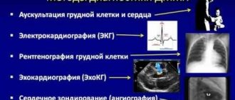
Methods for diagnosing congenital heart disease
Diagnosis of congenital heart disease is based on collecting medical history data (presence of developmental defects, including congenital heart defects, genetic diseases in close relatives; information about pregnancy and the presence of etiological factors in parents).
When collecting complaints, attention is paid to the child’s developmental delay, poor weight gain, poor appetite, sluggish sucking from the breast or bottle, breast refusal, cyanosis, and frequent respiratory infections.
Physical examination
During a physical examination, pay attention to the color of the skin, determine pulse and blood pressure (on the right arm and either leg), perform auscultation of the heart and lungs, pay attention to the presence of peripheral edema, perform pulse oximetry, and determine diuresis.
Instrumental diagnostics
However, the leading role in the diagnosis and confirmation, differential diagnosis of congenital heart disease is played by instrumental examination methods: x-ray examination of the chest organs, electrocardiography, echocardiography, MRI, CT, catheterization of the cardiac cavities.
Sign up for diagnostics To accurately diagnose the disease, make an appointment with specialists from the Family Doctor network.