Nature gave blood a special property - to coagulate; without this, even minor tissue damage would be fatal - a person would simply die from blood loss. But in some pathologies this process is disrupted. By taking a blood clotting test and learning how the hemostasis system works, you can judge the presence of many diseases in the body.
APPOINTMENT WITH ANY DOCTOR BASED ON TEST RESULTS—RUB 500. CLICK TO SIGN UP
Determination of blood clotting ability is called a coagulogram (hemostasiogram). The study is prescribed to identify a number of pathologies (hemophilia, cancer, thrombosis, liver disease, kidney disease, etc.) and before surgery or childbirth.
Normally, hemocoagulation should take place only outside the body, otherwise the resulting clots will travel through the vessels to the heart, lungs or brain, forming blood clots and causing ischemia, heart attacks and strokes. By donating blood for clotting, you can determine its ability to coagulate (clotting). This helps the doctor assess the risk of bleeding or developing clots in the blood vessels.
Types of laboratory tests for clotting
The content of the article
The blood clotting test includes the following indicators:
- Fibrinogen levels
This substance is a protein produced by the liver. In a healthy person, fibrogen is 2-4 g/l. - Antithrombin III.
This is a hemocoagulation regulator. The antithrombin index is 75-125% in adults, 80-120% in children older than one month, and 30-80% in newborns. - Prothrombin index (PTI)
. The percentage ratio of hemocoagulation time in a particular person with a similar indicator in the control sample. Normally, PTI is 93-107%. - Thrombin time.
In healthy people it ranges from 14 to 20 seconds. During this period, prothrombin must be transformed into thrombin. The results may vary with the use of certain medications, such as aspirin or warfarin. The thrombin time (PT) test shows how well a person's blood clots. - Activated partial thromboplastin time (aPTT)
. The indicator is used to monitor the hemocoagulation process in patients prescribed heparin and in the diagnosis of DIC syndrome. Normally, this figure is 29-39 seconds. - Period (time) of bleeding.
Indicates how quickly the blood will stop if the integrity of small vessels is violated. To do this, a puncture is made on the patient's finger and the time is noted. Normally it is 2-3 minutes. - Clotting time.
Determined by the time interval between blood sampling and clot formation. In healthy people, this process takes from 2 to 5 minutes. - D-dimer
. This is a substance formed as a result of the decomposition of fibrin. Normally, this figure should be no more than 250 mcg/l (0.25 mcg/ml).
Why is the rate of blood clotting determined, what does this indicator mean?
A coagulogram is important for assessing the state of hemostasis. This functional system is an evolutionarily developed protective reaction of the body aimed at preventing lethal blood loss due to vascular damage.
Hemostasis is a sequence of physiological processes that ensure a sufficient volume of hematological fluid for normal life and maintain it in an optimal state of aggregation.
Blood clotting according to Sukharev (the norm is estimated as the time interval between the collection of biological material and the beginning of its thickening) is determined for:
- making a diagnosis;
- clarification of the clinical picture;
- many other therapeutic purposes.
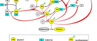
The coagulation mechanism is associated with physiological reactions and the chemical composition of the molecular plasma fraction of the hematological fluid. The leading role in the blood clotting process is assigned to fibrinogen, a colorless protein contained in the blood that is broken down by thrombin.
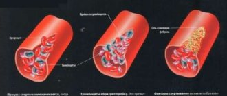
The process is automatically activated when the vascular walls are damaged. As a result, fibrin is formed - a high-molecular protein compound of a non-globular type. The substance is the finest insoluble thread-like structures that form a dense network.
A complex of biological transformations is aimed at the formation of clogging blood clots. They have a compacted structure that stops bleeding and heals wounds. The more platelets the material taken for laboratory analysis contains, the higher the coagulability of the hematological fluid.
Such diagnostic information characterizes the functional state of the hematopoietic system and is required:
- before operations;
- in the treatment of numerous diseases;
- to control gestation.
How to donate blood for clotting
Determination of bleeding time is carried out using capillary blood; for other clotting tests, material must be taken from a vein.
The tests are carried out on an empty stomach; you can only drink water before the analysis. If you are taking any medications, you should tell your doctor about this, as some medications may affect the results obtained.
By regularly taking hemostasiogram tests, you can promptly diagnose many dangerous ailments.
Increased blood clotting and thrombus formation
Many factors can lead to increased blood clotting, restriction or blockage of blood flow, and as a result, blood clots. Blood clots can travel through arteries and veins, causing serious consequences including sudden death from embolism.
Indications for examination
Normal blood clotting is 2-5 minutes (according to Sukharev). An analysis to determine this indicator (coagulogram, hemostasiogram) is prescribed for:
- diseases of internal organs;
- suspected hereditary hemostatic pathologies;
- pregnancy;
- varicose veins, thrombus formation;
- diabetes mellitus;
- prescribing coagulants;
- during the preoperative and postoperative period.
Risk factors
Increased hemocoagulation often occurs when:
- increased number of blood cells and hemoglobin, radiation, cancer;
- hyperfunction of the spleen, acidification and dehydration of the body, most often associated with poor bowel function;
- increased consumption of sugar and carbohydrates;
- overweight, pregnancy, prolonged bed rest, sedentary lifestyle and sedentary work;
- lack of specific hormones and enzymes, use of birth control pills or hormone replacement therapy.
Sometimes the tendency to hyperclotting is congenital. This pathology is called thrombophilia. It is caused by a congenital decrease in the level of anticoagulants C and S, antithrombin III, coagulation factor VII, heparin cofactor II, dysfibrinogenemia, sickle cell anemia, hyperlipoproteinemia, increased activity of Hageman factors, Rosenthal factors and antihemophilic globulin. With this pathology, patients report similar cases among close relatives. Sometimes hereditary thrombophilias cause miscarriages in women.
Blood thickening in old age threatens to disrupt brain activity, and in pregnant women it can negatively affect the condition of the mother and fetus. Poor rheology leads to ischemia of organs and tissues, which negatively affects the condition of the entire organism.
How to suspect increased coagulability
- A sign indicating increased hemocoagulation is thrombus formation. If you notice painful bluish “vessels” or nodules on the limbs, this is a reason to consult a doctor and be examined for blood clotting indicators.
- Another symptom that should alert you is problems with the functioning of the heart. They should encourage you to get tested.
If the coagulogram indicators do not correspond to the norm, treatment must be started urgently to prevent thromboembolism.
What is the essence of Sukharev’s method?
A coagulogram is a preventive measure to prevent complications with an increased risk of massive blood loss or thrombosis when prescribing drugs that affect the rheological properties of hematological fluid.
Blood clotting according to Sukharev
Sukharev's test involves the use of capillary biological material obtained by piercing the skin of the terminal phalanx of the finger with a scarifier.
The blood is placed in a laboratory vessel, alternately tilting it in opposite directions at an angle of 40°. Simultaneously with the start of the hematological test, a stopwatch is started, counting the time until the biomaterial clots.
The analysis is considered extremely simple, well established, and widely applicable. It allows you to obtain an informative hemostatic picture about the time interval of coagulation.
The test does not determine the chemical composition of the blood or provide data on physiological reactions. The main advantages of such analysis are speed, simplicity and availability in any laboratory.
The vessel is tilted in a given monotonous rhythm without sudden movements or shaking the contents of the test tube. According to Sukharev’s method, the beginning of coagulation is considered to be the cessation of free movement of biological material inside the vessel.
To automate the process, a special apparatus called a Panchenko tripod is used. The first drop of blood released from the finger is blotted, since it contains particles of the epidermis that distort the measurement result. The rheological properties of the following portions of biological material are studied.
Blood clotting according to Sukharev (the norm is determined by the beginning of the formation of fibrin protein) is necessary:
- to assess the effectiveness of therapy;
- for injuries;
- for hemodynamic disorders;
- in case of hepatobiliary dysfunctions, fibrin is produced by liver cells.
The indicator plays an important role in the interpretation of the coagulogram by the attending physician. Coagulation time according to Sukharev’s method is used to predict ischemic conditions, the risk of heart attack or stroke.
Diseases accompanied by increased blood clotting
- Atherosclerosis. With atherosclerosis, plaque forms inside the arteries. Over time, the plaque can rupture and platelets form clots at the site of injury.
- Vasculitis causes inflammation of blood vessels, platelet aggregation and, as a result, an increase in coagulogram parameters.
- Diabetes causes plaque to form in the arteries, which is why nearly 80 percent of people with the disease eventually die from causes related to blood clots and ischemia.
- In heart failure, this organ cannot pump enough blood to meet the body's needs, its circulation slows down, it thickens and blood clots appear.
- High blood clotting is often observed in patients with varicose veins. In this case, blood clots have to be “liquefied” with the help of special drugs to prevent gangrene and necrosis associated with poor blood flow.
- This figure also increases in some infectious diseases, especially those accompanied by fever. In this case, the blood becomes viscous, so patients are prescribed plenty of fluids and intravenous infusions of solutions.
Elevated coagulogram values are very dangerous, as this condition can lead to serious complications and serious consequences.
Factors influencing the test result
During the active menstrual cycle, minor deviations from the standard are observed. Among the physiological factors influencing the result of a hematological test, the gestation period is distinguished. Hormonal changes in the body change the rheological properties of the blood.
Clotting according to Sukharev's method distorts the use of oral contraceptives. Small deviations from the established norm are acceptable when using birth control pills or capsules. The patient's age is considered to be a factor influencing the test result.
In newborns, blood clots faster than in adults due to the absence in the infant’s body of some biochemical compounds involved in the hemostasis system and the immaturity of the protective mechanism of thrombus formation. In elderly patients, on the contrary, the concentration of fibrin protein is increased, which reduces coagulation time.
The test result is affected by:
- the current physiological state of the body;
- diet;
- drinking alcohol;
- smoking;
- deficiency or oversaturation of vitamins, minerals, microelements;
- completing a course of drug therapy;
- concomitant pathologies of internal organs.
Tumor processes, inflammation, and hyperactivity of the spleen distort the results of laboratory diagnostics. When the body dehydrates, the blood becomes more viscous, and when a large volume of drinks is consumed before the procedure, it becomes thinner.
How to suspect deterioration of coagulation hemostasis
The first signs indicating poor blood clotting
Prolonged bleeding occurs with minor skin injuries or after injections. Normally, cuts or injections should not bleed for more than 3-5 minutes, but if there is pathology, this time can increase significantly. Sometimes such people have hemorrhages under the skin.
Another symptom that indicates this condition is prolonged nosebleeds that are difficult to stop. Women with hemocoagulation disorders may experience menorrhagia and metrorrhagia. Sometimes traces of blood may even be present in urine and feces.
If these symptoms appear, it is recommended to donate blood for a coagulogram. Research conducted by our specialists will help identify disorders of coagulation hemostasis. All analyzes are carried out using modern equipment and reagents.
Possible reasons for deviation of analysis indicators from the norm
The standard coagulation time is modified by pathological or physiological factors. Any test results cannot serve as a basis for making a final diagnosis. Reduced or increased blood clotting time is a reason for in-depth diagnosis.
Typical pathological causes of coagulation disorders:
- autoimmune processes;
- DIC syndrome;
- genetic disorders;
- cardiac diseases;
- vascular pathologies;
- infectious lesions;
- chronic or acute inflammation.
Disorders of hemostatic function are provoked by recent injuries, hepatobiliary pathologies, and vitamin K deficiency. Most pathological causes require medical intervention and the selection of adequate treatment tactics depending on the etiological factor, the nature and severity of the blood clotting disorder.
Downgrade parameters
Hypocoagulopathic conditions are divided into congenital and acquired. Hereditary causes of deterioration in blood clotting are due to morphological changes in the vascular walls, abnormalities in platelet synthesis, and inhibition of plasma factors.
Acquired forms of hypocoagulopathy are often associated with immune, infectious-toxic, and dysmetabolic diseases. Impaired platelet hemostasis leads to a deterioration in the coagulability of hematological fluid. Coagulation is reduced by renal failure or splenic dysfunction.
Coagulability according to Sukharev (the norm depends on the age of the patient, which affects the parameters of fibrinolysis) slows down with bone marrow metastases, anemic conditions, eclampsia in pregnant women - a severe manifestation of late toxicosis. Septic and necrotic processes worsen coagulation.
The main reasons for a decrease in hemostatic parameters are considered to be:
- congenital or hereditary defects of vascular walls caused by collagen abnormalities;
- immune factors;
- inflammatory processes with the release of large amounts of prostaglandins into the blood;
- thrombocytopenia or thrombocytopathy – quantitative and qualitative changes, respectively;
- hemophilia types B and C;
- deficiency of plasma factors of the prothrombin complex associated with hepatitis, cirrhosis, dysbacteriosis;
Hypocoagulopathic conditions are particularly dangerous for patients over 40 years of age and women during gestation or menopause. In complex treatment, corticosteroids, immunosuppressants, synthetic antihemophilic globulin or thrombolytic concentrate are used.
Increasing parameters
This condition is called hypercoagulopathy or hyperviscose syndrome.
Increased blood density accompanies:
- hypoxia – a decrease in oxygen concentration in the blood, biological tissues, anatomical structures;
- food poisoning;
- diabetes;
- toxic liver damage;
- antiphospholipid syndrome is an autoimmune disease caused by the production of antibodies to fatty compounds, which serve as the basis of cell membranes;
- leukemia;
- polycythemia – an acute tumor proliferative myelopathic disease of the hematopoietic system;
- thrombophilia – a genetically determined recurrent predisposition to vascular thrombosis.
Increased blood clotting parameters are often associated with Waldenström macroglobulinemia, a B-cell lymphoplasmacytic lymphoma. The pathology is characterized by the entry into the hematological fluid of an excess volume of monoclonal immunoglobulin of the LgM type.
Metabolic disorders - amyloidosis, metabolic syndrome and others - contribute to blood thickening. Hyperquagulopathy occurs against the background of functional insufficiency of the adrenal cortex. Among the reasons for increased blood clotting parameters are thermal burns, varicose veins, and pancreatitis.
Reasons causing deterioration of hemocoagulation
Medications
- Coumarin group drugs reduce coagulation because they are vitamin K antagonists. These drugs are used to protect against thrombosis after major operations, but if prescribed incorrectly, these substances can significantly worsen blood clotting.
- Aspirin, often used for colds and heart pathologies, taken in high doses, can also cause a decrease in clotting rates.
- In some cases, this effect is achieved by a combination of several drugs that are not recommended to be taken together.
In such cases, after determining the clotting time and duration of bleeding, the patient is prescribed vitamin K, which plays an important role in the process of hemostasis. Patients are recommended to eat green vegetables (cabbage and spinach), eggs, milk and cereals, which eliminate the deficiency of this vitamin.
Hereditary diseases
- Hemophilia . This disease is hereditary and affects only boys. In patients, symptoms of decreased blood clotting appear in early childhood. With this disease, various coagulation factors may be absent, so the disease may be more or less severe. Patients are treated by a hematologist for life. Replacement therapy, blood transfusions and restriction of physical activity are carried out.
- Von Willebrand syndrome is also characterized by decreased clotting, but it affects both men and women. To treat patients, tranexamic acid, desmopressin are used, and blood transfusions are performed.
Other pathologies
A decrease in the number of platelets (thrombocytopenia) occurs in various anemic conditions, leukemia, after radiation therapy, viral infections, taking antibiotics and some other drugs. This condition can be caused by diseases of the liver, kidneys and spleen. This condition is treated by administering special blood products.
Another reason for impaired hemocoagulation is liver disease, which disrupts the synthesis of substances responsible for proper hemostasis. A decrease in PTI and an increase in blood clotting time are considered as an indirect sign of hepatitis.
A decrease in coagulogram parameters may be observed after operations accompanied by heavy blood loss. This condition does not pose a threat to human health and goes away on its own.
Properly selected treatment to reduce hemocoagulation will help avoid severe blood loss and serious complications.
Preparing for the test
Blood sampling for study by laboratory methods is carried out in the morning on an empty stomach. The procedure is extremely simple and quick. No special preparation is required for the test. Before the test, you are allowed to drink a small amount of liquid without sugar or gases.
3 days before the scheduled date, you must stop taking medications that affect the rheological properties of the blood. Women are not recommended to take the test during menstruation. 24 hours before taking blood from your finger, you should not eat fried, salty, or hard-to-digest foods.
To obtain an accurate result, you should not take the test against the background of heavy physical exertion, nervous strain, or strong emotional excitement. Within 30 min. You should not smoke before the procedure.
Blood clotting indicators during pregnancy
Every pregnant woman registered at the antenatal clinic is necessarily referred by a gynecologist for an analysis called a hemostasiogram. The results of a hemostasiogram make it possible to determine the presence of disorders in the blood coagulation system, if any.
The role of hemostasis during pregnancy
Hemostasis acts as a “thickener” of blood; thanks to this system, a person avoids significant blood loss in cases of vascular damage. In tandem with the coagulation system, an anticoagulant acts in the body - a “thinner” of the blood. When the balance is disturbed, the systems malfunction, as a result, the blood can become too viscous, which leads to the formation of blood clots, or too liquid, in both cases the blood clotting time changes.
What are the dangers of impaired hemostasis for pregnant women?
Increased blood clotting during pregnancy can provoke disseminated intravascular coagulation syndrome, in which thickening blood in the vessels causes disruption of placental blood flow. The condition is dangerous for the baby, as he cannot receive adequate nutrition, which immediately affects his development. In severe cases, pregnancy may stall, and ultimately the fetus dies. Timely testing for hemostasis gives a great chance to avoid such complications.
Disorders of the coagulation system can cause premature separation of the placenta, even if it is in a normal location.
When is it necessary to take a blood clotting test?
It is recommended to perform a hemostasiogram before the expected pregnancy. In case of poor performance, you can undergo a course of therapy, which the doctor will select individually. The risk group that requires mandatory preliminary screening for coagulation includes women:
- having relatives who have suffered a stroke, heart attack, thrombosis, varicose veins;
- suffered a miscarriage or frozen pregnancy;
- athletes and those working in heavy work.
During pregnancy, it is also necessary to undergo diagnostics, especially if the doctor diagnoses:
- recurrent miscarriage - the presence of two or more unsuccessful pregnancies (miscarriage, fading);
- gestosis - swelling of the extremities, protein in the urine, high blood pressure;
- threat of miscarriage.
If there is gestosis during pregnancy, blood clotting increases in 70% of cases, which aggravates treatment. You can also prevent the situation by taking a timely test and undergoing therapy.
Methods for treating hemostasis disorders in pregnant women
With timely diagnosis of hemostasis disorders, many complications of pregnancy can be avoided. In European medical practice, DIC syndrome is treated with low molecular weight heparins, which are absolutely safe for the fetus. Treatment can be done at home with the permission of a gynecologist, subject to control tests (every 2 weeks). At the same time, doctors prescribe antioxidants, aspirin-containing drugs, folic acid, and vitamin B.
If the patient does not have the opportunity to buy expensive low molecular weight heparins, the specialist can replace them with regular heparin. In this case, strict monitoring of the results and condition of the pregnant woman is necessary, since the dose of an unfractionated drug is very difficult to select immediately; you will have to visit the clinic twice a week. For control, you need to periodically take a blood clotting test.
If you find an error, please select a piece of text and press Ctrl+Enter
Indications for testing
The time interval for blood clotting is determined in preparation for surgery on internal organs.
The following pathologies are considered indications for a hematological test:
- drug, chemical or alcohol intoxication of the liver;
- hepatitis;
- cirrhosis;
- stomach ulcer;
- gastritis;
- erosive-perforative processes in the intestines;
- varicose deformation of venous beds;
- tendency to thrombosis;
- diabetes;
- endocrine disorders;
- metabolic and metabolic disorders.
This study is prescribed before using anticoagulant drugs.
Determination of blood clotting is indicated for women:
- For menstrual irregularities. The test is prescribed as part of differential diagnosis.
- Before childbirth. Diagnostic information about the rheological properties of blood allows you to avoid numerous complications.
- For the procedure of artificial abortion. Surgical intervention is traumatic and associated with massive blood loss.
- Before a caesarean section. Determination of hemostatic characteristics plays a significant role in ensuring the safety of mother and child.
- For hypermenorrhea. Dysfunctional-pathological uterine bleeding requires clarification of the cause and selection of adequate treatment tactics. A coagulogram using the Sukharev method is carried out as part of a comprehensive laboratory and hardware diagnostics.
- In preparation for artificial conception. Hemostatic disorders can provoke bleeding during in vitro fertilization, during embryogenesis, and subsequent labor. Excessive thrombus formation causes blockage of the blood vessels of the placenta, which leads to fetal death and miscarriage.
A coagulogram using the Sukharev method is used in the selection of oral contraceptives, many of which affect the rheological properties of blood. The study is indicated for patients taking Heparin for a long time.
A hematological test is carried out to clarify the clinical picture of DIC syndrome - a pathological process with nonspecific physiological characteristics, which is characterized by the formation of disseminated blood clots. Among the numerous indications for laboratory testing of blood clotting time are autoimmune diseases, cardiovascular dysfunctions, and genetic pathologies.
The test is performed if excessive viscosity of the hematological fluid is suspected, due to excessive concentration of fibrin protein or increased secretion of platelets. A coagulogram is prescribed during a comprehensive examination of patients prone to spontaneous nosebleeds.