The term “hydropericardium” refers to the accumulation of more than 50 milliliters of fluid in the pericardial cavity. Its normal amount is approximately 30 ml. Inflammation in the pericardium begins with pain and an audible pericardial friction noise. Next, the amount of fluid in the cardiac membrane increases.
- Etiology
- Pathogenesis
- Symptoms and diagnosis
- Treatment
- Complications and prognosis
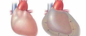
The effect of pericardial effusion on blood dynamics significantly depends on the rate of its accumulation and the extensibility of the outer layer of the pericardium. If fluid accumulates at a rapid rate, severe hemodynamic changes may occur. And a gradual increase in the amount of fluid over a long time may not manifest itself in any way. Due to pericardial effusion, the filling of the heart with blood becomes more difficult, its inflow decreases, and stagnation occurs in the systemic circulation and, partially, in the small circulation.
Cardiac tamponade occurs when a large amount of fluid accumulates, which limits the filling of the ventricles and atria, congestion begins in the veins of the systemic circulation and a decrease in cardiac output until the blood circulation stops completely. Exudative pericarditis with cardiac tamponade is divided into two types: acute and subacute.
Reasons for the development of the condition
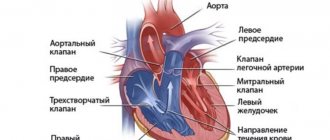
Normally, between the layers of the pericardium there is a small amount of fluid, which acts as a lubricant and a protective barrier.
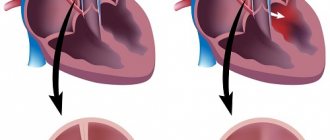
If, as a result of some pathological process, its volume increases or blood enters the cavity, the pressure increases and the heart chambers are compressed. Blood does not flow into the atria and ventricles, which can cause the heart to stop. As already mentioned, pathology can develop chronically and acutely.
In the first case, the liquid gradually fills the pericardial cavity, it stretches and can accommodate a significant volume (more than 1 liter). In this case, chronic heart failure develops.
This option occurs due to exudative pericarditis, the causes of which may be:
- infectious diseases;
- autoimmune pathologies;
- oncological neoplasms;
- endocrinopathies.
If the condition develops quickly (for example, with hemotamponade due to a penetrating wound), the pericardium cannot adapt and acute heart failure syndrome occurs.
Etiology
The causes of cardiac tamponade can be:
- tuberculosis
- acute pericarditis (viral or idiopathic)
- influence of radiation
- malignant tumors
- systemic connective tissue diseases (SLE, rheumatoid arthritis)
- injury
- Dressler syndrome
- postpericardiotomy syndrome
Any disease that affects the pericardium can lead to the accumulation of fluid in its cavity. Acute cardiac tamponade can occur as a result of trauma, including iatrogenic injury during pacemaker installation, aortic rupture during dissection of its aneurysm, or cardiac rupture during myocardial infarction (MI). Subacute cardiac tamponade, as a rule, appears as a consequence of pericarditis (idiopathic or viral), with uremia or a tumor in the pericardium. In most cases, doctors cannot determine the cause of exudative pericarditis, even when surgery is performed.
Clinical manifestations and typical symptoms
Symptoms depend on the pathogenesis of the disease. In the case of chronic inflammation of the pericardium, the clinical picture develops slowly, against the background of the underlying disease. The most typical signs are:
- discomfort, feeling of heaviness in the chest;
- dyspnea;
- weakness;
- Heart rate increases, but the pulse is weak and barely palpable;
- cyanosis, sweating.
The pronounced clinical picture of tamponade is manifested by the so-called Beck's triad:
- low blood pressure;
- swelling of the veins of the neck;
- dull tones on auscultation.
With hemopericardium, the clinic develops quickly, the symptoms are pronounced:
- sharp, acute pain behind the sternum (especially when the heart muscle ruptures);
- Beck's triad;
- disappearance of pulse when inhaling;
- disturbance of consciousness (up to fainting)
- pale skin and cyanosis of the face.
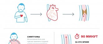
Symptoms of cardiac tamponade
The condition of impaired cardiac function, that is, tamponade, is defined by cardiologists as a life-threatening condition. Early recognition of the symptoms of this heart disease prevents cardiogenic shock, cardiovascular arrest, and even death. Based on the time of onset of symptoms of cardiac tamponade, two types are distinguished:
- subacute tamponade - symptoms appear no earlier than after a few hours, and sometimes after 4-5 days,
- Acute tamponade - symptoms appear a few minutes after fluid or blood begins to appear in the pericardial cavity.
If fluid or blood collection occurs quickly and rapidly, the heart cannot adapt to these conditions. The faster the fluid accumulates, the faster the so-called circulatory decompensation, usually called circulatory failure, occurs. The main symptoms of cardiac tamponade include:
- shortness of breath, especially severe when lying down,
- fainting or pre-fainting states,
- increased heart rate,
- sudden cough
- tachycardia (cardiac tachycardia)
- dilation of the jugular veins,
- elusive heartbeat, consisting of a decrease in its tone,
- hypotension (low blood pressure),
- cold, pale skin,
- rapid and frequent fatigue,
- the pulse is “strange”, which is definitely lower during inspiration.
Cardiac tamponade should be suspected in people who have suffered a strong blow to the chest area, especially the sternum. Symptoms of tamponade can lead to so-called venous stasis (blood cannot flow to the heart) and a drop in blood pressure, as well as ischemia of other organs. Doctors identify a classic triad of symptoms characteristic of cardiac tamponade, called Beck's triad. This consists of:
- hypotension, i.e. systolic blood pressure up to 90 mm Hg.
- quiet (muffled) heart sounds,
- filling of the jugular veins.
The presence of these three symptoms together clearly indicates cardiac tamponade. In a hospital setting, this is confirmed by the presence of serous fluid or blood in the pericardial cavity.
About
Diagnostics
On physical examination, the following signs are observed:
- Beck's triad;
- expansion of the borders of the heart (percussion);
- weakening or complete disappearance of the heartbeat.
The ECG shows a rather nonspecific picture - decreased voltage, arrhythmias, tachycardia, electrical alternans.
X-ray of the chest organs - enlarged borders of the heart.
The main method for diagnosing this condition is echocardiography. This method allows you to:
- detect fluid in the pericardial cavities and determine its amount;
- measure cardiac output, which makes it possible to judge the severity;
- examine the myocardium and large vessels for the presence of traumatic injuries;
- detect right ventricular diastolic collapse.
In some cases, transesophageal echocardiography is performed.
Symptoms of cardiac tamponade and first aid
The symptoms of cardiac tamponade are caused by the accumulation of fluid in the pericardium. This is a very serious disease that threatens human life. However, timely diagnosis of cardiac tamponade and taking appropriate measures will prevent cardiac arrest and death. Therefore, knowledge about it is very important and useful. It is also worth knowing what first aid should be for cardiac tamponade.
Every person's heart is contained within a sac-like membrane called the pericardial sac. Its job is to protect the heart from injury. The pericardial cavity is located between the outer and inner plates of the pericardium. This is where a small amount of serous fluid accumulates. But if it is exceeded, cardiac tamponade occurs. Then the work of this most important organ of every person is disrupted and requires rapid medical intervention.
Emergency care and treatment algorithm
Cardiac tamponade is a life-threatening condition that requires immediate intensive care and surgical intervention.
To maintain hemodynamics and water-electrolyte balance, the patient is given an infusion of crystalloids, albumin or plasma. In case of significant respiratory failure, artificial ventilation of the lungs is indicated.
The main treatment method is pericardiocentesis - puncture (puncture) of the heart sac and removal of accumulated fluid. This allows you to relieve the heart and restore normal blood circulation. The selected contents are sent for pathological examination. The procedure is usually carried out under the control of ECHO-CG.
After stabilization of the condition, drainage is introduced into the pericardial cavity to drain exudate and prevent relapses, continuing infusion therapy.
Further treatment is carried out depending on the cause. For inflammatory and infectious processes, the following drugs are indicated:
- antibiotics;
- glucocorticosteroids;
- non-steroidal anti-inflammatory drugs.
In more complex cases, open surgery is performed - pericardiotomy. In this case, holes are made in the wall for cavity inspection and drainage.
In case of hemotamponade, elimination of the source of bleeding into the pericardium is a prerequisite. The operation consists of suturing wounds of the myocardium or large vessels.
Treatment
Exudative pericarditis with cardiac tamponade is treated in a hospital. It is advisable to take into account the nature of the disease. Non-steroidal anti-inflammatory drugs prescribed in moderate dosages are an effective treatment. This treatment is also carried out for dry pericarditis. Glucocorticosteroids are also sometimes used. Prednisolone is effective in a dose of up to 60 mg/day, it is taken for 5 to 7 days, then the dose is slowly reduced. Thanks to this drug, the effusion quickly resolves. If during 2 weeks of treatment with glucocorticosteroids (GC) there is no expected effect, a large amount of effusion is present, then a pericardial puncture is performed with the introduction of GC into the cavity of the cardiac sac. The management of the patient also depends on the volume of fluid in the pericardial cavity. If the amount of fluid is small, no therapy is required.
To improve hemodynamics in case of hypotension, liquid is administered - plasma, colloidal or saline solutions in an amount of 400-500 ml intravenously. The effectiveness of these measures is monitored by increasing systolic pressure and systolic ejection. Whatever type of cardiac tamponade is detected, it is necessary to puncture the pericardium in a timely manner. Often this is what helps to significantly improve the patient’s condition.
conclusions
Cardiac tamponade, regardless of its cause, is a life-threatening condition that requires immediate hospitalization and intensive treatment. Compression of the atria and ventricles by accumulated fluid leads to a significant decrease in the ejection fraction and, as a consequence, circulatory disorders and cardiac arrest.
If any signs of acute heart failure are detected, such as shortness of breath, chest pain and cyanosis, you should immediately call an ambulance. With timely treatment, the prognosis for life is favorable.
Pathogenesis
According to the norm, between the layers of the pericardium there can be from 20 to 50 ml of fluid, which makes it easier for them to slide relative to each other. This liquid corresponds to blood plasma in its electrolyte and protein composition. If there is more than 120 ml of fluid there, this increases intrapericardial pressure, reduces cardiac output and provokes arterial hypotension.
Clinical manifestations correlate with the rate of fluid accumulation, its quantity, and characteristics of the pericardium. Symptoms of cardiac tamponade occur if fluid quickly accumulates and reaches an amount of 200 ml. If the rate of accumulation is slow, then symptoms may not appear, even if the fluid is already 1-2 liters. Increased intrapericardial pressure with fluid accumulation up to 5-15 mm Hg. is considered moderate, and more than this amount is considered pronounced. Diastolic filling of the ventricle due to increased pressure inside the pericardium is accompanied by an increase in pressure in the chambers of the heart and pulmonary artery, a decrease in stroke volume of the heart and hypertension.
First aid for cardiac tamponade
Cardiac tamponade is a medical emergency. The patient cannot help himself without a doctor or other medical assistance. Further prognosis and treatment options depend on timely and correct diagnosis. The only effective first aid method for cardiac tamponade is puncture of the pericardial sac and evacuation of fluid. This procedure is called pericardiocentesis and looks like this:
- performed or under the supervision of an experienced physician in a sterile room,
- the puncture is performed under local anesthesia with a special needle,
- once the sac is reached, a catheter is inserted to remove excess fluid or blood,
- The drainage process itself is done in small portions so as not to cause too much disturbance.
- sometimes the catheter is left in place for several days
- The patient is monitored during the procedure.
The exception to allowing a puncture to be performed outside a sterile environment is when the procedure must be performed immediately for life-saving reasons, such as during resuscitation. Each patient undergoing puncture should be monitored by echocardiography to prevent complications. The most serious complications after puncture of the pericardial sac:
- damage to the heart muscle
- coronary vessel damage,
- heart rhythm disturbances ,
- repeated tamponade,
- pneumothorax.
The second method of treating cardiac tamponade is surgery under general anesthesia in the operating room. This is how recurrent tamponade is usually treated. The choice of treatment method depends on the doctor and the patient's condition. Early symptoms of cardiac tamponade allow immediate treatment and restoration of normal heart function.
Vorobyova Marina
Neurologist of the highest qualification category (work experience 14 years), doctor of neurofunctional diagnostics (work experience 12 years); author of scientific publications on vertebroneurology; participant of scientific conferences on neurology and functional diagnostics of all-Russian and international significance.
Causes
Most often, cardiac tamponade is caused by the following factors:
- hemopericarditis due to damage to the integrity of the sternum and heart;
- rupture of dissecting aortic aneurysm;
- heart rupture due to myocardial infarction;
- hemorrhages during cardiac surgery;
- long-term course of chronic diseases (pericarditis, hemopericarditis, lymphoma, myxedema, systemic lupus erythematosus, cancer of the breast, lungs, etc.);
- renal failure developing during hemodialysis;
- taking anticoagulants;
- irradiation, etc.