Description of the procedure
Speaking about the fact that this is defibrillation, it should be noted that the procedure is characterized by the passage of a discharge through the ventricles of the heart in order to restore rhythm and normal functionality. That's what a defibrillator is for.
If the goal is to save the patient's life, the procedure is performed by the ambulance crew on duty.
Please note that defibrillation is not effective in cardiac arrest.
First, it is necessary to perform indirect cardiac massage and artificial respiration, and only after restoration of functions and independent contractile movements can one resort to this procedure. The cardiologist, rheumatologist and emergency physician are familiar with the operation of the device.
If the procedure is performed while the electrical activity of the heart is stable, contractility may be impaired, leading to cardiac arrest. And in the case of asystole, clinical death occurs.
Further tactics of assisting the patient after an attempt to restore rhythm
If defibrillation is successful, the patient requires observation and care. Often, applying an electrical discharge through the conduction system can cause the development of arrhythmia. Neurological disorders associated with temporary cerebral hypoxia are also possible.
Main goals of further actions:
- diagnosis and treatment of the causes of cardiac arrest;
- minimizing negative consequences on the nervous system.
To do this, the following conditions must be met:
- conducting an ECG in 12 leads;
- conducting encephalography in cases of impaired consciousness (coma) and in patients with epilepsy;
- emergency reperfusion (restoration of blood flow) if the cause of cardiac arrest is myocardial infarction;
- maintaining stable blood pressure and blood glucose levels;
- oxygen supply;
- control of body temperature (within 35-36 ˚C);
- consultation with a neurologist.
Resuscitation actions are stopped:
- if resuscitation measures carried out within 30 minutes are ineffective;
- when declaring death based on irreversible cessation of brain function.
Types of defibrillation
Electrostimulation of the heart is divided into:
- Defibrillation is when an activity is performed to normalize the ventricular rhythm.
- Cardioversion, in which the manipulation is associated with the restoration of atrial rhythm, and all actions are monitored by an ECG.
In the first case, the procedure is performed urgently if the ordered heart rhythm is disturbed. The patient is unconscious. Initially, the discharge is 200 joules, later - 360 joules.
Cardioversion can be elective or urgent. This procedure is usually prescribed for a specific period of time, but requires written consent from the patient. During the procedure, the patient is conscious under the influence of sedatives.
What is happening is shown on the monitor and everything is synchronized with the QRS rhythm. Cardioversion voltage is lower than defibrillation and delivers a shock of 50-200 J.
Both methods are performed externally on the chest, doctors use 2 defibrillator electrodes. A third alternative is also possible if the cardioverter-defibrillator is placed inside the chest. It is able to prevent arrhythmia by restoring the function of the generating medium.
What does the heart need to beat?
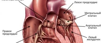
On average, the human heart contracts 60 to 100 times per minute. This occurs due to the work of special stimulating cells in the upper wall of the right atrium (the so-called sinoatrial node). Thanks to them, an electrical differential is created between the outer and inner sides of the cell membrane. At some point, they send an impulse through the entire heart muscle to its lower part, causing the muscle to contract. It would seem that since the heart works from the impulses sent, then what is wrong with electrical stimulation from the outside? To understand this, let's move on.
The electrical differential in the sinoatrial node is created for a reason, but thanks to the presence of electrolytes potassium, sodium and calcium. The electric charge from them passes through the cell walls through special channels (each has its own). An instant before the heart muscle contracts, potassium is contained inside the cells, and calcium and sodium are outside. When sodium enters the cell, it begins to squeeze potassium out, thereby creating an electrical potential. Then channels for calcium open and it also breaks through. This creates the charge necessary for the impulse. Then the impulse from the sinoatrial node goes to the atrium, and then a pulse originates in another node (atrioventricular). Thanks to this complex circuit, blood is pumped from the upper part of the heart to the lower part, and the impulse spreads to other parts of the heart muscle. And only the correct operation of this entire mechanism can create a heartbeat.
If the system fails, different consequences are possible. But we are now interested in the state of fibrillation. This happens if the sinoatrial node does not produce the impulse necessary for the heart. Then the cells of the heart muscle try to create the necessary impulse themselves for some time, but in this case, the contraction of different parts of the heart occurs at odds (fibrillation begins) and the muscle loses its ability to pump blood. It is clear that this cannot continue for long and cardiac arrest soon occurs. But while the muscle is still in a state of fibrillation, there is hope for a defibrillator.
Defibrillation in emergencies
In critical situations, defibrillators can be used to stop ventricular arrhythmias. The patient in this case is unconscious.
There are several pathological conditions for which this treatment is indicated:
- Ventricular fibrillation, the rhythm in this case is accelerated and disrupted.
- Trembling as a rhythm, ordered but accelerated.
- Tachycardia when drug treatment is ineffective.
Sometimes, against the background of these disorders, arterial hypotension or circulatory failure is observed.
The clinical picture in this case develops according to the following scenario:
- The man loses consciousness.
- Heartbeats quickly increase.
- The pulse is not felt.
- The patient is considered clinically dead.
CPR should be performed within the first few minutes. If medical attention is not provided immediately, biological death may occur and electrical defibrillation will no longer be effective.
Execution technique
The algorithm for resuscitation by defibrillation, approved by the American Heart Association in 2015, is as follows:
- Make sure there are indications for defibrillation (assessed using ECG data).
- Initiate cardiopulmonary resuscitation (CPR) with oxygen support.
- First category.
- Continue CPR immediately for five cycles (one cycle consists of 30 chest compressions and 2 breaths). You need to make 100 clicks per minute. Accordingly, the CPR break will take exactly 2 minutes.) Place an intravenous catheter and endotracheal tube. Start ventilation at a rate of 10 breaths per minute. Do not check your pulse and rhythm until this time is over.
- Check pulse and heart rhythm.
- If the rhythm is restored, defibrillation is stopped and the patient is placed under observation.
- If the first shock does not give the desired effect, it is worth repeating the shock.
- Repeat the “rhythm check-defibrillation-CPR” cycle.
The steps listed above are performed only by medical personnel.
External automated defibrillators (EADs) are now in widespread use. They can be found at airports, train stations, shopping centers and other crowded places. They are designed so that every person, without prior preparation, can save someone’s life with the help of audio prompts.
- If you witness a person losing consciousness, make sure that he is not responding to external stimuli - speak to him loudly, jog lightly.
- Call 911 or have someone else do it.
- Check if the victim is breathing and if there is a pulse. If these signs are absent or irregular, begin CPR. Have someone prepare the VAD.
- Before using the VAD, make sure that you and the victim are on a dry surface and there is no spilled water nearby.
- Turn on the device. He will guide your further actions.
- Free the victim's chest from clothing and underwear. If it is wet, wipe it off. Apply the adhesive electrodes as shown on the screen (above the right nipple and towards the armpit from the left nipple).
- Deliver a shock. Do not touch the victim at this moment. The human body conducts current, and electrical defibrillation of a healthy heart will incapacitate the resuscitator.
- Perform CPR for 2 minutes. Use a VAD to check your heart rhythm. If he recovers, continue CPR.
- If fibrillation does not stop, repeat the shock.
- Continue the cycle of actions until medical help arrives.
Treatment of cardiac pathologies with a defibrillator
Cardioversion may be performed as an emergency treatment for sudden tachycardia or as routine treatment for tachyarrhythmia when it cannot be controlled with medication. Urgent treatment is necessary when arrhythmia can turn into fibrillation, when the patient is in a pre-infarction state, heart failure is diagnosed, or a drop in blood pressure.
In planned treatment, you can combine electrical stimulation techniques and medications.
In atrial fibrillation, cardioversion is used for:
- There is no adequate effect from pharmacological treatment.
- In the presence of paroxysmal arrhythmias in Wolff-Parkinson-White syndrome.
- Medicines for hypertensive arrhythmia.
- Recurrence rate of paroxysmal arrhythmias.
- Pharmacological treatment of persistent atrial fibrillation is ineffective.
Contraindications
In defibrillation, the patient's life is most important, all other factors are ignored. The only contraindication is complete cardiac arrest. However, during planned cardioversion, manipulations cannot be performed if:
- The patient is taking cardiac glycosides. Otherwise, it may cause ventricular fibrillation.
- Chronic course of heart failure in the stage of decompensation.
- During the procedure, the patient suffers from an acute infectious disease.
- There are contraindications to the use of anesthesia.
- Electrolyte disturbances were detected.
- There are blood clots in the atria.
- Multiple atrial or sinus tachycardia is diagnosed.
- Ventricular hypertrophy or dystrophy is observed.
Physiology
Heart cells have their own automaticity and excitability. There are many reasons why impulse conduction and contraction of cardiomyocytes can become chaotic.
A short (tens of milliseconds) electrical discharge of a defibrillator with a voltage of 6000-7000 V and a power of 200-360 J causes excitation of most cardiomyocytes and their synchronization by refractoriness (the period when the cells are immune to electrical signals), after which it is possible to restore normal heart rhythm and contractile activity ventricles and blood circulation.[1]
Types of defibrillators and principles of their operation
A defibrillator is a device that delivers electrical impulses. It can be stationary or portable.
The device consists of three blocks:
The following types are also distinguished:
- A two-phase device that conducts current in one direction.
- Single-phase device. The operating principle of the defibrillator is based on alternating current energy, which is transmitted from one electrode to another and back.
Manual defibrillators are difficult to use but inexpensive. They are difficult to use as transport is not possible due to their size, so these devices are more common in clinics.
The advantage of automatic defibrillators is the ability to detect arrhythmias and the ability to adjust the power of the defibrillator depending on the specific situation.
This type of defibrillator is not complicated and can be used even by a beginner. However, the cost is quite high, and the choice of additional settings leaves much to be desired. There are also universal devices that combine both types.
Depending on the type of defibrillator, its maximum power also varies. Usually it is 5000-7000V.
What is a defibrillator
A defibrillator is a medical device designed to apply electric current to the heart muscle in order to restore and synchronize its rhythm. The procedure uses high voltage (about 1000 volts). During “shock therapy,” the patient’s heart receives approximately 300 J of electricity (approximately the same amount is used by a 100-watt lamp in 3 seconds).
The defibrillation method was first used in 1899. This was a scientific study on animals. Two physiologists from the University of Geneva discovered that applying a small electrical discharge to the heart can cause ventricular fibrillation, while a more powerful current, on the contrary, eliminates this process.
The first person to experience the effect of the electric discharge procedure was a 14-year-old boy. In 1947, professor of surgical sciences Claude Bec used electric current to restore normal heart rhythm in a child. In the Soviet Union, electric current treatment was initiated by V. Eskin and A. Klimov. In 1959, Bernard Lown and Baruch Berkowitz determined the optimal procedure time for various cases of arrhythmias.
The first portable defibrillator was created in 1965. The device was invented by a professor from Northern Ireland, Frank Pantridge.
The doctor was prompted to create the device by the fact that in the 1960s, defibrillators could only be used in medical institutions, but many patients with heart disease died on the way to the hospital. Pantridge's invention was very different from modern portable devices. The device weighed about 70 kilograms, and the “irons” in it were huge lead plates. But even such a device could already be transported in ambulances, and this was its huge advantage.
Using a defibrillator in childhood
Ventricular tachycardia, pulseless ventricular fibrillation, is rare in children. However, if this happens, the same rescue measures apply as for adults.
The main difference between defibrillation in children is the choice of electrodes and the device itself, which is based on:
- Size. It is important that the elements cover the correct area of the sternum, but do not touch each other. If the baby weighs less than 10 kg, use pads.
- Device model and child's age. An automatic defibrillator should not be used on children under 8 years of age or weighing less than 25 kg because the defibrillator shock cannot be adjusted. The child's weight is important in determining the correct defibrillator setting. For every 1 kg, 2 J are measured. If there is no effect, the dose is doubled, to 4 J / kg.
When to use a defibrillator
A defibrillator is a device used in medicine to eliminate the condition of fibrillation, that is, uneven, rapid, arrhythmic and unproductive contraction of the heart muscle, atria or ventricles.
The state of fibrillation on the cardiogram will look like a curved line with many small jumps up and down (and not like the smooth straight line from the movies). This graph indicates that different parts of the heart contract at different rates and with their own rhythm. And just the electric discharge gives a chance to restore the correct rhythm of contractions. Exposure to an electric current that is more powerful than the contraction of the heart muscle allows you to even out this process and force different parts of the heart to work in unison again.
The “miracle” of the defibrillator is that the electric current activates the electrolytes and they again begin to flow through the channels in their “schedule”. But that same straight line on heart monitors is asystole. She says that the electrolytes necessary to create an impulse are absent in the cells. The task of doctors is to apply defibrillation before the patient experiences asystole. Later, all the defibrillator can do is burn the heart with heat from the shock.
Medical indications for defibrillation:
- ventricular fibrillation (chaotic contraction at a speed of 200-300 beats per minute);
- ventricular flutter (contraction is rhythmic, but at a speed of about 300 beats per minute);
- atrial flutter (rhythmic but rapid contraction up to 240 beats per minute);
- atrial fibrillation (chaotic contraction, 300 beats per minute).
In case of ventricular fibrillation, so-called emergency defibrillation is performed (what, in fact, is shown in films).
In case of atrial rhythm disturbances, the procedure can be done as planned. In such cases we talk about cardioversion.
Carrying out the procedure
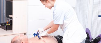
Emergency electrical stimulation consists of the following stages:
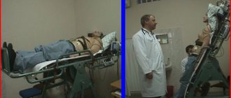
- The patient should be in a horizontal position on a flat surface.
- Remove excess clothing to leave your chest free.
- The electrodes should be coated with a special conductive gel. If it is not available, use gauze soaked in 7% sodium chloride solution.
- After selecting the power, the electrodes are charged.
- It is important to pay attention to the correct placement of the electrodes. The right electrode should cover the subclavian area near the sternum, the left electrode should be applied to the apex of the heart. In another embodiment, the left electrode is placed in the fifth intercostal space near the sternum, and the right electrode is placed under the scapula at the level of the second electrode. If the patient has a pacemaker, the left contact should not be closer than 8 cm from the device.
- The discharge is applied to the electrodes touching the body with a force of 10 kgf.
- The result is checked. You may notice a pulse or ECG change.
- If there is no effect, the device will charge with increasing power.
After four unsuccessful attempts, it is concluded that there is no chance of saving the person. Manipulations may include chest compressions and artificial respiration between discharges. It is important that no one touches the patient or his body while the shock is being administered.
Cardioversion should be performed after special preparation:
- Performing an ECG.
- EchoCG. This is a patient's esophageal test to rule out the presence of blood clots in the chambers of the heart.
- Blood test for potassium.
- Stop taking glycosides 3 days before the procedure.
- Refusal from food and water 4 hours before the procedure.
Planned cardioversion requires prior patient consent for the procedure. This is done according to the following algorithm:
- Saturation of the patient's body with oxygen.
- Placing the patient under anesthesia.
- Prepare the electrodes as in the previous case.
- Pressure monitoring and ECG setup.
- Once the shock is delivered, it should be aligned with the QRS complex or R wave to prevent ventricular arrhythmia.
Notes
- Pugovkin, Evlakhov, Rudakova.
Introduction to cardiac physiology. - SpetsLit, 2021. - P. 242. - 311 p. — ISBN 9785299010435. - In Russian literature also Hooker, Donald Russell Hooker
- D. R. Hooker et al., The effect of alternating electrical currents on the heart, Am. J. Physiol., 103
, 1933, pp. 444-454. - N. L. Gurvich, G. S. Yunyev, [On the restoration of normal activity of the fibrillating heart of warm-blooded animals through a capacitor discharge], BEBiM, VIII
(1), 1939, P. 55-58 - P. M. Zoll et al., Termination of ventricular fibrillation in man by externally applied electric countershock, NEJM, 254
(16), 1956, pp. 727—732 - The exact date is not known, but this happened in the mid-50s, according to an article in the journal “Soviet Health Care of Kyrgyzstan”, a description of which is available in the PubMed database: VI Lysenko et al., Some results with the use of the DPA-3 defibrillator ( developed by V. Ia. Eskin and A. M. Klimov) in the treatment of terminal states, Sov Zdravookhr Kirg. 4
, 1965, pp. 23-25. - V. Ya. Eskin, A. M. Klimov, Defibrillator “DPA-3” (portable defibrillator with an electrocardioscope and autonomous power supply)
, Vopr.
electropathology, electrical traumatism and electrical safety, 3
, 1962, pp. 75-85 - K. A. Azhibaev, V. Ya. Eskin, Experimental testing of a self-powered defibrillator
, I All-Union Conference on the Prevention and Treatment of Electrical Injuries, Academy of Sciences of the Kyrgyz SSR, Frunze, 1962 - I. V. Venin et al., History of defibrillation in the USSR, Russia and Ukraine: technology in the service of medicine Archival copy dated July 15, 2015 on the Wayback Machine, Archive of the history of defibrillation in the USSR, Russia and Ukraine (MIET), 2012
Complications after the procedure
During defibrillation, the main complications are burns, and less commonly, thromboembolic disorders of arterial blood. Burns are caused by strong discharge and are treated with corticosteroid ointments. Thromboembolism is much more difficult to treat; thrombolytic and anticoagulant drugs are used, sometimes requiring urgent surgery.
But these complications justify the goal - saving the patient's life in emergency cases. When deciding on elective cardioversion, possible negative consequences should be carefully assessed.
And here the same consequences are possible:
- Ventricular fibrillation. Occurs rarely, usually due to non-compliance with the technique. Treated with multiple blows.
- A sharp drop in blood pressure. This resolved spontaneously or with vasopressors.
- Atrial and ventricular extrasystoles.
- Pulmonary edema. It does not appear immediately, but after a few hours. Treated with diuretics, antispasmodics, oxygen inhalations.
It is impossible to restart the heart if it has been stopped completely by a defibrillator. Manipulation can only normalize the rhythm. If there is no contraction, cardiopulmonary resuscitation followed by defibrillation is used.
Conversely, cardioversion is useful for supraventricular arrhythmias and some forms of atrial fibrillation when ventricular synchronization is necessary.
A defibrillator does not restart a stopped heart. Defibrillator starting a stopped heart
If the heart has stopped, it can be started again using a defibrillator. Such scenes in Hollywood movies always end well. The hero lies on a hospital bed without moving and only rhythmic sound signals notify that all is not lost. And then, suddenly, the signal gets stuck on one note, and an ominous straight line appears on the monitor. Doctors burst in. One of them constantly shouts: “Defibrillator! We are losing him! And here are a few discharges, dramatic music, certainly someone’s cry “LIVE, DAMMIT YOU!”, and miraculously the heart begins to beat. The hero is saved!
And everything would be fine, but... the problem is that a defibrillator cannot be used to restart a stopped heart. Alas.
In medicine, a straight line on the monitor is called asystole and means the absence of heart contractions. The idea that these contractions can be restored by electric shock seems absolutely sound.
In order to understand why this is not so, we must first understand how the heartbeat occurs.
The heart typically receives 60-100 beats per minute from stimulating cells in the upper wall of the right atrium (sinoatrial node). These specialized cells create an electrical differential between the inside and outside of the cell membrane. At a certain moment, an impulse is sent down the heart muscle, causing it to contract. This electrical signal travels throughout the heart.
If someone goes into cardiac arrest and has no pulse, an electric shock may be needed, depending on how the conduction system works. During cardiac arrest, there may be several types of electrical rhythms.
The most common heart rhythm during cardiac arrest is called ventricular fibrillation (arrhythmic contraction of the muscle fibers of the atrium). When the sinoatrial node fails to fire, many other heart cells try to do so. As a result, multiple areas of the heart are shaken simultaneously from different directions. Instead of measured beats, we see a heart attack.
Fibrillation
With this rhythm, the heart cannot pump blood through itself. The only way to get all these different areas of the heart to work in unison again is to deliver an electrical shock more powerful than the ones they create.
When you run that kind of electricity through those cells, it activates all the electrolytes from the cells at the same time. The only hope (and this is really just a hope) is that the normal functioning of cardiac electrolytes, flowing through cell membranes in an orderly manner, will resume.
In a state of asystole, a person does not have such an electrical differential that can be shown by a heart monitor. In reality, there are simply no electrolytes inside the cell that can create an impulse. In such a situation, the discharge will not help. Thus, if asystole (the complete absence of ventricular contractions) occurs before you can use a defibrillator, all you can do is burn the heart with a high temperature from the discharge.
