Magnetic resonance imaging is one of the highly informative methods for examining internal organs. Its main advantage is the ability to identify pathology at an early stage and select effective treatment. Many patients are interested in whether cardiac MRI is performed. It is this diagnostic method that allows you to obtain the most specific and reliable information about the condition of the organ of interest.
Diagnosing the heart using a tomograph has a number of advantages over other examination methods. The main ones include:
- absence of any discomfort during the examination;
- the ability to identify pathology at the very beginning of its development;
- no radiation exposure and harmlessness to the body;
- the opportunity to examine the organ in as much detail as possible;
- obtaining the most accurate idea of the state of the organ.
No other diagnostic method, including ultrasound, has so many advantages.
Make an appointment now!
Indications for diagnosis
When it is necessary to diagnose heart vessels, specialists often prefer magnetic resonance imaging. Compared to ultrasound or other diagnostic methods, MRI allows you to more clearly visualize the clinical picture, especially if the study is carried out using contrast agents. As a rule, the study is prescribed by a cardiologist. Reasons for performing MRI of cardiac vessels may be:
- Congenital anomalies of the heart;
- The patient complains of pain or pressure in the chest area;
- Cardiomyopathy;
- Regular surges in blood pressure;
- Suspicion of vascular thrombosis;
- Suspicion of a heart attack;
- Monitoring the condition of large vessels of the lungs or abdominal cavity;
- Diagnosis of arterial atherosclerosis;
- Suspicion of aortic aneurysm.
Magnetic resonance imaging of the heart vessels is mandatory after cardiac surgery.
Types of equipment
There are several types of equipment on which diagnostics are carried out. There are open and closed devices. Tomographs are also divided into:
- low-floor - power up to 1 Tesla, usually 0.3 - 0.4 T;
- high-field - power 1.5 - 3 Tesla.
Some experts also highlight 1 Tesla mid-field devices.
The higher the magnetic field strength, the better the quality of the resulting images. Open devices have low voltage, so choosing such a tomograph is not always advisable. Always consult your doctor before going for an examination so as not to waste extra money and time redoing the tomography using other equipment.
Contraindications
Patients who have metal products in their bodies are not allowed to undergo diagnosis. These can be pins, prostheses, implants, pacemakers. For such patients, a different diagnostic method is recommended. Also, an MRI may not be allowed in the following cases:
- First trimester of pregnancy. Most often, an ultrasound examination is performed during pregnancy. An MRI is sent only if a dangerous pathology is suspected, and ultrasound does not provide a complete clinical picture.
- Deviations in the functioning of the nervous system. These include diseases accompanied by involuntary movements or the inability to control oneself, as well as claustrophobia.
- Allergy. Most often, MRI of cardiac vessels is performed using contrast agents. An allergic reaction to the administered drugs should first be ruled out.
- Kidney failure. Contrast agents are eliminated through the kidneys. Experts do not recommend doing an MRI with contrast if there are serious abnormalities in kidney function.
Magnetic resonance imaging does not have specific age restrictions, but it is difficult to perform an MRI on very young children, since it is very difficult to get them to remain still. For this reason, a different type of examination is recommended for young children.
We can talk about the complete safety of MRI only if the patient has no contraindications to this procedure. Be sure to consult your doctor before diagnosis.
Contrast tomography
Tomography using contrast is used to improve the differentiation of tissues and organs. This way the picture becomes clearer and more informative, and it is much easier for the doctor to navigate the resulting images. The vast majority of MRI contrast agents contain gadolinium. It enters the body intravenously, spreads through the bloodstream, staining all organs in its path.
Cardiac MRI uses an intravenous injection method. Gadolinium interacts with hydrogen protons, which increases the contrast of the three-dimensional image. The substance is considered completely safe for the human body and is excreted naturally through the kidneys.
The decision to perform contrast tomography is made by the doctor. In most cases, standard diagnostics without the use of auxiliary manipulations is sufficient.
Moreover, contrast agents are associated with a high risk of side effects.
Do I need to prepare for an MRI of cardiac vessels?
When going for magnetic resonance imaging of the heart vessels, you should consider several aspects:
- The diagnostic procedure is carried out on an empty stomach, so it is forbidden to eat for 4-5 hours;
- When diagnosing with contrast, it is necessary to drink tea, preferably sweet, a couple of hours before the examination, in order to avoid nausea and dizziness when the substance is injected into a vein;
- A few days before magnetic resonance imaging, it is forbidden to drink alcohol; a couple of hours before the diagnosis, stop smoking;
- If you are worried about the diagnosis or have fear due to confined spaces, you can take herbal sedatives a few days before the MRI (you should consult your doctor before using them);
- Ask the specialists at the clinic in advance if they will give you disposable clothing for the duration of the diagnosis. If this is not provided, you need to take clothes without metal inserts from home.
Preparation
No special preparatory procedures are required for cardiac MRI. Only tomography with contrast is performed strictly on an empty stomach.
Before the examination, the patient removes all jewelry, watches and other metal objects that may affect the operation of the device. Also, the doctor conducting the examination must make sure that the patient does not have implants containing steel (prostheses, catheters, etc.).
The subject will be asked to wear a special uniform made of cotton fabric and use earplugs or headphones to protect their hearing from the various noises of the operating device.
Patients suffering from fear of confined spaces or other nervous disorders should take a mild sedative before the procedure, and patients with acute pain should take a painkiller.
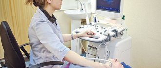
How is magnetic resonance imaging performed?
The diagnostic study is carried out in a closed-type apparatus. In this case, the human body is completely in a magnetic field, which increases the information content of the procedure. When entering the office, you must turn off your mobile phone, change clothes and take off your shoes. For the examination, the patient lies down on a special retractable table, and a few minutes before the diagnosis, a contrast agent is injected into the vein. The duration of magnetic resonance imaging of the heart vessels is 30-40 minutes.
The doctor must first inform the patient about the rules for conducting the examination. The main requirement is a still state and calm breathing. You can talk only if necessary: if you need to tell the doctor something or answer his question (the tomograph is equipped with a speakerphone). There is lighting and ventilation inside the tomograph cabin. There is absolutely nothing to worry about, since in case of unforeseen situations the device has a panic button.
Order of conduct
The procedure requires calm, measured work of the heart, because The faster it beats and the more movements the chest makes, the more noise there is in the image. To even out the functioning of the heart, the doctor may prescribe appropriate medications before the procedure.
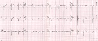
During the examination, electrodes (3 pieces) are glued to the patient’s chest to take ECG readings. To do this, men often have to shave the areas of skin to which the electrodes are attached.
The examination is performed in the supine position. The examination time depends on the diagnosis and takes from 30 minutes to an hour. When using contrast, the examination time increases.
During the examination, the patient receives commands to take in air and not breathe for 10 - 30 seconds, for a better scan.
MRI of cardiac vessels with contrast
Magnetic resonance imaging of the heart vessels is almost always performed using contrast agents. In most cases, drugs are used for intravenous administration. MRI with contrast enhances the staining of blood vessels, which allows the doctor to see the full clinical picture. This makes it possible to identify possible pathological processes, including life-threatening ones, at an early stage.
After administration, the contrast agent quickly and evenly spreads throughout the vessels in just a few minutes. Don't worry, such products are completely safe for your body and usually do not cause negative effects.
Indications
The main indication for the study is the need for detailed visualization of the circulatory organs in the event of an inaccurate result of cardiac ultrasound. The method can also be used as an alternative to multislice computed tomography (MSCT).
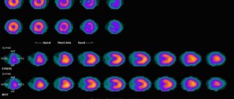
In cardiological practice, it is important to correctly calculate the mass of the myocardium, chamber volumes, ejection fraction, and also to establish the cause of the development and progression of heart failure, which is clearly shown by cardiac MRI.
The high resolution of tomography allows for optimal assessment of the contractile function of the heart. Breath-hold techniques help track the location of the coronary artery. The method makes it possible to evaluate the operation of the valve and indicate backflow of blood or stenosis. Tomography is the gold standard for assessing pericardial thickness. The method allows you to obtain clear images of the aorta, pulmonary arteries and veins, and assess myocardial perfusion.
Indications for cardiac MRI are formed if the following conditions are suspected:
- ventricular aneurysm and pseudoaneurysm;
- dilated and hypertrophic cardiomyopathy;
- congenital and acquired heart defects;
- myocarditis;
- cardiac fibrosis;
- arrhythmogenic myocardial dysplasia;
- hemochromatosis is an infiltrative cardiomyopathy in which iron accumulates in the myocardium;
- cardiac remodeling;
- amyloidosis;
- sarcoidosis;
- Chagas disease;
- Tumors in the heart are also easily detected, which helps to recognize the disease in the early stages.
Information content
The information content of the method directly depends on the type of device used, its power and settings.
An open (low-field) tomograph is recommended for examining patients with claustrophobia and other conditions that complicate the process. The power of the device usually ranges from 0.23 to 0.5 Tesla. Whether to do cardiac MRI with such low settings is a question of rationality, because the quality of the images is low.
High-field installations have a power of 1-1.5-3 Tesla, which provides thin slices and, therefore, a more detailed picture. In addition, this type of device works much faster.
Decoding the results
The results of the diagnostic procedure are given to the patient; the images can be printed on film or electronically on a digital medium. As a rule, a qualified cardiologist interprets the results.
If no ailments are detected during magnetic resonance imaging of the heart vessels, the conclusion will include approximately the following information:
- The structure of the heart vessels without pathological changes.
- The walls of the vessels are not changed, homogeneous, the lumen is clean.
- No inflammatory or infectious processes were detected.
- Blood circulation is normal, without deviations.
Sometimes during diagnosis it is possible to identify the following pathologies:
- Blockage of blood vessels;
- Congenital defects of heart vessels;
- Myocardial infarction;
- Traumatic damage to the arteries;
- Cardiac vascular stenosis;
- Thrombosis;
- Presence of cholesterol plaques;
- Benign or malignant neoplasms.
Only a doctor can evaluate the diagnostic results, so do not try to decipher the examination results yourself. Contact your doctor promptly and take care of your health!
Cardiac MRI results
After the examination is completed, the results are given to the patient, who hands them over to the doctor who ordered the MRI.
The doctor, by analyzing layer-by-layer images of the heart and blood vessels, can assess the state of coronary blood flow, study the functional characteristics of cardiac activity and note changes in the functioning of the heart chambers. In addition, MRI of the heart allows you to identify defects of the interventricular and interatrial septa, various space-occupying formations and congenital malformations, as well as assess the localization and degree of development of atherosclerosis.
Where to do MR coronary angiography in Moscow
MRI of cardiac vessels is performed in diagnostic medical centers. They can be found in any district of Moscow. You can contact us and we will sign you up for the procedure at the clinic you like.
Any additional information can be found by calling. The telephone is available 24 hours a day. The operator will provide information about the features of various MRI machines, tell you the details of the procedure, and tell you the address of the required diagnostic center. You can also make an appointment for an examination by calling this number.
Contraindications to MRI with contrast:
- history of polyvalent (to all medications) allergies;
- any previous adverse reactions to a contrast agent (containing gadolinium) for MRI in previous studies;
- chronic and acute renal failure (decrease in glomerular filtration from 30 ml/min/1.73 m and below);
- severe liver failure;
- severe form of bronchial asthma;
- pregnancy;
- MRI can be performed no earlier than 36 hours after any previous contrast study (MRI, CT, radiography, etc.).
After contrast enhancement, breastfeeding can be resumed after 36 hours.
results
The study is interpreted by a diagnostician - a radiologist. The conclusion is issued on the day of the examination, but a description of the images may take the doctor several hours.
The doctor examines the obtained images and compares them with normal values:
- size and location of organs, symmetry;
- presence/absence of abnormal formations;
- presence/absence of an aneurysm, free fluid, blood clots in the blood stream;
- signs of infection and inflammation;
- stagnation in the ducts;
- traumatic injuries.