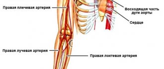
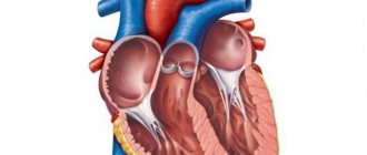
Deep femoral artery
The deep femoral artery, starting from its posterior wall, approximately 3-4 cm lower than the inguinal ligament, passes through the pectineus and iliopsoas muscles, is directed initially outward, and then downward, located behind the femoral artery.
This is its largest branch. Afterwards, the artery follows between the adductor muscles and the vastus medialis femoris, and its end is approximately the lower third of the thigh between the long and adductor muscles with a transition to the perforating artery. These are the numerous branches of the femoral artery.
Circling around the femur, the medial artery, moving away from the deep and behind the femoral artery, goes inward, penetrating transversely into the thickness of the pectineus and iliopsoas adductor muscles, then bends around the neck of the femur from the medial side.
A.V. MAKSIMOV, E.A. GAISINA, A.K. FEISKHANOV, V.V. GLINKIN
Republican Clinical Hospital of the Ministry of Health of the Republic of Tatarstan, Kazan
Kazan State Medical Academy
Maksimov Alexander Vladimirovich
— Candidate of Medical Sciences, Head of the Department of Vascular Surgery, Associate Professor of the Department of Cardiology, Endovascular and Cardiovascular Surgery
420064, Kazan, Orenburgsky tract, no. 138, tel. 2-686-987, e-mail:
False aneurysms of the great arteries in the structure of reconstructive operations on the great vessels range from 0.5% to
8-9%.
Their surgical treatment with the modern development of vascular surgery has been developed quite well. Therefore, the observation of large aneurysms in these localizations today is casuistry. The article presents a clinical case of successful surgical treatment of a giant false aneurysm of the femoral artery. Key words: giant false aneurysm, deep femoral artery replacement.
AV MAKSIMOV, EA GAYSINA, A K. PHEYSKHANOV, VV GLINKIN
Republican Clinical Hospital of the Ministry of Health of the Republic of Tatarstan, Kazan
Kazan State Medical Academy
Experience of surgical treatment of pseudoaneurysm of the deep femoral artery (medical case)
Pseudoaneurysm of the magistral arteries within corrective surgery on great vessels is from 0.5% to 9.8%. Their operative treatment in course of the current development of vascular surgery is worked out good enough. Therefore, the observation of large aneurysms of these locations today is casuistry. The article presents a medical case of successful surgical treatment of a giant pseudoaneurysm of femoral artery.
Key words:
giant pseudoaneurysm, prosthesis of the deep femoral artery.
False aneurysms of the great arteries account for from 0.5% to 8-9% in the structure of reconstructive operations on the great vessels [1, 2]. The most well-known causes of their occurrence are catheterization of the femoral artery during diagnostic and therapeutic procedures, trauma to the vessel, failure of the vascular anastomosis, or arrosive bleeding due to a purulent-inflammatory process.
Diagnosis of aneurysms of superficial arteries (femoral, brachial, carotid) is not difficult. Their surgical treatment with the modern development of vascular surgery has been developed quite well. Therefore, observations of large aneurysms in these localizations today are casuistry. In this regard, we present a clinical case of successful surgical treatment of a giant false aneurysm of the femoral artery.
Patient Z., 69 years old, was admitted to the department of vascular surgery of the Republican Clinical Hospital of the Ministry of Health of the Republic of Tatarstan with complaints of the presence of a painful dense formation in the left groin area and the upper third of the thigh. According to the patient, in 1988 (24 years ago) reconstructive surgery was performed on the left femoral artery. After discharge, he notes a slow growth of formation in the area of the postoperative wound. The patient did not seek medical help regarding this matter. The reason for this patient's visit was the appearance of pain.
Laboratory data on admission: Hb - 127 g/l, Er - 3.09 x 10¹²/l, L - 6.7 x 10⁹/l, total protein - 64 g/l; creatinine - 64 umol/l; potassium - 0.6 mmol/l; sodium -137 mmol/l, AST - 26 U/l, ALT - 2-2 U/l, PTI - 73%; APTT - 32.5 s.
On examination: the left lower limb is pale, cool on palpation, the veins are empty. In the groin area, along the anterior surface of the thigh, in the projection of the femoral artery, a dense, painful, weakly pulsating formation with a diameter of 18 cm, rising above the skin by 20 cm, is determined. The skin above the formation is thinned, tense, with areas of cyanosis and developing necrosis. Tactile sensitivity and active movements in the limb are preserved. There is no pulsation in the popliteal artery on both sides (Fig. 1).
Rice. 1.
Giant aneurysm of the left femoral artery
Distal arteriography and CT angiography were performed (Fig. 2), and the diagnosis was made: Atherosclerosis. Obliterating atherosclerosis of the arteries of the lower extremities.
Rice. 2.
Arteriography and CT angiography of patient Z
Occlusion of the superficial femoral artery on both sides. Condition after reconstructive surgery on the left femoral artery (1988). Giant false aneurysm of the left femoral artery. Chronic arterial insufficiency of both lower extremities, degree 2B (according to Pokrovsky A.V.). IHD. (PEAKS?). Hypertension of the 1st degree. Common psoriasis, acute stage.
On December 18, 2012, an operation was performed: resection of a giant false aneurysm of the femoral artery on the left. External-iliac-deep-femoral extra-anatomical prosthetics on the left.
Progress of the operation: under epidural anesthesia, from the retroperitoneal approach according to Pirogov, the external iliac artery was isolated on the left and clamped. A longitudinal incision was made to open a false aneurysm in the groin area. The aneurysm cavity is filled with parietal thrombotic masses and tissue detritus from muscles and surrounding tissues with a volume of up to 2-2.5 liters. At the bottom of the aneurysm, the orifice of the deep femoral artery is visualized. The femoral artery, as an anatomical structure, is absent. From a separate access, lateral to the opened cavity of the aneurysm, the unchanged distal segment of the deep femoral artery was isolated. Extra-anatomical ilio-deep femoral prosthesis was performed using an Intergard-Silver 8 mm prosthesis, which was placed extra-anatomically outside the aneurysm cavity through the lacuna musculorum. Thrombotic masses and tissue detritus from the aneurysm cavity were removed, altered tissue of the aneurysm bottom and excess fasciocutaneous flap were excised. The aneurysm cavity was actively drained and sutured in layers. Blood loss was 250 ml.
In the postoperative period - the formation of marginal necrosis of the wound. Healing by secondary intention. Blood flow in the lower limb is compensated. The ankle-brachial index at discharge was 0.7. Discharged with recovery.
LITERATURE
1. Webber GW, Jang J., Gustavson S., Olin JW Contemporary management of postcatheterization pseudoaneurysms // Circulation. - 2007. - Vol. 115. - P. 2666-2674.
2. Righini M. Post-catheterization false femoral aneurysms // Rev. Med. Suisse. - 2007. - Vol. 3 (97). - P. 341-345.; Tisi PV, Callam MJ Treatment for femoral pseudoaneurysms // Cochrane Database Syst. Rev. - 2009; (2): CD004981.
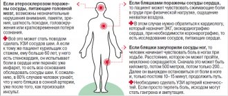
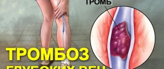
How dangerous is a gap?
Many patients, unaware of the presence of an aneurysm, continue to lead a normal lifestyle. The formation and growth of this formation can last more than 3 years, accompanied by minor symptoms. Under the influence of physical overload, during pregnancy or childbirth, with a sharp increase in pressure, a rupture of the wall of the femoral artery can occur. If the patient is not operated on in a timely manner, intense bleeding is life-threatening.
In addition to rupture, the presence of an aneurysm increases the risk of the following complications:
- blockage of an artery by a blood clot;
- movement of parts of a blood clot with embolism of the branches and gangrene of the lower extremities;
- suppuration of a hematoma (false aneurysm) with phlegmon of surrounding tissues;
- trophic disorders (dermatitis, ulcers) due to lack of blood flow.
Main branches
A series of connections extend from the main vessel. Each of them provides blood supply to a separate area and performs certain functions:
- Superficial epigastric artery. Transports blood to the external oblique muscle of the abdomen and the skin of the anterior wall of the peritoneum. It goes from the bottom of the inguinal ligament up the anterior abdominal wall to the umbilical ring. Near the navel it connects with the superior epigastric artery.
- Superficial femoral. Responsible for nourishing the groin muscles, lymph nodes and skin. Departs from the epigastric or from the outer wall of the femoral artery. It runs along the inguinal ligament to the anterior iliac spine.
- External genital arteries. Their number varies from 2 to 3. They are directed medially, bending around the anterior and posterior periphery of the femoral vein. They also include a large number of smaller branches that are located in the scrotum in men, the labia in women and above the pubis.
- Inguinal branches. Provides a flow of nutrients and blood to the lymph nodes and skin. They originate from the external genital arteries in the form of small stems. Then they pass through the fascia lata of the thigh.
- Deep artery of the femur. The largest of all branches, which consists of a whole network of vessels. It starts 3-4 cm below the inguinal ligament and ends in the lower third of the thigh, between the long and large adductor muscles. Arteries depart from it - lateral, medial, perforating, as well as small capillaries. They promote normal blood circulation in muscles, joints, and deep layers of the epidermis.
- Descending knee. A long vessel that can arise either directly from the femoral artery or from the lateral one. It ends in the thickness of the knee muscles and the capsule of the knee joint. It has branches - articular and subcutaneous.
Since the deep femoral artery is the main element of the blood circulation of the femoral artery, the peculiarities of its structure should be taken into account. Several more vessels depart from each of its branches:
- Medial artery. Its continuation is the ascending, transverse, deep branches and the branch of the acetabulum.
- Lateral. It arises from the outer wall of the deep artery and divides at the intersection with the trochanter of the femur. There the ascending, descending and transverse branches depart from it.
- Perforated arteries. Located at different levels from the main artery. In the area where the adductor muscles attach to the femur, they move to the back of the thigh. They supply the adductor, semimembranosus, semitendinosus, and biceps muscles.
Disruption of blood flow in at least one channel is fraught with serious consequences for the entire vascular system. Ligaments, external genitalia, and lower limbs also suffer due to lack of oxygen and nutrients.
The Scarpian or femoral triangle is formed by the superficial epigastric, superficial and genital arteries. Its height is 15-20 cm.
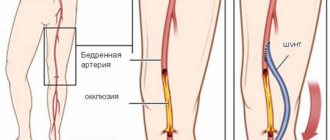
Treatment of femoral artery aneurysm
Conservative treatment in the presence of an aneurysm is not carried out. With the help of medications, you can only delay the rupture of the artery wall by using antihypertensive drugs. The optimal treatment option is surgery. The main types of surgical interventions are presented in the table.
- Where is the carotid artery located and what is its role in the human circulatory system?
| Types of surgery | Short description |
| Prosthetics | A section of the vessel with the aneurysmal sac is excised, and in its place a prosthesis made of synthetic material or the patient’s vein is installed. |
| Bypass surgery | After removing the fragment of the artery with the aneurysm, a bypass path is created to feed the tissue; the patient’s own vessel (usually a peripheral vein) is used for the shunt. |
| Stenting | It is considered a less traumatic technique, since it does not require open access; a metal frame is inserted into the vessel, which strengthens the walls of the femoral artery, preventing its rupture and thrombosis. |
If the size of the operation is small and the risk is high, a wait-and-see approach may be chosen. In this case, physical overexertion is contraindicated for the patient; medications are prescribed to maintain normal blood pressure and prevent blood clots. Such patients should be regularly examined and observed by a vascular surgeon.
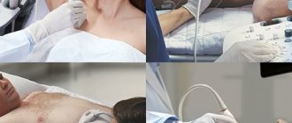
How to prepare
Before the procedure, you must undergo an examination. This will allow you to determine the exact degree of stenosis. In this case, both standard and specific studies are prescribed. Their list includes:
- blood test (for general indicators, for HIV, for markers of hepatitis C and B, for coagulation, for syphilis);
- urine analysis for general indicators; duplex color scanning;
- X-ray contrast angiography;
- Magnetic resonance imaging;
- plethysmography (assessment of arterial blood flow);
- ankle-brachial index (an indicator of damage to the arteries of the legs);
- coronary angiography;
- Cardiography (ECG).
In addition, other types of examinations may be needed, depending on the presence of certain diseases. This approach gives a detailed picture of the preoperative state of the body and allows you to correct some aspects in advance. 7 days before the intervention, stop taking certain medications, 2-3 days before - alcohol and tobacco, 1 - food. Before the operation, an enema cleansing of the intestines is performed.