How does the heart apparatus work?
The main organ of the cardiovascular system is the heart; its key task is to move blood fluid through the vessels. The movement of blood is carried out in one direction - from the veins it enters the cavity of the atria, then into the ventricles, after which it moves to the large arterial vessels. With the help of special valves, one-way continuous blood flow occurs.
Important! The muscles begin rhythmic contraction in the atrium cavity, then they contract in the ventricles, after which there is a pause. This cycle repeats itself constantly.
“Main” circulation
The large circle originates in the left ventricle.
The only arch of the aorta coming from it is the left one, and not the right one, like in birds. Branches from it carry blood throughout the body, saturating organs and tissues with oxygen and other necessary substances.
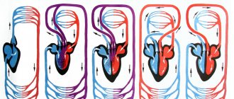
structure of the circulatory system of mammals photo
From them it takes in carbon dioxide and metabolic products.
Venous blood, saturated with carbon dioxide, is directed through the veins to the right atrium. Two vena cavae flow into it, the first of which carries blood from the head and forelimbs, and the second from the back of the body.
Vascular and nervous apparatus
The connection of the vessels takes place using anastomosis. Both vessels of the same type and vessels of different types communicate. There is the following classification of anastomoses:
- arterial;
- venous;
- arterial-venous.
Through anastomoses, longitudinal capillaries, networks, and collectors are formed. The heart also contains the autonomic nervous system. With the help of sympathetic nerve fibers, cardiac function is stimulated. And thanks to parasympathetic nerve fibers, cardiac activity slows down. Autonomic nerve fibers are in close communication with the neuromuscular system of the cardiac apparatus.
Lymphatic system
The lymphatic system is closely connected with the circulatory system and is an intermediary between it and tissues in the exchange of nutrients.
It consists of blood plasma and lymphocytes.
It is noteworthy that mammals do not have “lymphatic hearts,” unlike reptiles and amphibians, which is the name given to areas of lymphatic vessels that can contract: lymph in mammals, which lead a much more active lifestyle, moves due to contraction of the skeletal muscles.
Mammals also have lymph nodes that cleanse the lymph from harmful microorganisms.
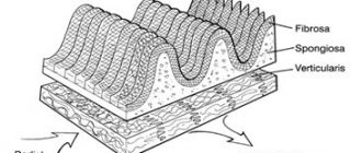
The structure of the walls of the heart
The walls of the organ consist of the following capsules - endocardium, myocardium and epicardium.
The inner layer is the endocardium. The layer thickness is different in different areas. On the left it is thicker, and in the area where the tendon threads are located it is somewhat thinner. There are three layers of the endocardium:
- endothelium is the bottom layer;
- subendothelial zone - its basis is loose veins of connective tissue;
- Muscular-elastic zone - this is where muscle tissue connects.
The middle layer is the myocardium. It is a very thick muscular layer. The difference between the myocardium and skeletal muscle tissue is that there are intercalary crossbars between certain myocardial fibers. This layer forms muscle tissue; its cells are directly involved in nerve impulses and contraction of the heart muscles.
The outer layer is the epicardium. It is the outer serous membrane. Its surface is covered by mesothelium, underneath there is connective tissue consisting of loose fibers.
If any disruptions occur in the functioning of the heart and blood vessels, a disruption of metabolic processes and the functions of other organs occurs. Therefore, it is very important for every farmer to know how diseases manifest themselves and how to treat them.
How does the heart work in animals?
Why is it so important?
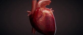
The heart is a vital organ with a complex structure. The most important muscle in the entire living organism. Very often the entire circulatory system (or, in other words, the cardiovascular system) is compared to a plumbing system, and the heart in it acts as a pump, only it pumps blood instead of water. Blood gives oxygen and nutrients to all tissues and organs, and takes away carbon dioxide and metabolic products.
In you and me, and in our cats and dogs, like all mammals, the heart is almost equally tripled. Outwardly, it looks completely different from what you imagine. The organ looks more like an inverted cone. It is a hollow organ consisting of chambers and muscular walls. There are only 4 chambers. By analogy, the heart can be compared to a two-story house, the first floor represents the ventricles, right and left, the second - the right and left atrium. The right and left halves of the heart do not communicate in healthy animals. Thanks to this, the blood is clearly divided into arterial, oxygen-rich, and venous, oxygen-poor. The right half of the heart is venous; it collects oxygen-poor blood from the entire body, and then it is sent through the vessels to the lungs, where it is enriched with oxygen. Then from the lungs, through the pulmonary veins (they are called veins, but they carry arterial, that is, oxygen-rich blood), the blood enters the left atrium, then into the left ventricle and then spreads throughout the body through the aorta.
The atria, ventricles and main vessels are separated by valves. They are designed like doors that open only in one direction. They consist of valves.
The division of blood into systemic and pulmonary circulation (arterial and venous blood) is a fundamental feature of the structure of the mammalian heart.
The heart itself is supplied with blood by special vessels, which are called coronary vessels, because they form a network like a “crown”. They arise from the aorta at its very beginning.
What ensures constant contractions of the heart muscle?
The heartbeat, which is its rhythm, is provided by electrical impulses that are formed by the heart muscle itself. These impulses are formed in the right atrium, and then go through the interventricular septum and further through all the ventricles, causing them to contract.
Despite the fact that the heart of animals and humans is very similar, the diseases still differ . For example, animals almost never have myocardial infarctions, since there is no coronary heart disease or atherosclerotic plaques. Their coronary vessels are structured differently and fat metabolism occurs differently. Therefore, you should never self-medicate animals.
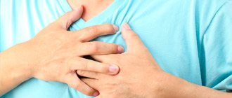
In order to suspect heart disease, it is necessary to detect various clinical manifestations, which you can read about in our article - WHEN CAN YOU SUSPECT HEART DISEASE IN AN ANIMAL?
Pericarditis
The development of pericarditis occurs against the background of previous diseases of infectious etiology. In some cases, injury to the pericardium causes inflammation. The main provoking factors for the development of the disease include a poor diet, which causes metabolic disorders. The main signs of pericarditis include:
- general weakness;
- periodic hyperthermia;
- loss of appetite;
- lack of chewing gum;
- deterioration in quantitative indicators of products;
- pulse increases to 120 beats;
- swelling appears in the neck, chest or abdomen;
- breathing becomes rapid.
A sick cow becomes restless, moans, and takes a body position in which the chest is higher than the hip area. There is weakness of heart beats, and murmurs are clearly audible on auscultation. If traumatic pericarditis develops, treatment will not be effective. In this case, the cow must be culled. If non-traumatic pericarditis develops, the animal needs to be kept at rest and fed light food.
The doctor prescribes antibacterial drugs, cold compresses and applications to the heart area. The doctor also prescribes medications intended to restore cardiac activity: camphor, glucose, caffeine, calcium chloride.
To prevent the development of this disease, it is recommended to promptly treat diseases that can cause its development.
Dropsy of the cardiac sac
The development of this disease is characterized by the accumulation of fluid in the sac, which is located near the heart. Often, the development of dropsy can be triggered by other diseases or insufficiency of chronic blood microcirculation. The main features include:
- weakness;
- deterioration in milk yield;
- the appearance of swelling in the space between the jaws;
- sudden jumps in blood pressure.
Therapeutic measures involve getting rid of the underlying disease. For the cow, you need to provide the most balanced nutrition possible and give a large amount of liquid.
Important! To reduce the amount of accumulated fluid, the doctor prescribes a course of cardiac, diuretic, diaphoretic and iodine medications.
Composition of mammalian blood
The blood of mammals consists of liquid plasma, which contains a full set of so-called formed elements:
- Red blood cells are carriers of the iron-containing substance hemoglobin, they carry oxygen;
- Platelets are bodies responsible for blood clotting and serotonin metabolism;
- Leukocytes are white bodies responsible for immunity.
Red blood cells and platelets of mammals, unlike other groups of animals, do not contain nuclei.
Platelets are actually “blood platelets”; the absence of nuclei in red blood cells is explained by the need to accommodate a larger amount of hemoglobin.
Also, red blood cells do not have mitochondria, so they synthesize ATP without the use of oxygen, making them the most effective carriers of it.
Myocarditis
This disease is characterized by inflammation of the myocardium, which then develops various types of disturbances in the functioning of the heart. The organ's ability to contract deteriorates. Most often, this illness is a complication of previous infectious diseases or poisoning. The main symptoms include:
- hyperthermia;
- loss of appetite;
- development of tachycardia or extrasystole;
- increased heart rate;
- increased blood pressure;
- rapid breathing;
- blue discoloration of the mucous membranes and nasolabial triangle;
- swelling in different parts of the body.
With the development of this pathological process, the functions of other internal organs are disrupted. Therapy is determined by the severity of the disease. Initially, you should get rid of the reason that provoked its appearance. A sick animal must be transferred to a clean, dry, warm room. Give food in small portions, drink warm water. The doctor prescribes medications to reduce inflammation and reduce stimulation of the heart muscles.
Myocardosis
This is a pathological process, which is characterized by the development of dystrophic changes in the walls of the myocardium. Often a complication of myocarditis. The clinical picture is manifested by the following symptoms:
- weakness;
- refusal to eat;
- reduction in muscle tone;
- a sharp decrease in blood pressure;
- deterioration of the elasticity of the epidermis;
- blue discoloration of the mucous membranes and nasolabial triangle;
- swelling in different parts of the body;
- disruptions in heart contractions.
First you need to get rid of the cause that could cause the development of the disease. The sick animal is isolated and transferred to a dry and warm room. The diet should be balanced, portions should be small. The veterinarian prescribes special medications that will help prevent further degenerative changes in the walls of the myocardium.
Myocardiofibrosis
This disease is the result of dystrophic or degenerative changes in the heart muscles, and is characterized by poor circulation. The clinical picture is manifested by the following symptoms:
- Auscultation reveals muted tones;
- arrhythmia or tachycardia develops;
- the pulse is faintly audible;
- swelling occurs;
- breathing quickens.
This pathological process can develop over a long period of time, and can last for several months or even a year. A sick cow should be moved to a warm room and given balanced feed in small portions. Additionally, the doctor prescribes medications to improve blood circulation and prevent further development of the pathology.