I34 Non-rheumatic lesions of the mitral valve
Excludes: mitral (valve): disease (I05.9), insufficiency (I05.8), stenosis (I05.0), lesions specified as rheumatic (I05.), with an unknown cause, but with mention of: aortic valve disease (I08.0), mitral stenosis or obstruction (I05.0)
I34.0 Mitral (valve) insufficiency
I34.1 Prolapse (prolapse) of the mitral valve
Excludes: Marfan syndrome (Q87.4)
I34.2 Non-rheumatic mitral valve stenosis
I34.8 Other non-rheumatic lesions of the mitral valve
I34.9 Non-rheumatic disease of the mitral valve, unspecified
I08 Lesions of several valves
Included: cases specified or unspecified as rheumatic
Excluded:
rheumatic diseases of the endocardium, valve unspecified (I09.1)
endocarditis, valve unspecified (I38)
I08.0 Combined lesions of the mitral and aortic valves
I08.1 Combined lesions of the mitral and tricuspid valves -
new
I08.2 Combined lesions of the aortic and tricuspid valves
new
I08.3 Combined lesions of the mitral, aortic and tricuspid
chat valves
I08.8 Other multiple valve diseases
I08.9 Multiple valve lesions, unspecified
Classification
According to this international classification, ICD 10 code lists all existing heart defects.
There are quite a few of them. Conventionally, all defects can be divided into two groups, each of which has its own subgroups:
- White. Heart diseases in which mixing of venous and arterial blood does not occur.
- defect of the interatrial and interventricular septum;
- pathologies caused by depletion of the pulmonary circulation;
- defects associated with depletion of the systemic circulation;
- pathologies not associated with hemodynamic disorders.
- Blue. Venous and arterial blood mix.
- defects caused by enrichment of the small circle;
- pathologies caused by depletion of the small circle.
Each of the above groups and subgroups has its own specific code, which helps to determine the type of disease as accurately as possible.
Rheumatism in children: current state of the problem
Rheumatism , or acute rheumatic fever
according to the International Classification of Diseases (ICD), is a systemic inflammatory disease of connective tissue with a predominant localization of the process in the cardiovascular system, developing in connection with acute streptococcal infection in persons predisposed to it, mainly at the age of 7–15 years. While not a widespread disease, rheumatism nevertheless represents a serious problem in cardiorheumatology due to the frequent formation of heart defects and the development of temporary and permanent disability. In accordance with ICD-10, acute rheumatic fever is classified as a disease of the circulatory system (class IX) under the codes:
100 – Rheumatic fever without mention of cardiac involvement;
101 – Rheumatic fever with cardiac involvement;
102 – Rheumatic chorea.
Rheumatism was known already in the 5th century BC. Hippocrates in his work “The Four Books of Diseases” wrote: “With arthritis, fever appears, acute pain seizes all the joints of the body, and these pains are sometimes sharper, sometimes weaker, affecting first one or the other joint.” In ancient times, doctors believed that inflammation in the joints was caused by some toxic liquid spreading throughout the body. This is where the name of the disease came from - “rheumatism” (from the Greek “rheum” - flow). Damage to the cardiovascular system was considered a complication of articular syndrome. Only after the publication of the outstanding works of the French doctor Buyot (1836) and the Russian doctor I.G. Sokolsky (1838), rheumatism was identified as an independent disease involving heart damage.
Over more than a century and a half of studying this serious, often disabling disease, the connection between its development and streptococcal infection has been determined, and a system of diagnosis, treatment and prevention has been developed and implemented. This contributed to a widespread decline in the incidence of rheumatism by the middle of the twentieth century. However, in recent years, due to a number of negative socio-economic processes, there has been a tendency towards an increase in the incidence of rheumatism in all age groups, and more intensely in children
. This trend is also due to the presence of secular rhythms characteristic of aggressive streptococcal infections and a decrease in the sensitivity of streptococci to penicillins. The study of the dynamics of the epidemic process shows that in the last decade, streptococcal infection has appeared and is growing, which is an analogue of that of past times, which can contribute to the increase and severity of rheumatism. Therefore, in the future, the problem of rheumatism will not lose its relevance.
Etiology and pathogenesis
The development of rheumatism is closely related to a previous acute or chronic nasopharyngeal infection caused by group A beta-hemolytic streptococcus
.
A special role is given to M protein
, which is part of the streptococcal cell wall. Of the more than 80 known varieties of M protein, M-5, M-6, M-18 and M-24 are considered so-called rheumatogenous. In this case, a stable hyperimmune response to various streptococcal antigens is determined with the formation of antibodies - antistreptolysin O (ASL-O), antistreptohyaluronidase (ASH), antideoxyribonuclease, etc.
Genetic factors play an important role
, which is confirmed by the more frequent incidence of the disease in children from families in which one of the parents suffers from rheumatism.
The importance of genetic factors is to a certain extent evidenced by the results of studying the association of histocompatibility antigens, which revealed, in particular, the frequent occurrence of Dr5–Dr7, Cw2–Cw3 and a number of others in patients with different forms of rheumatism. The genetic marker of this disease, according to a number of researchers, is the B-lymphocyte alloantigen
, determined using monoclonal antibodies D8/17, which is found with high frequency both in patients with rheumatism and in their close relatives. It is associated with a hyperimmune response to streptococcal antigen.
In the pathogenesis of rheumatism, the direct or indirect damaging effects of streptococcal components and its toxins on the body with the development of immune inflammation are of no small importance. Anti-streptococcal antibodies that cross-react with heart tissue (molecular mimicry)
.
Morphological changes
reflect systemic disorganization of connective tissue, especially the cardiovascular system with specific necrotic-proliferative reactions (Ashoff-Talalaev granulomas) and nonspecific exudative manifestations. The latter are more distinct in childhood, which determines the greater (compared to adults) severity and activity of the process, the severity of carditis and other manifestations of rheumatism.
Clinical picture
Rheumatism in children is characterized by a variety of clinical manifestations and variability of course. As a rule, it occurs at school age, less often in preschoolers
and practically does not occur in children under 3 years of age.
In typical cases, the first signs of rheumatism are detected 2–3 weeks after a sore throat or pharyngitis
in the form of fever, symptoms of intoxication, articular syndrome, carditis and other manifestations of the disease. It is also possible for the disease to have asymptomatic onset with the appearance of fatigue, low-grade fever, in the absence of noticeable disorders of the joints or heart, which can be mistakenly regarded as residual effects of a previous infection.
Joint syndrome
One of the earliest signs of rheumatism is articular syndrome (arthritis or arthralgia), detected in 60–100% of affected children. Rheumatoid arthritis is characterized by an acute onset, involvement of large or medium-sized joints (usually knee, ankle, elbow) in the form of mono- or oligoarthritis, volatility of the lesion, and rapid reversal of the process. Articular syndrome relatively rarely develops in isolation at the onset of the disease; more often it is accompanied by cardiac disorders.
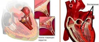
Heart damage
Symptoms of heart damage ( carditis
) are determined in 70–85% of cases at the onset of the disease and somewhat more often during subsequent attacks, depending on the predominant localization of the process in the myocardium, endocardium, and the degree of involvement of the pericardium. Due to the difficulty of identifying signs of damage to one or another lining of the heart, the term “rheumatic carditis” is used in practice.
Complaints of a cardiac nature (pain in the heart, palpitations, shortness of breath) are observed in children mainly with severe cardiac disorders. More often, especially at the onset of the disease, various asthenic manifestations (lethargy, malaise, increased fatigue) are observed.
The first objective signs of rheumatic carditis are: disturbance of heart rate (tachycardia, less often - bradycardia); increase in heart size, predominantly to the left; muffled heart sounds, the appearance of systolic murmur.
Character of systolic murmur
, its localization is determined by the degree of involvement of the myocardium and endocardium in the process.
With myocarditis
, the murmur is usually weak or moderate and rarely extends beyond the heart.
In case of endocarditis
with the most characteristic lesion of the mitral valve in rheumatism (valvulitis), a prolonged blowing systolic murmur is heard with a maximum at the apex and at Botkin's point, intensifying on the left side and during exercise, conducted outside the heart. It is mitral valve valvulitis that plays the main role in the formation of valvular heart defects, the development of which can be established no earlier than 6 months from the onset of the disease. The development of rheumatic carditis may be accompanied by circulatory failure, usually not exceeding stage I.
On ECG
with rheumatic carditis, rhythm disturbances are often detected (tachy- or bradyarrhythmia, migration of the pacemaker, sometimes extrasystole, atrial fibrillation),
slowing of atrioventricular conduction
, mainly of the first degree, disturbances in ventricular repolarization, prolongation of electrical systole.
The phonocardiogram (PCG) records a decrease in the amplitude of the first tone at the apex and an increase in the amplitude of the third and fourth sounds. In case of myocarditis, FCG reveals a systolic murmur that is not associated with the first sound, variable in different cardiac cycles, and having a medium-amplitude, medium-frequency character. Mitral valve valvulitis is manifested by high-frequency pansystolic or protosystolic murmur of varying amplitude.
X-ray
in addition to the not always pronounced enlargement of the heart, signs of decreased tonic and contractile function of the myocardium, mitral (with valvulitis of the mitral valve) or aortic (with damage to the aortic valve) configuration of the heart are determined.
When performing an echocardiography, thickening, “shaggy” echo signal from the leaflets of the affected valves, a decrease in their excursion, signs of impaired myocardial contractile function and a number of other symptoms are revealed. With rheumatic carditis in children, the development of pericarditis is also possible, the clinical manifestations of which are determined much less frequently than instrumental signs on the ECG and especially EchoCG.
Currently, with timely treatment, primary rheumatic carditis ends in recovery in most children. The formation of valvular heart defects, often with the development of mitral insufficiency, is determined in 15–18% of cases during the first attack, mainly in severe, protracted or latent course of the disease.
With repeated attacks, which, according to the recommendation of the American Heart Association (AHA), are considered as a new episode of acute rheumatic fever, and not a relapse of the first, the formation of heart defects, often in the form of combined and/or combined valvular lesions, reaches 100%. Due to rheumatic carditis, the formation of prolapse of the mitral (less often aortic) valves
, development
of myocardiosclerosis
with rhythm and conduction disturbances (extrasystole, atrial fibrillation, complete atrioventricular block), as well as
adhesive pericarditis
. Severe rheumatic carditis, its relapses, the presence of myocardiosclerosis, and heart defects contribute to the development of persistent heart failure, leading to disability in patients and possible death.
Small chorea is characteristic of childhood rheumatism
, occurring
in 12–17% of cases
, mainly in early puberty and in girls. Classic symptoms of minor chorea: hyperkinesis, hyper- or hyporeflexia, muscle hypotonia, impaired coordination of movements, changes in psychological state and various autonomic disorders.
The disease begins acutely or, more often, gradually. Children develop emotional lability, irritability, changeable mood, tearfulness, absent-mindedness, memory loss, and deterioration in academic performance. An objective examination reveals involuntary twitching of the muscles of the face and limbs with grimacing, jerky, awkward movements; slurred, unclear speech, changes in handwriting, gait, which makes feeding, dressing, and learning difficult. Hyperkinesis is often bilateral in nature, intensifies with excitement, weakens during sleep until it stops completely.
Often, minor chorea precedes the development of rheumatic carditis or proceeds without clear disturbances in cardiac activity, and is characterized by a torpid course and a tendency to relapse.
Other symptoms
Rarer symptoms of rheumatism include annular rash and rheumatic nodules
.
Annular rash
(ring-shaped erythema) - pale pink, dim rashes in the form of a thin ring-shaped rim, which do not rise above the surface of the skin and disappear with pressure. It is found in 7–10% of children with rheumatism, mainly at the height of the disease and is usually unstable.
Subcutaneous rheumatic nodules
– round, dense, inactive, painless, single or multiple formations localized in the area of large and medium-sized joints, spinous processes of the vertebrae, in tendons, on the aponeurosis. Currently, they are rare, mainly in severe forms of rheumatism, lasting from several days to 1–2 months.
Abdominal syndrome, damage to the lungs, kidneys, liver and other organs due to rheumatism in children is now extremely rare, mainly in severe cases.
Laboratory indicators
Laboratory indicators in patients with rheumatism reflect signs of streptococcal infection, the presence of inflammatory reactions and an immunopathological process. In the active phase, the following are determined: leukocytosis with a shift to the left, an increase in ESR, and often anemia; increased levels of seromucoid, diphenylamine reaction; dysproteinemia with hypergammaglobulinemia; increased titers of ASH, ASLO, increased immunoglobulins class A, M and G; C-reactive protein (CRP), circulating immune complexes, anticardiac antibodies.
Diagnostics
As is known, there are no specific tests for diagnosing rheumatism. In practice, the syndromic method of assessing the status of the patient
, the principle of which was proposed in 1940 by the domestic pediatrician A.A. Kisel.
The author identified such syndromes pathognomonic for rheumatism as migratory polyarthritis, carditis, chorea, annular erythema, and rheumatic nodules as the main diagnostic criteria. Later, A.I. Nesterov made additions to the criteria, and for a long time, when establishing the diagnosis of rheumatism, doctors used the Kisel-Jones-Nesterov criteria. Subsequently, this scheme was repeatedly modified, and currently, in accordance with WHO recommendations, the Jones diagnostic criteria
, revised by the AKA in 1992, are used as international criteria for acute rheumatic fever (Table 1).
The detection of two major or one major and two minor criteria in a patient, combined with data documenting a previous infection with group A streptococci, indicates a high probability of acute rheumatic fever. However, in the early stages, nosological diagnosis in conditions of vague and nonspecific symptoms presents certain difficulties.
The final diagnosis is often established after a thorough differential diagnosis and dynamic observation of the patient with assessment of the results of the therapy.
Differential diagnosis
The differential diagnosis of rheumatism is carried out with a large group of conditions and diseases occurring with articular syndrome (reactive arthritis, juvenile rheumatoid arthritis, Lyme disease, leukemia, neoplastic processes); with a number of inflammatory cardiac diseases (non-rheumatic carditis, infective endocarditis); congenital and acquired disorders of the heart structure (mitral valve prolapse, congenital mitral valve insufficiency or anomaly of its development); autonomic dystonia syndrome. The diagnosis of minor chorea requires, first of all, the exclusion of thyroid hyperkinesis in children, Tourette's syndrome, and chorea in systemic lupus erythematosus.
The formulation of the diagnosis and, accordingly, treatment is carried out on the basis of the classification developed by A.I. Nesterov and the nomenclature of rheumatism (Table 2).
Determining the degree of process activity
(I, II, III) is carried out taking into account the severity of clinical and laboratory manifestations.
The acute course
reflects the rapid development of rheumatism, polysyndromic nature, and vivid clinical and laboratory manifestations (duration – 2–3 months).
The subacute course
may (at its onset) resemble an acute one or is characterized by a slower onset of the disease, less pronounced clinical and laboratory manifestations (duration – up to 6 months).
The protracted course is characterized by moderate signs of activity, torpidity to the therapy (duration - more than 6 months). With a relapsing course,
polysyndromic nature, severity of clinical manifestations, and relapses are noted. The latent course is characterized by the progression of heart disease in the absence of signs of process activity.
Treatment
Treatment of rheumatism in children is based on the early administration of complex therapy aimed at suppressing streptococcal infection and the activity of the inflammatory process, preventing the development or progression of heart disease. The implementation of these programs is carried out according to the principle of stages: 1st stage - inpatient treatment, 2nd stage - follow-up treatment in a local cardio-rheumatological sanatorium, 3rd stage - dispensary observation in a clinic.
Stage 1
At the 1st stage in the hospital, the patient is prescribed drug treatment (antibacterial, antirheumatic and symptomatic), nutritional correction and physical therapy, which are determined individually taking into account the characteristics of the disease and, above all, the severity of carditis. Due to the streptococcal nature of rheumatism, etiotropic therapy is carried out with penicillin
.
Antirheumatic therapy involves one of the nonsteroidal anti-inflammatory drugs (NSAIDs
), which are prescribed alone or in combination with
glucocorticosteroid hormones (GCS)
, depending on the indications (Table 3).
Antibacterial therapy
penicillin is carried out for 10–14 days. In the presence of chronic tonsillitis, frequent exacerbations of focal infection, the duration of treatment with penicillin is increased, or another antibiotic is additionally used - amoxicillin, macrolides (azithromycin, roxithromycin, clarithromycin), cefuroxime axetil, and other cephalosporins in an age-specific dosage.
NSAIDs
apply for at least 1–1.5 months until signs of process activity are eliminated.
Prednisolone
in the initial dose is prescribed for 10–14 days until a clinical effect is obtained, then the daily dose is reduced by 2.5 mg every 5–7 days under the control of clinical and laboratory parameters, and subsequently the drug is discontinued.
Duration of treatment with quinoline drugs
for rheumatism, it ranges from several months to 1–2 years or more, depending on the course of the disease.
Chronic foci of infection are also sanitized in a hospital setting.
, in particular, tonsillectomy, carried out 2–2.5 months from the onset of the disease in the absence of signs of process activity.
Stage 2
The main task at the second stage is to achieve complete remission and restore the functional capacity of the cardiovascular system of children with rheumatism. In the sanatorium, the therapy started in the hospital is continued, foci of chronic infection are sanitized, and the appropriate treatment and health regimen is carried out with differentiated physical activity, physical therapy, and hardening procedures.
Stage 3
The third stage of complex therapy for rheumatism involves the prevention of relapses and progression. For this purpose, long-acting penicillin
, mainly bicillin-5, the first administration of which is carried out during the period of hospital treatment, and subsequently - 1 time every 2-4 weeks year-round. Regularly, 2 times a year, an outpatient examination is carried out, including laboratory and instrumental methods; prescribe the necessary health measures and physical therapy. For children who have had rheumatic heart disease and have valvular heart disease, bicillin prophylaxis is carried out until they reach the age of 21 years or more. For rheumatism without cardiac involvement, bicillin prophylaxis is carried out for 5 years after the last attack. In the spring-autumn period, along with the administration of bicillin, a monthly course of NSAIDs is indicated.
Prevention of rheumatism is divided into primary and secondary.
Primary prevention
is aimed at preventing rheumatism and includes:
1. Increasing immunity (hardening, alternating exercise and rest, good nutrition, etc.).
2. Detection and treatment of acute and chronic streptococcal infections.
3. Preventive measures for children predisposed to the development of rheumatism: from families in which there are cases of rheumatism or other rheumatic diseases; often suffer from nasopharyngeal infection; having chronic tonsillitis or having had an acute streptococcal infection.
Secondary prevention
is aimed at preventing relapses and progression of the disease in children with rheumatism under conditions of clinical observation (see earlier: “Third stage of therapy”).
Key Concepts
Rheumatism is a disease that develops in connection with a streptococcal infection, mainly at the age of 7–15 years. It is characterized by polymorphism of clinical manifestations and variability of course.
The clinical picture of the disease is primarily characterized by heart damage, articular syndrome and minor chorea; Its other manifestations are less common. Cardiac symptoms are caused by the predominance of endo- or myocardial damage with the possible, but more rare, development of pericarditis. A characteristic feature of rheumatic carditis is the development of mitral valve valvulitis. Articular syndrome is often determined already at the onset of the disease in the form of unstable arthritis or arthralgia. The priority of rheumatism in children is minor chorea, which has quite characteristic clinical symptoms.
Primary rheumatism with timely treatment usually ends in recovery. The development of heart defects, as a rule, is manifested by mitral insufficiency with the possibility of subsequent addition of other valvular disorders. Repeated attacks of rheumatism much more often lead to the formation of heart defects.
Treatment of children with rheumatism is carried out on the basis of the principles of staged therapy, depending on the nature of the existing pathology, the degree of activity of the process and the characteristics of its course. For these purposes, antibiotics, NSAIDs, and glucocorticosteroids are used.
An integral part of preventive measures for rheumatism is primary and secondary prevention, aimed at preventing the development of the disease, the occurrence of relapses and the progression of the pathological process.
Literature
1. Belov B.S. “Modern aspects of diagnosing acute rheumatic fever in adolescents.” Det. rheumat. 1996; 2.
1. Belov B.S. “Modern aspects of diagnosing acute rheumatic fever in adolescents.” Det. rheumat. 1996; 2.
2. Belov B.S. “Modern aspects of acute rheumatic fever” Lecture for doctors. Moscow, 1998.
3. Dolgopolova A.V., Kuzmina N.N. “Primary rheumatic carditis in children.” M., Medicine, 1978.
4. Kuzmina N.N. “The problem of rheumatism in children at the present stage.” Det. Rheumat., 1996; 2.
5. Nasonova V.A., Kuzmina N.N., “Rheumatism” in the book. “Rheumatic diseases” (a manual for doctors edited by Nasonova V.A., Bunchuk N.V.). M., “Medicine”, 1997.
6. Nesterov A.I. "Rheumatism". M., Medicine, 1973.
| Applications to the article |
| Risk. In type I diabetes mellitus, diabetic retinopathy occurs in 70% of cases (proliferative retinopathy - 45%, blindness - 10%), in type II diabetes mellitus - in 39% of cases. |
| cost $10.5 million from 1972 to 1982. The study generated a net benefit of $231 million to society and provided patients with a total of 279,000 years of vision. |
| Therapeutic treatment of myopia includes drugs that affect accommodation, improve hemodynamics of the eye, and stimulate metabolic processes in the tissues of the eye. |
| Patients with primary dyslexia (the inability to read with intact intelligence, unimpaired visual functions and normal motivation) have a defect in the visual memory center, which makes it difficult for the child to retain visual information about symbols. |
| Fact. The number of nerve fibers in the optic disc varies from 800,000 to 1,200,000 and gradually decreases with age. |
| Possible oligosymptomatic onset of rheumatism with the appearance of fatigue and low-grade fever |
| The formation of valvular heart defects is determined in 15–18% of cases during the first attack |
Symptoms
When diagnosing congenital heart disease, the pediatrician and cardiologist must first conduct a conversation with the child’s parents.
In this conversation, it is necessary to establish how the child developed, when the first signs of the defect began to appear, etc. A congenital defect leaves its mark not only on the functioning of the heart, but also on many other signs:
- Body type. Few defects can cause changes in physique. For example, if the functioning of the aorta of the heart is impaired, the child experiences accelerated development of the shoulder girdle. As a rule, such children do not gain weight well, and this is reflected in their physique.
- Covers of the skin. If a patient is diagnosed with a white type defect, his skin is too pale. With a blue defect, cyanosis of the integument is clearly manifested.
- Respiratory system. Due to increased blood flow in the lungs, patients with congenital heart disease often suffer from shortness of breath and have difficulty breathing.
- Digestive organs. The disease also affects the digestive system. For example, there is an increase in the size of the liver. Often, such patients experience vomiting with increased physical activity, which is accompanied by severe pain in the abdominal area.
All these symptoms can appear from an early age, or they can begin to remind themselves at the age of 5–6 years.
Publications in the media
Cardiac myxoma is a benign intracavitary tumor that accounts for up to 50% of all primary cardiac tumors. In 75% of cases, the tumor affects the left atrium, in 20% - the right. In other cases, it is located in the ventricles of the heart, sometimes the valve apparatus is involved.
Frequency • Myxomas make up 50% of all benign heart tumors in adults and 15% in children • Myxomas in newborns - casuistry • Sporadic myxomas in women occur 4 times more often than in men, hereditary forms - with the same frequency in both sexes • The highest the frequency of myxomas is detected at the age of 30–60 years • 94% of sporadic myxomas are solitary, 22% of hereditary forms are multilocular • About 75% of myxomas occur in the left atrium, 15–20% in the right atrium, 6–8% in the ventricles • 21– 76% of hereditary forms recur after surgical treatment, sporadic forms almost never recur • About 20% of patients with hereditary forms have concomitant oncological diseases (adrenocortical nodular dysplasia, Sertoli cell tumors, pineal tumors, multiple myxoid fibroadenomas of the mammary gland, cutaneous myxomas, multiple nevi of the face and lips) • According to pathological autopsies, the detection rate ranges from 0.0005 to 0.02%.
Genetic aspects . Carney syndrome. Etiology • In 80% of cases, myxomas occur in people with a normal genome • In 5% of cases, it is possible to establish the family nature of the pathology, in this case, 20% of patients have various genetic abnormalities (most often - tetraploidy) • Apparently, there is a relationship between heart injuries , previous VSD repair, transseptal puncture, percutaneous balloon dilatation of the mitral valve and subsequent development of myxoma.
Pathogenesis • The presence and severity of the stenotic effect and the nature of circulatory disorders depend on the size and location of the tumor • The nature of hemodynamic changes - see Mitral valve stenosis; Aortic valve stenosis; Tricuspid valve stenosis; Pulmonary valve stenosis.
Morphology • Cardiac myxoma in appearance resembles a polyp of jelly-like consistency on a short stalk, fixed to the interatrial septum • Dimensions vary from 5 to 12 cm in diameter, weight reaches 250 g • Morphologically, hereditary and sporadic forms are identical.
Clinical picture and diagnosis. The main clinical signs are identified that allow one to suspect or confirm the presence of cardiac myxoma • Sudden appearance of clinical signs of circulatory failure, depending on a change in the patient’s body position • Rapid development of cardiovascular failure for no apparent reason and against the background of appropriate therapy • Short duration of the disease compared to rheumatic defects heart • The occurrence of embolism of peripheral vessels or pulmonary vessels against the background of sinus rhythm, especially in young people; in this case, embolectomy with histological examination of the embolus can facilitate the diagnosis of the tumor • Shortness of breath or short-term loss of consciousness that occurs suddenly • Variability of noises when the patient’s body position changes • Infective endocarditis, tolerant to antibacterial therapy. The complaints of patients are caused by three pathological mechanisms - embolism, obstruction of blood flow and constitutional signs (the cause of the latter is unknown, most likely multiple small emboli or an autoimmune reaction). A relatively rare and late complication of myxoma is infective endocarditis (see Infectious endocarditis).
• Symptoms of embolism •• Occur in 30–40% of patients •• Since most myxomas are localized in the left parts, about 50% of embolisms occur in extra- or intracranial arteries of the brain, as well as in the vessels of the lower extremities, less often - in the intestinal vessels, coronary and renal arteries •• Emboli in right-sided myxomas are usually asymptomatic and lead only to moderate pulmonary hypertension detected instrumentally. • Symptoms of obstruction of blood flow - see Mitral valve stenosis; Aortic valve stenosis; Tricuspid valve stenosis; Pulmonary valve stenosis. • Constitutional symptoms •• Weight loss weakness, low-grade fever, nausea •• Laboratory changes - leukocytosis, increased ESR, hemolytic anemia, thrombocytopenia, increased levels of CRP, IgG and IL-6 •• Raynaud's syndrome •• Arthralgia •• Myalgia • • Erythematous rash •• Lethargy (rare). Physical examination • See Mitral valve stenosis; Aortic valve stenosis; Tricuspid valve stenosis; Pulmonary valve stenosis • For myxomas localized on the free leaflet of the AV valves, it is possible to listen to the “tumor clap” - the late diastolic sound of the tumor contacting the ventricular endocardium. ECG and chest x-ray: no specific changes. EchoCG • Highly sensitive diagnostic method • Visualization of a space-occupying lesion, measurement of its size, clarification of localization, mobility and place of attachment • Assessment of intracardiac hemodynamic disorders • All patients undergo transesophageal echocardiography to diagnose small myxomas of the left atrium and multiple myxomas of other localizations • Using transesophageal access it is possible to diagnose tumors measuring 1–3 mm.
CT and MRI • Allow additional information regarding the extent of the process and tumor invasion into the wall of the heart (differentiation from malignant tumors) • Using CT and MRI, tumors measuring 5–10 mm can be diagnosed • Typically, CT and MRI are performed only in case of diagnostic difficulties, as well as with tumors of the right atrium spreading into the vena cava or right ventricle. Coronary angiography. Performed on all candidates for surgical treatment over 40 years of age. Cardiac catheterization. Performed for the purpose of tumor biopsy and preoperative verification of its benign nature.
Angiocardiography - identifying a filling defect with smooth contours.
TREATMENT. Conservative treatment is carried out in terms of preoperative preparation; see Heart failure. Surgery . • Indications: All patients with cardiac myxoma. • Methods of surgical treatment : excision of the tumor and its attachment site.
Prognosis: • Myxomas do not metastasize • Their growth pattern is expansive • Tumors previously considered as myxomas with infiltrative growth are in fact primary malignant tumors (usually sarcomas) followed by myxomatous degeneration • The duration of the disease varies and depends on the severity of obstruction and severity embolism • Risk of sudden death - 8-30% • Hospital mortality after removal of atrial myxomas - less than 5%, ventricular myxomas - less than 10% • Recurrence rate of familial forms of myxoma without genetic anomalies - 1-4.7%, with genetic anomalies - 12- 40% • The most common relapses are within 4–6 months after surgery • The course is usually progressive - in the natural course of the disease, the average life expectancy after the first symptoms of cardiac myxoma rarely exceeds 2 years • Causes of death - occlusion of the heart valves or tumor embolism.
ICD-10 • D15.1 Benign neoplasm of the heart
Application. Carney syndrome (*160980, 2p16, CNC gene, ). Clinically: atrial myxoma, diffuse pigmentary changes in the skin (moles, freckles, lentiginous rashes on the face and mucous membranes, hyperpigmentation of the lips), hemangiomas, mucous liposarcomas, subcutaneous neurofibromas, primary nodular adrenocortical dysplasia, virilizing adrenocortical carcinoma, giant cell tumor of Sertoli cells, prolactin secretion adenoma pituitary gland, nasopharyngeal schwannoma, Cushing's disease, acromegaly.
Diagnostics
The more methods were used to diagnose congenital heart disease code according to ICD 10, the more accurate and detailed the picture of the disease will be visible.
The following methods are used for diagnosis:
- Electrocardiogram. This method gives excellent results, even in the initial stages of pathology development. Based on the interpretation of the cardiogram results, the cardiologist can accurately determine the intensity of the development of the disease and its danger to the child’s life.
- X-ray. When conducting an X-ray examination in 3 projections, the condition of the lungs and the entire respiratory system can be determined.
- Echocardiography. Allows the cardiologist to obtain the most accurate picture of the patient’s condition.
- Angiography or catheterization. These techniques are more difficult to perform, but they allow you to determine the pressure in the lungs, as well as accurately determine in which direction the blood flow is directed (from the right atrium to the left or vice versa).
All of the above diagnostic methods are used in combination.
Only in this case can an accurate diagnosis be established and an effective course of treatment be chosen. Regardless of whether the defect is congenital or acquired, you must be attentive to your health and the health of your loved ones; if one or more signs appear, immediately contact a pediatrician and cardiologist. Be healthy!
Reasons for development
Factors that can provoke the birth of a child with a congenital pathology of the development and functioning of the heart can be divided into two large groups:
- Genetic. In this case, we are talking about point gene changes or specific chromosome mutations.
- Ecological. This group includes quite a few factors. Among the main ones are the influence of ion radiation on a pregnant woman and her contact with pesticides. Smoking and drinking alcoholic beverages also fall into this group. If a woman has suffered any infectious or viral diseases during pregnancy, they can also provoke the development of congenital heart disease in the child.
Often the factors that lead to heart pathology can be combined.
Symptoms
When diagnosing congenital heart disease, the pediatrician and cardiologist must first conduct a conversation with the child’s parents.
In this conversation, it is necessary to establish how the child developed, when the first signs of the defect began to appear, etc. A congenital defect leaves its mark not only on the functioning of the heart, but also on many other signs:
- Body type. Few defects can cause changes in physique. For example, if the functioning of the aorta of the heart is impaired, the child experiences accelerated development of the shoulder girdle. As a rule, such children do not gain weight well, and this is reflected in their physique.
- Covers of the skin. If a patient is diagnosed with a white type defect, his skin is too pale. With a blue defect, cyanosis of the integument is clearly manifested.
- Respiratory system. Due to increased blood flow in the lungs, patients with congenital heart disease often suffer from shortness of breath and have difficulty breathing.
- Digestive organs. The disease also affects the digestive system. For example, there is an increase in the size of the liver. Often, such patients experience vomiting with increased physical activity, which is accompanied by severe pain in the abdominal area.
All these symptoms can appear from an early age, or they can begin to remind themselves at the age of 5–6 years.
Diagnostics
The more methods were used to diagnose congenital heart disease code according to ICD 10, the more accurate and detailed the picture of the disease will be visible.
The following methods are used for diagnosis:
- Electrocardiogram. This method gives excellent results, even in the initial stages of pathology development. Based on the interpretation of the cardiogram results, the cardiologist can accurately determine the intensity of the development of the disease and its danger to the child’s life.
- X-ray. When conducting an X-ray examination in 3 projections, the condition of the lungs and the entire respiratory system can be determined.
- Echocardiography. Allows the cardiologist to obtain the most accurate picture of the patient’s condition.
- Angiography or catheterization. These techniques are more difficult to perform, but they allow you to determine the pressure in the lungs, as well as accurately determine in which direction the blood flow is directed (from the right atrium to the left or vice versa).
All of the above diagnostic methods are used in combination.
Only in this case can an accurate diagnosis be established and an effective course of treatment be chosen. Regardless of whether the defect is congenital or acquired, you must be attentive to your health and the health of your loved ones; if one or more signs appear, immediately contact a pediatrician and cardiologist. Be healthy!
PSYCHIATRIC
ICD-10 Preface.
In the early 1960s, the World Health Organization began active work on a program aimed at improving the diagnosis and classification of mental disorders.
At that time, WHO held a series of meetings at which representatives of various disciplines and schools of psychiatry from around the world summarized the then existing knowledge in this area. WHO stimulated and conducted research into classification criteria and diagnostic reproducibility. In addition, procedures for joint diagnostic assessments of clinical material based on the study of videotaped interviews with patients and other methods were developed and disseminated. As a result of numerous proposals to improve the classification of mental disorders, the 8th revision of the International Classification of Disease (ICD-8) was carried out during the widest consultations. A special glossary has been developed to define each category of mental disorder in ICD-8. Work on the above program also led to the creation of a team of individuals and a network of national centers dealing with the problems of improving psychiatric classification. The 1970s saw increased interest in improving psychiatric taxonomy throughout the world. This was facilitated by the expansion of international contacts, the organization of several international collaborative studies and the emergence of the possibility of new forms of therapy. The development of specific classification criteria has been encouraged in a number of countries to improve diagnostic reproducibility. In particular, the American Psychiatric Association developed and disseminated the 3rd revision of the Diagnostic and Statistical Manual, which included the use of operational criteria in its classification system.
In 1978, WHO entered into a long-term project with the US Administration on Alcohol and Drug Abuse to further improve the classification and diagnosis of mental disorders and alcohol and drug use problems. In a series of workshops that brought together scientists from different psychiatric traditions, knowledge in their respective fields was reviewed and recommendations for further research were developed. These recommendations were summarized at a major international conference in Copenhagen, Denmark in 1982.
Several major studies have been undertaken to implement the recommendations of the Copenhagen Conference. One of them, which involved centers in 17 countries, aimed to develop the Composite International Diagnostic Interview, an instrument that would be suitable for conducting epidemiological studies of mental disorders in general populations in different countries. Another major project focused on developing an assessment tool suitable for use by clinicians. Another study was devoted to the development of an instrument to assess personality disorders in different countries.
In addition, several more dictionaries with clear definitions of terms have been prepared and are being prepared. Work on these projects was fruitfully associated with the development of definitions of mental and behavioral disorders in the International Statistical Classification of Diseases and Related Health Problems (ICD-10). The translation of diagnostic criteria into diagnostic algorithms included in assessment tools has proven useful in identifying inconsistencies, points of contention, and repetition that can be eliminated. On the other hand, work on ICD-10 has helped in the development of assessment tools. The final result was the creation of a clear system of ICD-10 criteria and assessment tools that can provide the data needed to classify disorders according to the criteria included in Chapter V(F) of ICD-10.
The Copenhagen Conference also recommended that the positions of the various schools of psychiatry be presented in publications on the sources of the ICD-10 classification. As a result, several major publications appeared.
The first book among the publications compiled on the basis of Chapter V (F) of ICD-10 was the glossary “Clinical Descriptions and Diagnostic Guidelines”. It represents the culmination of the efforts of many people who have worked on it over a number of years. In the course of this work, several large projects were prepared, each of which came out after numerous consultations with groups of experts, national and international psychiatric associations and individual consultants. The 1987 project led to trials at 40 national centers, an unprecedented study of its kind aimed at improving psychiatric diagnosis. The results of these trials were used to prepare the final clinical guidelines.
The text presented in this book has also been widely reviewed. Researchers and clinicians from 32 countries participated. Subsequent publications will include a version for general practitioners, a multi-axis version of the classification, a series of publications detailing more specific problems (for example, on the assessment and classification of mental retardation), as well as reference materials allowing comparison of relevant terms in ICD-10, ICD-10 9 and ICD-8.
With the accumulation of experience and the expansion of our knowledge, it should be possible to further improve the classification of mental disorders. This task will be assigned primarily to those WHO centers that participated in the preparation of this classification.
There are numerous publications from national centers based on and related to ICD-10 research. A complete list of these and reprints of the articles can be obtained on request from the Department of Mental Health, WHO, 1211 Geneva 27, Switzerland.
Classification is a way of seeing the world at a certain time stage. Without a doubt, scientific progress and experience with the present research criteria will require their revision and updating. I hope that such a revision will be the result of the same cordial and productive international scientific cooperation as in the preparation of this classification.
Norman Sartorius
Director of the WHO Division of Mental Health