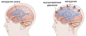
A description is given of the forms of hydrocephalus that can be incidental findings on MRI/CT of the brain - primary hydrocephalus and conditions with which a differential diagnosis must be made. Information about benign intracranial hypertension (BIH), normotensive and replacement hydrocephalus is presented to the extent that is necessary for a neurologist in outpatient practice to make decisions and explain to the patient the essence of his condition. The structure of the document reflects the tasks facing it. You can get acquainted with the question in more detail using the links in the relevant sections of the text. The clinical picture of intracranial hypertension consists of general cerebral and focal symptoms. Intracranial hypertension is characterized by the absence of pathognomonic disorders. In a typical clinical picture, a triad stands out: headache, vomiting, congestion in the fundus. Increased intracranial pressure is often associated with hydrocephalus, a disorder involving the accumulation of excess cerebrospinal fluid (CSF) in the cerebral ventricles and/or subarachnoid spaces, due to an imbalance between secretion and absorption, which is accompanied by dilation of the ventricles and/or subarachnoid spaces. The set of disorders in patients may be similar to the clinical picture of other lesions of the nervous system: - disseminated demyelinating focal lesions of the brain substance and cranial nerves, - focal lesions of the brain stem with involvement of the medial longitudinal fasciculus and the development of disorders of consciousness - space-occupying formations of the brain with general cerebral changes, focal symptoms and symptoms of damage to the cranial nerves, - conditions with diffuse brain damage - encephalopathy. — neuroinfections (congenital syphilis, cytomegalovirus infection, mumps, etc.) — thrombosis of the sinuses and veins of the brain. Asymptomatic cases of intracranial hypertension (ICH) are accompanied by nonspecific complaints - dizziness, fatigue. The variety of variants of the clinical picture, both in severity and in the set of symptoms, is the reason for the overdiagnosis of hypertensive-hydrocephalic syndrome, both in adult and pediatric neurological practice. An additional harmful effect is the high rate of false-positive data on dilation of the ventricular system when using echoencephaloscopy. Doctors can claim that a patient has ICH if rheoencephalography, duplex scanning of the brachiocephalic arteries, or transcranial Doppler sonography reveals venous drainage from the cranial cavity.
Imaging signs of hydrocephalus with increased ICP include: An increase in the size of the inferior horns of the lateral ventricles by more than 2 cm with the absence of visualization of the subarachnoid spaces of the convexital areas, interhemispheric and lateral fissures of the brain; Balloon-shaped dilatation of the anterior horns of the lateral ventricles (Mickey Mouse symptom) and the third ventricle; Periventricular decrease in tissue density seen on CT or increased T2 signal seen on magnetic resonance imaging (MRI) as a result of transependymal infiltration or migration of cerebrospinal fluid. In addition to structural changes, the clinical manifestations of hydrocephalus are taken into account. The last clarification is necessary, since taking into account only structural changes does not allow us to classify disorders of liquorodynamics without changing the configuration of the ventricles as hydrocephalus, for example, benign intracranial hypertension (BIH).
NORMOTENSIVE HYDROCEPHALUS
Normal pressure hydrocephalus (NPH) is associated with impaired CSF absorption, and dilatation of the cerebral ventricles develops in the presence of normal intracranial pressure.
EPIDEMIOLOGY
The prevalence of NG is 1-2 cases per 1,000,000 people. In the practice of a neurologist specializing in patients with extrapyramidal diseases, as a rule, no more than a dozen patients are recorded per year. This type of hydrocephalus is more common in older people. The condition should be excluded in persons over 60 years of age with a combination of cognitive impairment (dementia), pelvic organ dysfunction (usually urinary incontinence) and walking impairment (lower body parkinsonism) - the Hakim-Adams triad. Due to the fact that in a significant proportion of cases, bypass surgery in the early stages leads to improvement in walking, it is important to suspect and confirm this condition in a timely manner.
CLINIC
The structure of cognitive impairment is dominated by frontal-subcortical disorders: decreased activity, apathy, and behavioral disorders. Walking disorders have been described as magnetic gait, gait apraxia, and frontal ataxia. Patients experience the greatest difficulty when starting to walk. The area of support is increased, the length and height of the step is reduced, the smoothness of movements is impaired, and there is a progressive slowdown in walking with each step. Patients always have postural instability; often, upon questioning, you can find out that there have been falls before.
DIFFERENTIAL DIAGNOSIS
However, other diseases with cognitive and motor impairments may have a similar pattern of impairments. When carrying out differential diagnosis, the following are considered: vascular dementia (discirculatory encephalopathy stage III), Parkinson's disease with dementia and dementia with Lewy bodies, Alzheimer's disease. The main diagnostic method is MRI of the brain. The examination reveals the expansion of the lateral ventricles, the rounded shape of their anterior horns, and the smoothness of the relief of the cerebral cortex. It is important to use the Evans ventricular-hemispheric index, which is the ratio of the distance between the most distant points of the anterior horns of the lateral ventricles to the largest internal diameter of the skull. Ventriculomegaly is diagnosed if the index exceeds 0.31. NG is characterized by changes in the periventricular white matter, similar to leukoaraiosis (see above). Their severity correlates with the degree of cognitive impairment].
Figure 2 MRI signs of cerebral atrophy (A) in Alzheimer's disease and (B) normal pressure hydrocephalus
At first glance, the images are quite similar, but the image on the right shows the rounded shape of the horns of the lateral ventricles and the smoothness of the sulci of the cerebral hemispheres.
Figure 3 Gliotic and atrophic changes after traumatic brain injury, replacement hydrocephalus
Causes of intracranial pressure - description, factors
The main etiology is a violation of the outflow of fluid into the brain. The condition worsens if the brain
- blood from the veins arrived in a larger volume;
- a lot of spinal substance got in.
Intracranial compression exists in the form of:
1. Hypertension, when the pressure rises to 20 or 25 mmHg and fluid enters the brain in the walls of the skull:
- spinal;
- tissue due to brain tumors;
- blood with stagnation in the veins;
- foreign during the period of brain tumors.
The reasons that cause increased intracranial pressure in the patient are: situations of traumatic injury (TBI), tumors, effusion of blood inside the skull, meningoencephalitis (the brain is affected), hydrocephalus (“dropsy” due to cerebrospinal fluid), excess weight, excess fluid, as well as others factors when cerebrospinal fluid increases.
An increased degree of pressure on the brain (over 40 mmHg) threatens human life.
If pressure on the brain increases in adults, it is necessary to undergo examination and rule out a malignant tumor.
2. Intracranial hypotension, when the pressure on the brain, on the contrary, is reduced.
In this case, the prerequisites are:
- the state of destruction of the bones of the skull, membranous membranes, which are caused by injuries, combined with an outpouring of cerebrospinal fluid;
- taking dehydrating diuretic medications in larger quantities than necessary;
- loss of cerebrospinal fluid due to lumbar puncture or external drainage of the lateral ventricles of the brain.
REPLACEMENT HYDROCEPHALUS
The terms “replacement hydrocephalus” and ex-vacuo hydrocephalus denote expansion of the ventricles of the brain of a secondary nature, due to brain atrophy - a decrease in the volume of brain tissue. Replacement hydrocephalus is not accompanied by disturbances in cerebrospinal fluid dynamics, including increased intracranial pressure. Therefore, the relationship of this condition to hydrocephalus is disputed. Replacement hydrocephalus is more often encountered in elderly patients with chronic vascular and/or toxic damage to the brain, consequences of traumatic brain injury. In the last two cases, a selective decrease in the volume of individual brain regions is possible: atrophy of the cerebellum and frontal lobes, atrophy of brain regions in the projection of the bruise. Replacement hydrocephalus does not have a clear correlation with neurological disorders and does not require special treatment.
Against the background of terminological disagreements on the issue, there are several classifications that do not contradict each other:
- communicating and non-communicating;
- obstructive and aresorptive;
- congenital and acquired;
- genetic or associated with malformations of the central nervous system;
- isolated intraventricular-obstructive and extraventricular simple and complicated.
The term “compensated hydrocephalus” describes conditions without progressive enlargement of the ventricles of the brain; otherwise, uncompensated hydrocephalus is diagnosed. “External hydrocephalus” or “benign enlargement of the subarachnoid spaces” is an excessive accumulation of fluid, a condition quite often associated with familial macrocephaly (the size of the skull is larger than the size of the brain, while there are no disturbances in cerebrospinal fluid dynamics, including increased CSF pressure).
- The term "hydrocephalus" is used to refer to a group of conditions that are distinguishable from each other by CT or MRI of the brain.
- Judging hydrocephalus without CT or MRI data may be erroneous.
- In children, hydrocephalus is usually associated with increased intracranial pressure (ICP). In most cases, it is caused by excess production of CSF, which accumulates in the ventricles of the brain due to disturbances in its circulation (obstructive or non-communicating hydrocephalus). Less commonly, CSF accumulates due to malabsorption (communicating hydrocephalus).
- In adults, unlike children, forms of hydrocephalus without increased ICP are much more common.
- Since hydrocephalus can not only be an isolated condition, but also accompanies certain neurological diseases, the exact prevalence of the syndrome is not known.
- To date, no unified classification has been created that covers the causes of hydrocephalus in patients of different ages.
- Due to the high “stigmatizing” significance of the diagnosis of “hydrocephalus” for patients, before announcing the final diagnosis and prescribing treatment, it is necessary to conduct all the studies necessary to establish the nosological affiliation of the changes identified in the brain.
- The patient must have complete and adequate information about the disease, which is most important in relation to benign and treatable forms.
Osteopathy as an auxiliary method in the treatment of intracranial hypertension
In most cases, the condition can be alleviated by treatment with an osteopath, who, using manual techniques, returns the vertebrae to their natural position and relieves bone tension, and with it the symptoms. As a result, blood flow in the vessels of the brain is restored, stagnation of cerebrospinal fluid in the skull is eliminated and tension in the skull is relieved.
The use of techniques acts similarly to drugs with a diuretic effect, excess cerebrospinal fluid is eliminated naturally and intracranial pressure, its symptoms are normalized.
The duration of treatment and the duration of sessions are set by the doctor depending on the patient’s condition and related factors. As a rule, symptoms disappear or become significantly weaker after just a few treatments.
The combination of osteopathic therapy with other treatment methods can achieve stable remission and significantly improve the quality of life.
Indications and contraindications for osteopathic therapy
Here is just a small list of body ailments for which a specialist appointment is indicated:
- various injuries;
- diseases of the musculoskeletal system;
- dysfunction of childbirth;
- pain of various etiologies;
- pain in internal organs;
- states of the nervous system;
- curvature of posture;
- intracranial vascular disorder;
- increased intracranial hypertension;
- pregnancy planning.
Contraindications for manipulation:
- strokes, heart attacks;
- bleeding in the brain;
- cardiac, renal, hepatic, respiratory failure during decompensation;
- infections in the acute stage;
- open form of tuberculosis;
- untreated hematomas;
- mental illness;
- blood diseases;
- oncology, tumors.
The doctor will not be able to immediately cure high blood pressure and pain in adults and children, but after an acute period of the disease, during the rehabilitation stage, he will help to recover faster. In our Center, every visitor is treated with close attention and care. An individual approach and an atmosphere of trust between doctor and patient are proof of successful healing.
Treatment and prognosis
Therapeutic measures are prescribed based on diagnostic results. First of all, it is important to get rid of the cause that provokes excessive fluid pressure on the skull. If this phenomenon is caused by traumatic brain injury, the patient is advised to rest completely, take gentle nutrition, and take anti-inflammatory drugs. In some cases, surgical intervention is necessary (in the presence of hematomas between the membranes of the brain, as well as in the presence of injuries that require surgical treatment). Surgery is also prescribed when various tumors are detected in brain tissue that is prone to rapid growth.
A separate set of measures is carried out when diagnosing hydrofecal in a child. To remove excess fluid, a shunt is installed, through which it flows into the abdominal cavity, and the pressure is normalized. The operation is repeated as the child grows, and the patient is constantly monitored. In some children, the need for artificial fluid removal gradually disappears.
Drug therapy for ICP pathologies is secondary. However, drugs are prescribed to eliminate symptoms and make the patient feel better. The following medications may be helpful:
- hormonal anti-inflammatory drugs;
- neuroprotectors and substances for stimulating blood circulation in the brain - the effectiveness of this group has not been proven, despite its widespread use;
- loop diuretics (diuretics) - medications that stimulate the excretion of excess fluid;
- osmodiuretics - including reducing the production of cerebrospinal fluid.
The Clinical Brain Institute specializes in the diagnosis and treatment of diseases of the central nervous system. Increased intracranial pressure is not a separate disease, but a symptom that indicates a number of pathologies. Experienced specialists will accurately determine the cause of such a violation, as well as its severity and possible consequences. It is worth understanding that only timely seeking medical help can guarantee successful treatment and a return to your normal lifestyle without headaches.
Symptoms of increased intracranial pressure
The symptoms of increased blood pressure are so vivid and strong that they affect the entire life and activity of the individual:
- pressing pain spreading to all areas;
- pain that occurs in the morning and intensifies throughout the day, not relieved by conventional analgesic medications;
- signs of sleep disturbance;
- nausea, vomiting;
- slowdown in heart rate;
- sudden deterioration of vision;
- lack of ability to focus on small objects;
- decreased memory and attention.
If the headache occurs due to a significant pathology, the patient should maintain bed rest. Otherwise, dizziness, lapses of consciousness, and memory loss begin.
With reduced ICP, symptoms are observed:
- constant mood swings, lethargy, indifference, asthenia, dissatisfaction, fatigue;
- vertigo, sometimes fainting after minor sports activities;
- headache;
- with a general lack of fluid, blood pressure simultaneously decreases;
- decreased ability to see, cloudy spots in the eyes;
- lightheadedness, abdominal pain;
- pain in the heart area.